
Abstract
BACKGROUND AND PURPOSE: Selective venous sampling from the posterior portion of the cavernous sinus (CS) is recommended for the diagnosis of Cushing disease, because samples from the posterior portion yield higher adrenocorticotropic hormone (ACTH) levels than those from the anterior and middle portions. We prospectively assessed this intracavernous gradient of ACTH level to determine which site in the CS yields adequate sampling.
MATERIALS AND METHODS: In 5 patients with Cushing syndrome, cavernous sinography was performed to assess drainage pattern of the CS. Sampling was performed from the anterior, middle, and posterior parts of the CS, inferior petrosal sinus (IPS), and the peripheral vein. The ratio of the concentration in CS and IPS to that in peripheral blood plasma (C/P ratio) was calculated.
RESULTS: Cavernous sinography showed that the main drainage route was the IPS in 6 sides and that the pterygoid plexus (PP) was developed to the same extent as the IPS in 3 sides. In 1 patient, the CS drained mainly to the PP. In 1 patient with an ectopic lesion, no increase in ACTH level was detected. In 3 of 4 patients with Cushing disease, the highest C/P ratio was obtained from the posterior portion. In 1 patient whose main drainage route was the PP, the highest C/P ratio was obtained from the anterior portion. In this case, sampling data from the posterior portion and the IPS yielded false-negative results.
CONCLUSION: Understanding the drainage patterns of the CS is essential for interpretation of sampling data from the CS and avoiding false-negative results.
Measurement of adrenocorticotropic hormone (ACTH) levels in plasma from the inferior petrosal sinuses (IPSs) of patients with Cushing syndrome, with or without administration of corticotropin-releasing hormone (CRH), can distinguish adrenocorticotropin-secreting pituitary tumors (Cushing disease) from other causes of this syndrome.1 Although this is an invasive procedure and its rates of success and complications depend on operator skill and experience, it is the most direct way of demonstrating pituitary hypersecretion of ACTH and, thus, identifying patients who will benefit from pituitary surgery.2
In the treatment of patients with Cushing disease, accurate preoperative lateralization of microadenoma is particularly important, because it can lead to successful selective adenomectomy of ACTH-secreting tumors by transsphenoidal microsurgery with preservation of normal pituitary function. Bilateral simultaneous venous sampling from the IPS is a reliable means of diagnosing Cushing disease but is not reliable for lateralizing ACTH-secreting pituitary adenomas.3 Teramoto et al4 developed a method of selective venous sampling directly from the cavernous sinus (CS) as an alternative to sampling from the IPS.5 The advantage of this method is its higher rate of correct prediction of the cause of Cushing syndrome and lateralization of microadenomas than sampling from the IPS. Teramoto et al5 also reported that significant gradients of ACTH were detected among the anterior, middle, and posterior portions of the CS (intracavernous gradient).
We prospectively assessed this intracavernous gradient of ACTH level to determine which site in the CS yields adequate sampling. We discuss the importance of understanding the drainage pattern of the CS in interpretation of sampling data from the CS and IPS.
Materials and Methods
We prospectively studied 5 patients with Cushing syndrome to evaluate the diagnostic efficacy of multispot sampling in the bilateral CSs between 2001 and 2006. Informed consent was obtained from all of the patients before the procedure. All of the patients were women, whose ages ranged from 41 to 66 years. In 3 of the 5 patients, MR imaging study detected microadenomas, and a macroadenoma with suprasellar and lateral extension compressing the right CS was detected in 1 patient. One patient with microadenoma had previously undergone unsuccessful pituitary surgery.
In 4 of the 5 patients, transsphenoidal surgery was performed. In each patient, the pituitary gland was carefully inspected, and the location of the adenoma was recorded. All of the resected adenomas stained positively for ACTH. Postoperatively, ACTH level became normal in 1 of the 4 patients, and 2 patients became hypocortisolemic with remission of symptoms. In a patient with macroadenoma, though ACTH level remained high, medical conditions improved with remission of symptoms.
The patients’ plasma cortisol levels were high (18.1–59.1 μg/dL; mean, 39.4 μg/dL; normal range, 4–23.3μg/dL) and accompanied by elevated ACTH levels (19–828 pg/mL; mean, 260 pg/mL; normal range, 7–56 pg/mL). In all 5 of the patients, low-dose dexamethasone (2 mg/d) failed to suppress free urinary cortisol to less than 50% of the baseline value. A high dose (8 mg/d) of dexamethasone could suppress these levels in 4 patients.
Bilateral catheterization of the CS was accomplished through a percutaneous femoral-vein approach. All of the patients received systemic heparinization (50–60 U/kg). A microcatheter (Rapid Transit microcatheter; Cordis Neurovascular, Miama Lakes, Fla) was introduced through a guiding catheter (4F) placed in the internal jugular vein, and its tip was positioned at the orifice of the IPS. The tip of the microcatheter was then advanced into the anterior part of the CS with the aid of fluoroscopy, and anteroposterior and lateral venous angiograms (cavernous sinography) were obtained to assess the drainage pattern of the CS. Venous sampling from the CS was performed from the anterior, middle, and posterior parts of the CS bilaterally in order. Sampling from the bilateral IPS and a peripheral vein were also performed. ACTH levels in the samples were used to calculate the ratio of the concentration in plasma from the CS and IPS to the concentration in peripheral-blood plasma (C/P ratio).
Results
Cavernous sinography showed that the main drainage route was the IPS in 6 sides of 4 patients (Table 1). Figure 1 shows sinography in case 5, demonstrating that the main drainage route was the IPS bilaterally. In 3 sides of 2 patients, the pterygoid plexus was developed to the same extent as the IPS (Fig 2). In 1 patient (case 4), the right CS mainly drained to the pterygoid plexus (Fig 3).
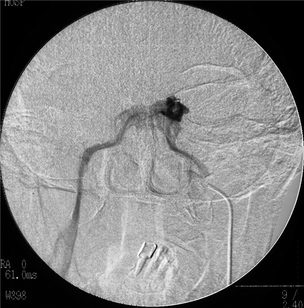
Anteroposterior view of cavernous sinography in case 5, demonstrating that the main drainage route was the IPS bilaterally.

Lateral view of right cavernous sinography in case 1, showing that the pterygoid plexus was developed to the same extent as the IPS. Note that the opacification of the pterygoid plexus is more than the one of the IPS, but this may be due to the rate and the force of injection of contrast material.

Anteroposterior view of right cavernous sinography in case 4, showing the tip of the microcatheter (arrow) in the CS (right) and that the main drainage route is via the pterygoid plexus (left). Note that the microcatheter is introduced through the IPS into the CS and that contrast material moves from the CS into the pterygoid plexus.
Intracavernous and petrosal sinus gradient of ACTH levels
Multispot sampling was successfully accomplished in 4 patients, with no major morbidity. In a patient with macroadenoma with suprasellar and lateral extension compressing the right CS (case 5), the microcatheter could not be advanced into the anterior and middle parts of the right CS, and venous sampling from the right CS was performed from the posterior part of the CS.
ACTH levels in blood samples from the anterior part of the CS ranged from 14 to 6960 pg/mL. Levels in the middle and posterior parts of the CS and the IPS ranged from 20 to 20,100 pg/mL, 18 to 56,100 pg/mL, and 15 to 1880 pg/mL, respectively.
The C/P ratios of ACTH in the anterior, middle, and posterior CS sampling points and the IPS are shown in Table 1. The intracavernous gradients ranged from 0.8 to 67.8 and from 0.8 to 7.0 in the IPS. In a patient with an ectopic lesion (case 2), no increase in ACTH level was detected in either sampling point (0.9–1.0). In the case of Cushing disease, the highest value in each patient exceeded 2. The highest C/P ratio of ACTH values in samples taken from the CS was obtained from the anterior portion in a patient whose main drainage route from the CS was the pterygoid plexus (case 4). In the other 3 patients, the highest C/P ratio of ACTH value in samples taken from the CS was obtained from the posterior portion.
The highest C/P ratio of ACTH level in samples taken from the IPS exceeded 2 in 3 of 4 patients. In 1 patient with a developed pterygoid plexus (case 4), the ratio was below 1, yielding a false-negative result.
The intercavernous and intrapetrosal gradients of ACTH in 4 patients with pituitary adenoma are shown Table 2. The intercavernous gradients ranged from 1.3 to 37.4. The intrapetrosal gradients ranged from 1.1 to 6.5. The lateralization of an adenoma was confirmed at the transsphenoidal surgery and was correctly predicted in all of the patients. The intercavernous and intrapetrosal gradients indicated false laterality in none of these cases.
Intercavernous and interpetrosal gradients of ACTH levels in 4 patients with pituitary adenoma
Discussion
Bilateral sampling of the IPS, performed without and with CRH stimulation, has been used to establish the cause of Cushing syndrome and to predict the side of the pituitary gland containing microadenoma in patients with Cushing disease.1,6–13 Sampling from the IPS is, in trained hands, a safe procedure and is the method with highest diagnostic sensitivity in confirming pituitary ACTH hypersecretion.7 An IPS gradient greater than 1.4:1 correctly indicates the side of the pituitary gland that contains microadenoma in approximately 85% of patients. This should improve the results of surgery with only patients with Cushing disease subjected to transsphenoidal exploration and by permitting surgeons to find smaller tumors or to remove the portion of the gland containing microadenoma if no tumor can be identified at surgery.11
The diagnostic accuracy of sampling from the IPS ranges between 90% and 100%.3 The main reason for sampling from the IPS to demonstrate pituitary hypersecretion is that ACTH can be expected to be found at higher levels close to the pituitary gland than in the peripheral veins, where significant admixture has already occurred.14 However, because ACTH secretion may be intermittent, there is a risk of sampling in the interval between 2 secretory phases and, thus, obtaining a false-negative IPS/peripheral blood plasma (IPS/P) ratio.14–16
CRF administration during IPS sampling should reduce the incidence of false-negative result in patients with Cushing disease.7,11 The safety and consistency of simultaneous bilateral sampling of the IPS with CRH stimulation and its high diagnostic accuracy have been reported.1 Threshold criteria for a pituitary source have been defined as an IPS/P basal ratio of 2:1 or greater without CRH administration or an IPS/P ratio of 3:1 or greater after CRH stimulation.1,15 Using this criterion, however, false-negative results were found in 5%–13% of cases in previous reports.7,12,13,15,17–22
As an alternative to sampling from the IPS, selective venous sampling directly from the CS has been developed.4,5 The advantage of this method is its higher rates of correct prediction of the cause of Cushing syndrome and lateralization of microadenomas compared with those yielded by sampling from the IPS. Teramoto et al5 reported that sampling should be performed from the posterior portion of the CS, because samples from the posterior portion of the CS yielded higher ACTH readings than did those from the anterior and middle portions. Kai et al23 also reported that multiple-site sampling of ACTH from the CS was valuable for lateralizing the adenoma in patients with Cushing disease. Some consistent patterns of distribution of the C/P ratio at multiple sampling points have been noted: the highest C/P ratios were obtained in the middle and posterior CS and the IPS, and no patients had the highest C/P ratio recorded in the anterior CS.23
In our multispot sampling of ACTH, in 3 of 4 patients with Cushing disease, intracavernous gradient of ACTH was assessed, and in 1 of these 3 cases, a paradoxical intracavernous gradient pattern of ACTH was observed: sampling from the anterior portion of the CS yielded a higher ACTH reading than those from the middle and posterior portions. The CS receives blood flow from the superior and inferior ophthalmic veins and the sphenoparietal sinus, anteriorly. Posteriorly, the CS drains into the petrosal sinuses and then into the internal jugular vein.24 In the patient exhibiting the paradoxical ACTH gradient pattern, the CS had a well-developed anastomosis with the pterygoid plexus by way of the sphenoid emissary veins via the foramen of Vesalius, foramen ovale, and foramen lacerum,24 and this route was the main drainage route from the CS. In this case, sampling data from the posterior portion of the CS and the IPS yielded false-negative results. This finding indicates that it is important to consider the venous drainage pattern in obtaining correct results of multispot sampling of ACTH.
A limitation of the present study was the small number of patients who underwent the multispot sampling in the CS and cavernous sinography. Because the cavernous sinography was obtained from injection through the microcatheter, there might be incomplete opacification of the CS, and this could lead to an erroneous depiction of the CS drainage. In conclusion, understanding of the drainage patterns of the CS in each patient with cavernous sinography is essential for interpretation of sampling data from the CS and avoiding false-negative results.
References
- Received March 6, 2007.
- Accepted after revision May 2, 2007.
- Copyright © American Society of Neuroradiology
In this issue


Jump to section
Related Articles
Cited By...
- No citing articles found.