Advertisement

Main menu

  • Home
  • Content
    • Current Issue
    • Accepted Manuscripts
    • Article Preview
    • Past Issue Archive
    • Video Articles
    • AJNR Case Collection
    • Case of the Week Archive
    • Case of the Month Archive
    • Classic Case Archive
  • Special Collections
    • AJNR Awards
    • ASNR Foundation Special Collection
    • Most Impactful AJNR Articles
    • Photon-Counting CT
    • Spinal CSF Leak Articles (Jan 2020-June 2024)
  • Multimedia
    • AJNR Podcasts
    • AJNR SCANtastic
    • Trainee Corner
    • MRI Safety Corner
    • Imaging Protocols
  • For Authors
    • Submit a Manuscript
    • Submit a Video Article
    • Submit an eLetter to the Editor/Response
    • Manuscript Submission Guidelines
    • Statistical Tips
    • Fast Publishing of Accepted Manuscripts
    • Graphical Abstract Preparation
    • Imaging Protocol Submission
    • Author Policies
  • About Us
    • About AJNR
    • Editorial Board
    • Editorial Board Alumni
  • More
    • Become a Reviewer/Academy of Reviewers
    • Subscribers
    • Permissions
    • Alerts
    • Feedback
    • Advertisers
    • ASNR Home

User menu

  • Alerts
  • Log in

Search

  • Advanced search
American Journal of Neuroradiology

American Journal of Neuroradiology

ASHNR American Society of Functional Neuroradiology ASHNR American Society of Pediatric Neuroradiology ASSR
  • Alerts
  • Log in

Advanced Search

  • Home
  • Content
    • Current Issue
    • Accepted Manuscripts
    • Article Preview
    • Past Issue Archive
    • Video Articles
    • AJNR Case Collection
    • Case of the Week Archive
    • Case of the Month Archive
    • Classic Case Archive
  • Special Collections
    • AJNR Awards
    • ASNR Foundation Special Collection
    • Most Impactful AJNR Articles
    • Photon-Counting CT
    • Spinal CSF Leak Articles (Jan 2020-June 2024)
  • Multimedia
    • AJNR Podcasts
    • AJNR SCANtastic
    • Trainee Corner
    • MRI Safety Corner
    • Imaging Protocols
  • For Authors
    • Submit a Manuscript
    • Submit a Video Article
    • Submit an eLetter to the Editor/Response
    • Manuscript Submission Guidelines
    • Statistical Tips
    • Fast Publishing of Accepted Manuscripts
    • Graphical Abstract Preparation
    • Imaging Protocol Submission
    • Author Policies
  • About Us
    • About AJNR
    • Editorial Board
    • Editorial Board Alumni
  • More
    • Become a Reviewer/Academy of Reviewers
    • Subscribers
    • Permissions
    • Alerts
    • Feedback
    • Advertisers
    • ASNR Home
  • Follow AJNR on Twitter
  • Visit AJNR on Facebook
  • Follow AJNR on Instagram
  • Join AJNR on LinkedIn
  • RSS Feeds

AJNR is seeking candidates for the AJNR Podcast Editor. Read the position description.

OtherINTERVENTIONAL

De Novo Cerebral Arteriovenous Malformation: Case Report and Literature Review

J. Stevens, J.L. Leach, T. Abruzzo and B.V. Jones
American Journal of Neuroradiology January 2009, 30 (1) 111-112; DOI: https://doi.org/10.3174/ajnr.A1255
J. Stevens
  • Find this author on Google Scholar
  • Find this author on PubMed
  • Search for this author on this site
J.L. Leach
  • Find this author on Google Scholar
  • Find this author on PubMed
  • Search for this author on this site
T. Abruzzo
  • Find this author on Google Scholar
  • Find this author on PubMed
  • Search for this author on this site
B.V. Jones
  • Find this author on Google Scholar
  • Find this author on PubMed
  • Search for this author on this site
  • Article
  • Figures & Data
  • Info & Metrics
  • Responses
  • References
  • PDF
Loading

Abstract

SUMMARY: We describe a rare case of a de novo cerebral arteriovenous malformation (AVM) in a 9-year-old girl. MR imaging at 6 years of age demonstrated band heterotopia. Follow-up MR imaging 3 years later demonstrated a new 3.5-cm AVM in the left parietol-occipital region, confirmed by conventional angiography. This report, along with limited previous reports, suggests that AVMs can be acquired lesions and that AVM development is a dynamic process extending into the postnatal period.

An arteriovenous malformation (AVM) is a vascular malformation characterized by arterio-venous shunt through a collection of tortuous vessels without an intervening capillary bed.1 The pathogenesis of AVMs is currently unknown. Although they are traditionally thought to be congenital in origin, with an initiating event occurring in early embryologic development, some have suggested a more dynamic developmental process.2,3 We present a case of de novo AVM development in a region of the brain without abnormal vascularity on prior MR imaging and briefly review the literature on de novo AVM development and pathogenesis.

Case Report

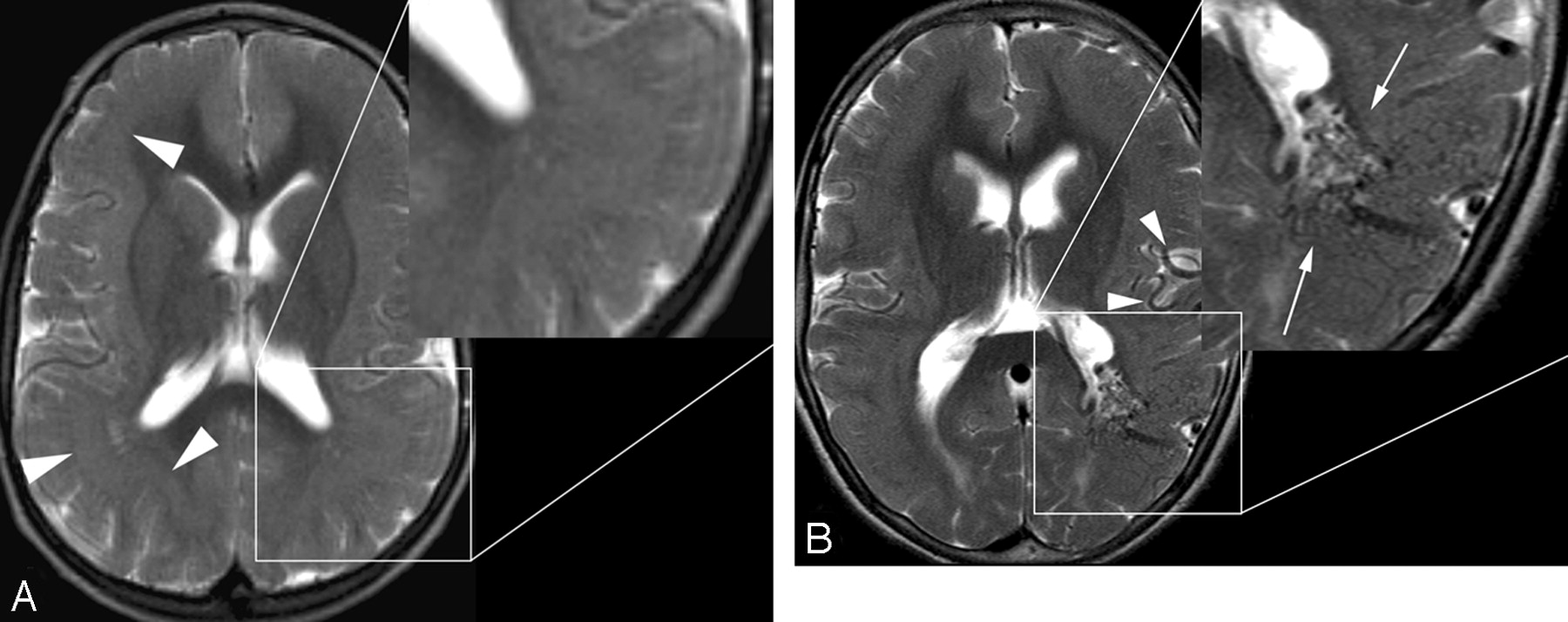
A 6-year-old girl was evaluated with MR imaging because of seizures and developmental delay. Imaging demonstrated continuous band heterotopia (Fig 1A). No vascular abnormality or abnormal signal intensity was demonstrated.

Fig 1.
  • Download figure
  • Open in new tab
  • Download powerpoint
Fig 1.

A, Axial T2-weighted MR imaging at 6 years of age demonstrates diffuse band heterotopia (arrowheads). Note the absence of enlarged vessels in the magnified segment. B, Axial T2-weighted image at the same level 3 years later demonstrates a new serpiginous grouping of signal-intensity voids in the left parieto-occipital region along the margin of the left lateral ventricle consistent with an AVM (arrow). Enlarged left middle cerebral artery feeding vessels are also noted (arrowheads).

Three years later the patient presented with increased seizure activity, behavioral changes, increased ataxia, and aphasia after a minor head injury. CT demonstrated no evidence of a skull fracture or hemorrhage. MR imaging (Fig 1B) revealed a tangle of serpiginous flow voids along the margin of the left lateral ventricle at the junction of the left temporal and occipital lobes, consistent with an AVM. Cerebral conventional angiography (Fig 2) delineated a 3.5-cm AVM nidus with predominant arterial supply from distal branches of the left middle and posterior cerebral arteries. Venous drainage was through the ipsilateral thalamostriate veins and also through parenchymal veins extending into the left transverse sinus. The patient is currently undergoing radiation therapy for this lesion.

Fig 2.
  • Download figure
  • Open in new tab
  • Download powerpoint
Fig 2.

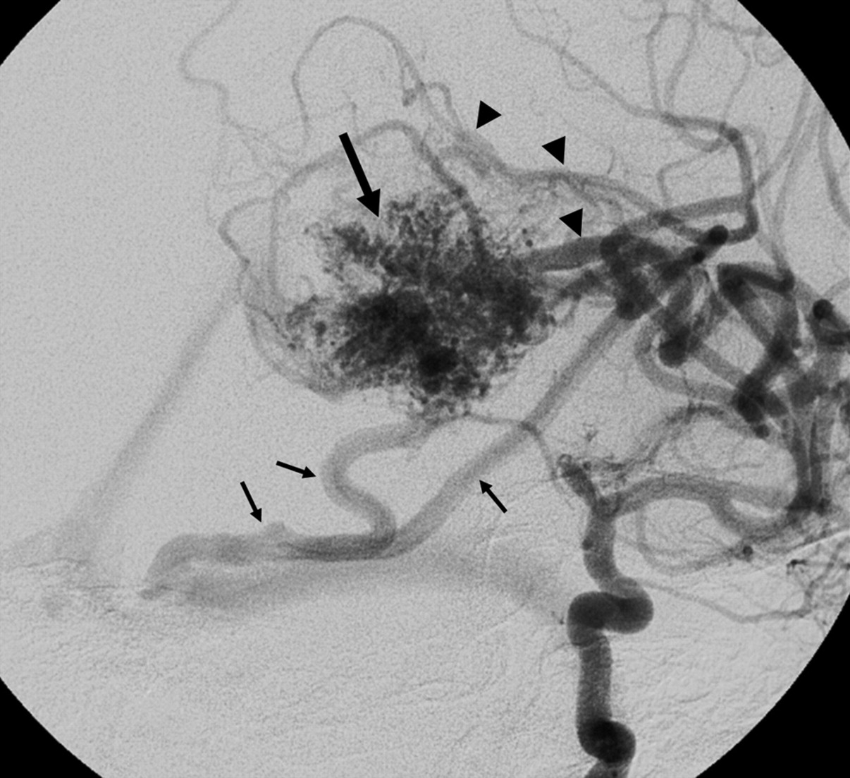
Oblique projection of a left internal carotid angiogram shows a 3.5-cm left parieto- occipital AVM nidus (large arrow) supplied by parietal middle cerebral artery branches (arrowheads), with venous drainage primarily to the medial transverse sinus near the torcular (small arrows). Additional deep venous drainage is not as well seen on this projection.

Discussion

We present a rare example of de novo AVM formation in a child with abnormalities of neuronal migration. A literature review identified 5 previously reported cases of de novo AVM development. Three of these cases are of patients with pre-existing vascular pathology who developed AVMs in or near pathologically altered brain.4–6 Only 2 previous reports7,8 document a cerebral AVM arising in brain parenchyma shown to be without vascular pathology on prior MR imaging. To our knowledge, no previously reported case has documented a de novo cerebral AVM developing in a patient with a neuronal migrational disorder.

The pathogenesis of AVMs is not completely understood and has largely been theorized. It has been suggested that cerebral AVMs are primarily congenital, originating at or before the 40- to 80-mm embryo length stage and may be related to a primary abnormality of primordial capillary or venous formation.9 However, excluding Galenic fistulas, we have found few reports of confirmed cerebral AVMs diagnosed in utero,10 and they are much more commonly diagnosed in adults than in children, challenging the assumption that all AVMs are congenital. Other concepts of cerebral AVM pathogenesis focus on the potential temporal vulnerability of susceptible vascular cellular elements to a physiologic trigger. This trigger could be mechanical, hormonal, thrombotic, hemodynamic, thermal, ischemic/hypoxemic, or inflammatory in nature.11,12

Vascular endothelial growth factor (VEGF) may be 1 link between insult and AVM formation. One study demonstrated significantly higher levels of VEGF in patients with AVMs compared with a control group.13 Patients with hereditary hemorrhagic telangiectasia (HHT) have an increased prevalence of AVMs and also have elevated levels of VEGF.14,15 Hypoxia is known to stimulate expression of VEGF. Hemorrhage and thrombogenesis can result in the release of VEGF from platelets. Infection can lead to the release of VEGF at the source of infection. Thus, in theory, insults such as stroke, brain contusion, and encephalitis could all potentially promote the development of AVMs.16

Cerebral AVMs are not static lesions: Growth and shrinkage of AVMs with time have been documented.17,18 Several cases of AVM recurrence in the same location following some form of intervention (ie, gamma knife, excision, embolization, etc) have been reported in pediatric patients and adults.19,20

Abnormalities of arterial21 venous,22 and microvascular23 structures have been described in relation to areas of abnormal neuronal migration. Abnormal vascular structures have been described in association with periventricular nodular heterotopia in filamin 1 gene mutation,23 suggesting 1 potential link between neuronal migrational disorders and vascular malformations.

Conclusions

This case report demonstrates postnatal development of an arteriovenous malformation in a patient with extensive band heterotopia. This case and the few previous reports of de novo AVM challenge the concept that cerebral AVMs are purely congenital lesions. AVMs appear to be dynamic lesions that have the potential to develop and grow with time. It is possible that acquired insults can provide the catalyst for the formation of AVMs later in life, and as suggested by this case, disorders of neuronal migration may share common genetic ground with abnormalities of endothelial proliferation.

Footnotes

  • Paper previously presented at: Annual Meeting of the American Society of Neuroradiology, June 7–14, 2007; Chicago, Ill.

References

  1. ↵
    Fleetwood IG, Steinber GK. Arteriovenous malformations. Lancet 2002;359:863–73
    CrossRefPubMed
  2. ↵
    Stapf C, Mohr JP. New concepts in adult brain arteriovenous malformations. Curr Opin Neurol 2000;13:63–67
    CrossRefPubMed
  3. ↵
    Nussbaum ES, Heros RC, Madison MT, et al. The pathogenesis of arteriovenous malformations: insights provided by a case of multiple arteriovenous malformations developing in relation to a developmental venous anomaly. Neurosurgery 1998;43:347–51
    CrossRefPubMed
  4. ↵
    Schmit BP, Burrows PE, Kuban K, et al. Acquired cerebral arteriovenous malformation in a child with Moyamoya disease: case report. J Neurosurg 1996;84:677–80
    CrossRefPubMed
  5. Rodriquez-Arias C, Martinez R, Rey G, et al. Recurrence in a different location of a cerebral arteriovenous malformation in a child after radiosurgery. Childs Nerv Syst 2000;16:363–65
    PubMed
  6. ↵
    Song JK, Niimi Y, Kupersmith MJ, et al. Postnatal growth and development of a cerebral arteriovenous malformation on serial magnetic resonance imaging in a child with hemangiomatosis. J Neurosurg 2007;106 (suppl 5):384–87
    CrossRefPubMed
  7. ↵
    Gonzalez LF, Bristol RE, Porter RW, et al. De novo presentation of an arteriovenous malformation: case report. J Neurosurg 2005;102:726–29
    PubMed
  8. ↵
    Bulsara KR, Alesander MJ, Villavicencio AT, et al. De novo cerebral arteriovenous malformation: case report. Neurosurgery 2002;50:1137–41
    PubMed
  9. ↵
    Mullan S, Mojtahedi S, Johnson DL, et al. Embryological basis of some aspects of cerebral vascular fistulas and malformations. J Neurosurg 1996 :85 ;1–8
    PubMed
  10. ↵
    DeCesare B, Omojola MF, Fogarty EF, et al. Spontaneous thrombosis of congenital cerebral arteriovenous malformation complicated by subdural collection: in utero detection with disappearance in infancy. Br J Radiol 2006;79:e140–44
    Abstract/FREE Full Text
  11. ↵
    Lasjaunias PL. Vascular Diseases in Neonates, Infants, and Children: Interventional Neuroradiology Management. Berlin, Germany: Springer-Verlag; 1997 :51–65
  12. ↵
    Yasargil MG. Pathological considerations. In Yasargil MG, ed. Microneurosurgery: AVM of the Brain, History, Embryology, Pathological Considerations, Hemodynamics, Diagnostic Studies, Microsurgical Anatomy. New York: Thieme Verlag; 1987 :49–211
  13. ↵
    Sandalcioglu IE, Wende D, Eggert A, et al. Vascular endothelial growth factor plasma levels are significantly elevated in patients with cerebral arteriovenous malformations. Cerebrovasc Dis 2006;21:154–58
    CrossRefPubMed
  14. ↵
    Du R, Hashimoto T, Tihan T, et al. Growth and regression of arteriovenous malformations in a patient with hereditary hemorrhagic telangiectasia. J Neurosurg 2007;106:470–77
    CrossRefPubMed
  15. ↵
    Matsubara S, Manzia JL, ter Brugge K, et al. Angiographic and clinical characteristics of patients with cerebral arteriovenous malformations associated with hereditary hemorrhagic telangiectasia. AJNR Am J Neuroradiol 2000;21:1016–20
    Abstract/FREE Full Text
  16. ↵
    Sure U, Butz N, Schlegel J, et al. Endothelial proliferation, neoangiogenesis, and potential de novo generation of cerebrovascular malformations. J Neurosurg 2001;94:972–77
    PubMed
  17. ↵
    Ezura M, Kagawa S. Spontaneous disappearance of a huge cerebral arteriovenous malformation: case report. Neurosurgery 1992;30:595–99
    PubMed
  18. ↵
    Minakawa T, Tanaka R, Koike T, et al. Angiographic follow-up study of cerebral arteriovenous malformations with reference to their enlargement and regression. Neurosurgery 1989;24:68–74
    PubMed
  19. ↵
    Gabriel EM, Sampson JH, Wilkins RH. Recurrence of a cerebral arteriovenous malformation after surgical excision: case report. J Neurosurg 1996;84:879–82
    PubMed
  20. ↵
    Kader A, Goodrich JT, Sonstein WJ, et al. Recurrent cerebral arteriovenous malformations after negative postoperative angiograms. J Neurosurg 1996;85:14–18
    CrossRefPubMed
  21. ↵
    Abe T, Singer R, Marks M, et al. Arterial vascular abnormality accompanying cerebral cortical dysplasia. AJNR Am J Neuroradiol 1997;18:144–46
    Abstract
  22. ↵
    Barkovich AJ. Abnormal vascular drainage in anomalies of neuronal migration. AJNR Am J Neuroradiol 1988;9:939–42
    Abstract/FREE Full Text
  23. ↵
    Kakita A, Hayashi S, Moro F, et al. Bilateral periventricular nodular heterotopia due to filamin 1 gene mutation: widespread glomeruloid microvascular anomaly and dysplastic cytoarchitecture in the cerebral cortex. Acta Neuropathol 2002 :104 ;649–657. Epub 2002 Jul 23
    PubMed
  • Received May 13, 2008.
  • Accepted after revision June 16, 2008.
  • Copyright © American Society of Neuroradiology
View Abstract
PreviousNext
Back to top

In this issue

American Journal of Neuroradiology: 30 (1)
American Journal of Neuroradiology
Vol. 30, Issue 1
January 2009
  • Table of Contents
  • Index by author
Advertisement
Print
Download PDF
Email Article

Thank you for your interest in spreading the word on American Journal of Neuroradiology.

NOTE: We only request your email address so that the person you are recommending the page to knows that you wanted them to see it, and that it is not junk mail. We do not capture any email address.

Enter multiple addresses on separate lines or separate them with commas.
De Novo Cerebral Arteriovenous Malformation: Case Report and Literature Review
(Your Name) has sent you a message from American Journal of Neuroradiology
(Your Name) thought you would like to see the American Journal of Neuroradiology web site.
CAPTCHA
This question is for testing whether or not you are a human visitor and to prevent automated spam submissions.
Cite this article
J. Stevens, J.L. Leach, T. Abruzzo, B.V. Jones
De Novo Cerebral Arteriovenous Malformation: Case Report and Literature Review
American Journal of Neuroradiology Jan 2009, 30 (1) 111-112; DOI: 10.3174/ajnr.A1255

Citation Manager Formats

  • BibTeX
  • Bookends
  • EasyBib
  • EndNote (tagged)
  • EndNote 8 (xml)
  • Medlars
  • Mendeley
  • Papers
  • RefWorks Tagged
  • Ref Manager
  • RIS
  • Zotero
0 Responses
Respond to this article
Share
Bookmark this article
De Novo Cerebral Arteriovenous Malformation: Case Report and Literature Review
J. Stevens, J.L. Leach, T. Abruzzo, B.V. Jones
American Journal of Neuroradiology Jan 2009, 30 (1) 111-112; DOI: 10.3174/ajnr.A1255
del.icio.us logo Twitter logo Facebook logo Mendeley logo
  • Tweet Widget
  • Facebook Like
  • Google Plus One
Purchase

Jump to section

  • Article
    • Abstract
    • Case Report
    • Discussion
    • Conclusions
    • Footnotes
    • References
  • Figures & Data
  • Info & Metrics
  • Responses
  • References
  • PDF

Related Articles

  • No related articles found.
  • PubMed
  • Google Scholar

Cited By...

  • Neonatal giant pial arteriovenous malformation: genesis or rapid enlargement in the third trimester
  • Crossref (52)
  • Google Scholar

This article has been cited by the following articles in journals that are participating in Crossref Cited-by Linking.

  • Renal Arteriovenous Shunts: Clinical Features, Imaging Appearance, and Transcatheter Embolization Based on Angioarchitecture
    Miyuki Maruno, Hiro Kiyosue, Shuichi Tanoue, Norio Hongo, Shunro Matsumoto, Hiromu Mori, Yoshiko Sagara, Junji Kashiwagi
    RadioGraphics 2016 36 2
  • Are parenchymal AVMs congenital lesions?
    Saul F. Morales-Valero, Carlo Bortolotti, Carmelo Sturiale, Giuseppe Lanzino
    Neurosurgical Focus 2014 37 3
  • De novointracerebral arteriovenous malformations and a review of the theories of their formation
    A. Dalton, G. Dobson, M. Prasad, N. Mukerji
    British Journal of Neurosurgery 2018 32 3
  • Pathogenesis and radiobiology of brain arteriovenous malformations: implications for risk stratification in natural history and posttreatment course
    Achal S. Achrol, Raphael Guzman, Monika Varga, John R. Adler, Gary K. Steinberg, Steven D. Chang
    Neurosurgical Focus 2009 26 5
  • De novo arteriovenous malformations: case report and review of the literature
    Amit Mahajan, Tejas C. Manchandia, Grahame Gould, Ketan R. Bulsara
    Neurosurgical Review 2010 33 1
  • Capillary malformation–arteriovenous malformation: a clinical review of 45 patients
    Margarita Larralde, María Eugenia Abad, Paula Carolina Luna, Mariana Viktoria Hoffner
    International Journal of Dermatology 2014 53 4
  • De novo cerebral arteriovenous malformation in a child with previous cavernous malformation and developmental venous anomaly
    Hortensia Alvarez, Victor Perry, Michael Solle, Mauricio Castillo
    Journal of Neurosurgery: Pediatrics 2012 9 3
  • Pediatric de novo cerebral AVM: report of two cases and review of literature
    Jared J. Y. Yeo, Sharon Y. Y. Low, Wan Tew Seow, David C. Y. Low
    Child's Nervous System 2015 31 4
  • The putative role of the venous system in the genesis of vascular malformations
    Mariam S. Aboian, David J. Daniels, Stylianos K. Rammos, Eugenio Pozzati, Giuseppe Lanzino
    Neurosurgical Focus 2009 27 5
  • Arteriovenous Malformations: Congenital or Acquired Lesions?
    Anastasia Tasiou, Christos Tzerefos, Cargill H. Alleyne, Edoardo Boccardi, Bengt Karlsson, Neil Kitchen, Robert F. Spetzler, Christos M. Tolias, Kostas N. Fountas
    World Neurosurgery 2020 134

More in this TOC Section

  • SAVE vs. Solumbra Techniques for Thrombectomy
  • Contrast-Induced Encephalopathy after NeuroIR
  • CT Perfusion&Reperfusion in Acute Ischemic Stroke
Show more Interventional

Similar Articles

Advertisement

Indexed Content

  • Current Issue
  • Accepted Manuscripts
  • Article Preview
  • Past Issues
  • Editorials
  • Editor's Choice
  • Fellows' Journal Club
  • Letters to the Editor
  • Video Articles

Cases

  • Case Collection
  • Archive - Case of the Week
  • Archive - Case of the Month
  • Archive - Classic Case

Special Collections

  • AJNR Awards
  • ASNR Foundation Special Collection
  • Most Impactful AJNR Articles
  • Photon-Counting CT
  • Spinal CSF Leak Articles (Jan 2020-June 2024)

More from AJNR

  • Trainee Corner
  • Imaging Protocols
  • MRI Safety Corner

Multimedia

  • AJNR Podcasts
  • AJNR Scantastics

Resources

  • Turnaround Time
  • Submit a Manuscript
  • Submit a Video Article
  • Submit an eLetter to the Editor/Response
  • Manuscript Submission Guidelines
  • Statistical Tips
  • Fast Publishing of Accepted Manuscripts
  • Graphical Abstract Preparation
  • Imaging Protocol Submission
  • Evidence-Based Medicine Level Guide
  • Publishing Checklists
  • Author Policies
  • Become a Reviewer/Academy of Reviewers
  • News and Updates

About Us

  • About AJNR
  • Editorial Board
  • Editorial Board Alumni
  • Alerts
  • Permissions
  • Not an AJNR Subscriber? Join Now
  • Advertise with Us
  • Librarian Resources
  • Feedback
  • Terms and Conditions
  • AJNR Editorial Board Alumni

American Society of Neuroradiology

  • Not an ASNR Member? Join Now

© 2025 by the American Society of Neuroradiology All rights, including for text and data mining, AI training, and similar technologies, are reserved.
Print ISSN: 0195-6108 Online ISSN: 1936-959X

Powered by HighWire