Advertisement

Main menu

  • Home
  • Content
    • Current Issue
    • Accepted Manuscripts
    • Article Preview
    • Past Issue Archive
    • Video Articles
    • AJNR Case Collection
    • Case of the Week Archive
    • Case of the Month Archive
    • Classic Case Archive
  • Special Collections
    • AJNR Awards
    • ASNR Foundation Special Collection
    • Most Impactful AJNR Articles
    • Photon-Counting CT
    • Spinal CSF Leak Articles (Jan 2020-June 2024)
  • Multimedia
    • AJNR Podcasts
    • AJNR SCANtastic
    • Trainee Corner
    • MRI Safety Corner
    • Imaging Protocols
  • For Authors
    • Submit a Manuscript
    • Submit a Video Article
    • Submit an eLetter to the Editor/Response
    • Manuscript Submission Guidelines
    • Statistical Tips
    • Fast Publishing of Accepted Manuscripts
    • Graphical Abstract Preparation
    • Imaging Protocol Submission
    • Author Policies
  • About Us
    • About AJNR
    • Editorial Board
    • Editorial Board Alumni
  • More
    • Become a Reviewer/Academy of Reviewers
    • Subscribers
    • Permissions
    • Alerts
    • Feedback
    • Advertisers
    • ASNR Home

User menu

  • Alerts
  • Log in

Search

  • Advanced search
American Journal of Neuroradiology

American Journal of Neuroradiology

ASHNR American Society of Functional Neuroradiology ASHNR American Society of Pediatric Neuroradiology ASSR
  • Alerts
  • Log in

Advanced Search

  • Home
  • Content
    • Current Issue
    • Accepted Manuscripts
    • Article Preview
    • Past Issue Archive
    • Video Articles
    • AJNR Case Collection
    • Case of the Week Archive
    • Case of the Month Archive
    • Classic Case Archive
  • Special Collections
    • AJNR Awards
    • ASNR Foundation Special Collection
    • Most Impactful AJNR Articles
    • Photon-Counting CT
    • Spinal CSF Leak Articles (Jan 2020-June 2024)
  • Multimedia
    • AJNR Podcasts
    • AJNR SCANtastic
    • Trainee Corner
    • MRI Safety Corner
    • Imaging Protocols
  • For Authors
    • Submit a Manuscript
    • Submit a Video Article
    • Submit an eLetter to the Editor/Response
    • Manuscript Submission Guidelines
    • Statistical Tips
    • Fast Publishing of Accepted Manuscripts
    • Graphical Abstract Preparation
    • Imaging Protocol Submission
    • Author Policies
  • About Us
    • About AJNR
    • Editorial Board
    • Editorial Board Alumni
  • More
    • Become a Reviewer/Academy of Reviewers
    • Subscribers
    • Permissions
    • Alerts
    • Feedback
    • Advertisers
    • ASNR Home
  • Follow AJNR on Twitter
  • Visit AJNR on Facebook
  • Follow AJNR on Instagram
  • Join AJNR on LinkedIn
  • RSS Feeds

AJNR is seeking candidates for the AJNR Podcast Editor. Read the position description.

Research ArticleInterventional

Intraoperative Angiography for Cranial Dural Arteriovenous Fistula

P. Pandey, G.K. Steinberg, E.M. Westbroek, R. Dodd, H.M. Do and M.P. Marks
American Journal of Neuroradiology June 2011, 32 (6) 1091-1095; DOI: https://doi.org/10.3174/ajnr.A2443
P. Pandey
  • Find this author on Google Scholar
  • Find this author on PubMed
  • Search for this author on this site
G.K. Steinberg
  • Find this author on Google Scholar
  • Find this author on PubMed
  • Search for this author on this site
E.M. Westbroek
  • Find this author on Google Scholar
  • Find this author on PubMed
  • Search for this author on this site
R. Dodd
  • Find this author on Google Scholar
  • Find this author on PubMed
  • Search for this author on this site
H.M. Do
  • Find this author on Google Scholar
  • Find this author on PubMed
  • Search for this author on this site
M.P. Marks
  • Find this author on Google Scholar
  • Find this author on PubMed
  • Search for this author on this site
  • Article
  • Figures & Data
  • Info & Metrics
  • Responses
  • References
  • PDF
Loading

Abstract

BACKGROUND AND PURPOSE: IA is a valuable adjunct during surgery for a variety of neurovascular diseases; however, there are no reported series describing IA for DAVFs. This study was undertaken to evaluate the safety and efficacy of IA for DAVFs.

MATERIALS AND METHODS: A retrospective review of DAVF surgical cases during a 20-year period was conducted, and cases with IA were evaluated. Clinical details, surgical and angiographic findings, and postoperative outcomes were reviewed. The incidence of residual fistula on IAs, the utility of the surgical procedure, and the incidence of false-negative findings on IA were also determined.

RESULTS: IA was performed in 29 patients (31 DAVFs) for DAVFs. The distribution of the fistulas was the following: transverse-sigmoid (n = 9), tentorial (n = 6), torcular (n = 3), cavernous sinus (n = 4), SSS (n = 4), foramen magnum (n = 3), and temporal-middle fossa (n = 2). Twelve patients had undergone prior embolization, while 6 patients had unsuccessful embolization procedures. Thirty-eight surgeries were performed for DAVF in 29 patients, and IA was performed in 34 surgeries. Forty-four angiographic procedures were performed in the 34 surgeries. Nine patients underwent multiple angiographies. In 11 patients (37.9%), IA revealed residual fistula after the surgeon determined that no lesion remained. This led to further exploration at the same sitting in 10 patients, while in 1 patient, further surgery was performed at a later date. False-negative findings on IA occurred in 3 patients (10.7%).

CONCLUSIONS: IA is an important adjunct in surgery for DAVF. In this series, it resulted in further surgical treatment in 37.9% of patients. However, there was a 10% false-negative rate, which justified subsequent postoperative angiography.

Abbreviations

AVM
arteriovenous malformation
CCF
carotid cavernous fistula
DAVF
dural arteriovenous fistula
ECA
external carotid artery
IA
intraoperative angiography
ICA
internal carotid artery
LECA
left external carotid artery
LVA
left vertebral artery
NPV
negative predictive value
PPV
positive predictive value
SSS
superior sagittal sinus

DAVF is an abnormal connection between an arterial feeder and a dural venous sinus or a leptomeningeal vein, with the nidus located within the dural leaflets. These lesions compose 10%–15% of all the intracranial vascular malformations.1 Although a large number of DAVFs are benign and do not bleed, the presence of leptomeningeal venous drainage predisposes them to hemorrhage and venous hypertension.2 Endovascular techniques from both arterial and venous accesses are used to treat a majority of DAVFs and have become the treatment of choice for most of these lesions.3–8 However, there is a small subset of patients who have DAVFs with anatomic features requiring surgical treatment.9–11 Because DAVFs do not have a compact nidus and are surgically treated by various means, including packing of the sinus, disconnection of the leptomeningeal veins, and disconnection of the dural feeders from the sinus,12–14 it is important to document complete resection of the DAVF and absence of any early draining veins intraoperatively to avoid the risk of hemorrhage postoperatively and to avoid a second surgery for the patient.

IA is an important tool for assessing the adequacy of surgical treatment of various vascular lesions of the brain, including aneurysms, AVMs, and DAVFs.15,16 Various authors have reported their experience in the use of IA in aneurysms and AVM surgery.15–24 IA can detect the residual neck of a aneurysm, other aneurysms, and unnoticed parent vessel occlusion in aneurysm surgery.15–18 It is used in AVM surgery to assess the presence of residual AVMs and early draining veins18–22; to localize small AVMs, determining whether the feeding vessel is a dedicated feeder or a vessel en passage; and during emergency surgery for AVM, for hematoma evacuation. Previous reports of IA for vascular malformations have included only a few DAVFs. To our knowledge, there is no report describing the use of IA during surgery for DAVF alone. We evaluate our experience with the utility of IA in surgery for cranial DAVFs.

Materials and Methods

A retrospective review was conducted of all patients with intracranial DAVFs treated at our institution by surgical resection between 1990 and 2010. All patients who had IA at the time of their resections were included in this study. Patient age, sex, location of the DAVF, clinical presentation, preoperative embolization, and preoperative clinical condition were documented. The operative notes and the angiographic images and report were used to record the results of the IA.

IA Procedure

Informed consent was obtained from all patients for IAs performed with the surgical procedure. After general anesthesia was induced, a right femoral sheath was introduced and was connected to a heparinized saline flush. During the positioning, the patient's head was fixed by using a radiolucent head frame to allow imaging. IA was performed after the surgeon thought that the fistula was obliterated. The vessels injected depended on the preoperative angioarchitecture of the DAVF. Hand injection of contrast was used in all the patients. The predominant angiographic finding suggesting a residual DAVF was the presence of an early draining vein. If there was a residual DAVF, the surgeon generally explored the fistula further; angiography was then repeated until early venous drainage was not seen.

Postoperative Angiography

A formal biplane postoperative angiogram was obtained in all patients in the angiography suite. In most patients, it was performed during the same hospital admission, within 1–3 days of surgery. In some patients, it was obtained after discharge for logistic reasons. Absence of an early draining vein or sinus was taken as a sign of angiographic cure of the DAVF. Residual fistulas were managed with repeat surgery, embolization, or a combination of these.

Statistical Analysis

Findings of the IA were compared with the surgical findings and postoperative angiography results. The sensitivity and specificity of IA and the positive predictive value were calculated. The relationship between the presence of residual fistula on IA and the location of the DAVF was evaluated by using a χ2 test.

Results

During the study period, 49 patients underwent surgery for cranial DAVFs. Of these, IA was performed in 29 patients, who composed the study population. IA was not performed in 20 patients for the following reasons: surgery for exposure of the sinus (n = 3), superficial temporal artery-middle cerebral artery bypass before sacrifice of the ICA for CCF (n = 2), emergency surgery for hematoma evacuation and planned partial excision (n = 3), surgeon preference (n = 8), patient undergoing surgery in the prone position (n = 3), and data not available (n = 1).

There were 17 women and 12 men, with ages ranging from 24 to 73 years (mean age, 50.8 years). These 29 patients had 31 DAVFs, with 2 patients having multiple DAVFs.

The distribution of the fistulas was the following: transverse-sigmoid (n = 9), tentorial (n = 6), torcular (n = 3), cavernous sinus (n = 4), SSS (n = 4), foramen magnum (n = 3), and temporal-middle fossa (n = 2). Presenting features were the following: hemorrhage related to the fistula (n = 13), hemorrhage not related to the fistula (n = 2), tinnitus (n = 7), nonhemorrhagic neurologic deficits (n = 3), ophthalmoplegia (n = 3), and intracranial hypertension. (n = 1). Twenty-two patients had cortical venous drainage (6 patients had venous aneurysms). Twelve patients underwent prior embolization, while 6 patients had attempted but unsuccessful embolization procedures. The Borden grades25 of the fistulas were the following: grade 1 (n = 7), grade 2 (n = 21), and grade 3 (n = 3).

IA

A total of 38 surgeries were performed for the DAVFs in 29 patients (31 DAVFs), and IA was used in 34 of those surgeries (Figs 1 and 2). It was not performed in 4 of the surgeries because of surgeon preference. A total of 44 angiographic procedures were performed in the 34 surgeries, with 9 patients undergoing multiple angiographies until fistula obliteration. IA was performed without moving patients from their surgical positioning. During their surgery, patients were positioned supine (n = 20 patients), lateral (n = 4), or three-quarter prone (n = 10). Postoperative angiography was performed in all patients to look for any residual fistula/early draining vein. In 26 patients, postoperative angiography was performed on postoperative days 1–3, while it was done after 1–2 months in 3 patients.

Fig 1.
  • Download figure
  • Open in new tab
  • Download powerpoint
Fig 1.

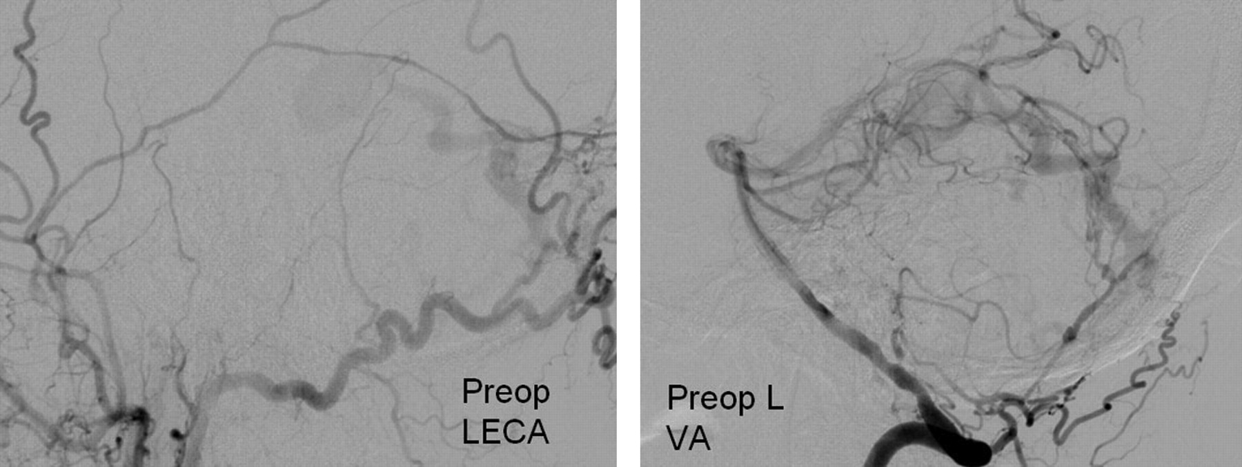
Preoperative angiogram with LECA and LVA injections showing a posterior fossa DAVF with feeders from the left occipital artery and the muscular/meningeal branches of the LVA, with a single large draining cortical vein. The patient underwent partial embolization of the fistula, and it was decided to treat the remaining portion of the fistula with surgery.

Fig 2.
  • Download figure
  • Open in new tab
  • Download powerpoint
Fig 2.

IA (left) showing no residual fistula. Postoperative angiogram, LECA and LVA injections, shows no residual fistula.

In 11 patients (37.9%), IA revealed residual fistula and an early draining vein after the surgeon determined that there was no residual fistula (Fig 3). The locations of these fistulas were the following: tentorial (n = 4), SSS (n = 3), cavernous sinus (n = 2), transverse-sigmoid (n = 1), and torcular (n = 1). Ten patients had immediate further exploration. One patient with a parietal parasagittal DAVF with bilateral dural and pial feeders had further surgical exploration at a later date. In 1 patient, a total of 3 intraoperative angiograms were obtained, while in another patient, 4 intraoperative angiograms were obtained. In all patients except 1, the final intraoperative angiogram was obtained before the surgeon closed. In 1 patient, however, the final angiogram was not obtained for logistic reasons. This patient had undergone 3 prior IAs, and the surgeon thought that there was no need for the fourth IA.

Fig 3.
  • Download figure
  • Open in new tab
  • Download powerpoint
Fig 3.

Preoperative left ICA injection, lateral projection showing a CCF. Following an unsuccessful attempt at endovascular treatment, it was treated surgically. The first IA shows evidence of residual fistula, after which the surgeon further packed the cavernous sinus. The second intraoperative angiogram and a postoperative angiogram show no evidence of residual fistula.

In all patients, IA could be performed in all the vessels intended. There was no complication related to the IA procedure. In 1 patient with a torcular DAVF, IA also identified a surgical complication in the form of occlusion of the posterior sss. Attempts to open up the sinus by both surgery and endovascular methods proved unsuccessful. In another patient with a cavernous sinus DAVF who underwent packing of the sinus, IA identified parent vessel (ICA) occlusion, which occurred during the packing of the sinus. This patient had excellent cross-flow through the anterior communicating artery and did well postoperatively and on follow-up. Postoperative angiography confirmed the parent vessel occlusion, as revealed by IA.

False-negative IA findings occurred in 3 patients (10.3%). In all these patients, IA revealed that there was no residual fistula, but a postoperative study revealed a residual one. The locations of these fistulas were the following: torcular (n = 1), transverse-sigmoid (n = 1), and temporal-middle fossa (n = 1). In 1 of these patients with a temporal-middle fossa DAVF, there was a doubtful residual fistula on IA; however, better visualization was not possible and hence it was reported as near-complete excision. All these fistulas had very slow flow. The 3 residual fistulas were treated respectively with repeat surgery, embolization followed by surgery, and embolization. At the end of the treatment, none of the patients had any residual fistula.

Of the 44 IAs, 41 correctly predicted (93.2%) either the presence or absence of residual fistula. In 3 of the 44 angiograms (6.8%), there was an incorrect finding attributable to the IA. There was no false-positive result, meaning that when the IA showed the presence of a residual fistula, it was a correct finding. Hence the sensitivity and specificity of IA were 82.3% and 100%, respectively, while positive and negative predictive values were 100% and 90%, respectively (Table). The incidence of residual fistula was higher in tentorial (4/6, 75%), SSS (3/4, 75%), and cavernous sinus (2/4, 50%) locations compared with others like torcular (1/3, 33%), transverse-sigmoid (1/9, 11%), middle fossa (0/3, 0%), and foramen magnum (0/3, 0%). However on χ2 testing, the difference between the location of the DAVF and the presence of residual fistula on IA was not statistically significant because of the low number in each group.

View this table:
  • View inline
  • View popup

Sensitivity, specificity, and predictive values

Discussion

Some DAVFs are preferably treated with surgery due to the anatomy and angioarchitechture of the DAVF. Surgery for vascular malformations is aimed at complete obliteration of the lesion, to protect against future hemorrhage risk. Partial excision/obliteration of AVMs or DAVFs does not offer protection against future hemorrhage risk and, in many reports, increases the risk of hemorrhage in the postoperative period.26,27,34,35 Various surgical techniques, including packing of the sinus, skeletonization of the sinus, disconnection of the feeders, as well as disconnection of the leptomeningeal venous drainage, have been described.9–11,13,36–40 Although it has been mentioned in many surgical series that routine IA for DAVF is recommended and is useful, there is no report specifically analyzing the role of IA in surgery for DAVF, to our knowledge.

Many authors have published their experience with IA in surgery for aneurysms and AVMs.15–16,18–22,24,28–33 IA has been used for many years to look for the presence or absence of a residual shunt before closure, particularly with AVMs.17,28,29 The advantage of performing an intraoperative study is that it alerts the surgeon to an unexpected finding, and the surgeon can re-explore to excise the residual malformation, thus avoiding a second surgery in many instances. This also serves to protect against the high risk of rebleed associated with a small residual vascular malformation.

Vitaz et al18 described a large series of 91 patients with intracranial aneurysms and 98 patients with AVMs. The initial angiographic findings caused the surgical procedure to be modified in 29% of the patients with an AVM. In all these patients, there was a small residual nidus not apparent on microscopic examination. Three or more intraoperative angiograms were obtained in 10% of the patients. Other authors have also reported similar findings, though their rates of residual AVM on intraoperative angiogram are lower.15,22,41 Yanaka et al24 reported their experience in 20 patients with vascular malformations, 2 of which were DAVFs, and found an unexpected residual nidus in 1 patient (5%). They also reviewed the literature and found a residual nidus confirmed by IA in 13.5% of patients. The false-negative rate of IA was 4.6%, while the complication rate related to IA was 2.1%.

IA in patients with DAVFs is especially challenging because the arterial feeders to the fistula can be bilateral, from both the ICA and ECA, as well as from the vertebral artery. The examination might warrant cannulation of multiple arteries for a complete study, depending on the angioarchitecture of the DAVF. In the present series, 13 patients (44.8%) underwent cannulation of multiple vessels for their IA.

We found a relatively high rate of residual DAVF after the surgeon's assessment of complete obliteration of the DAVF. Eleven patients (37.9%) had residual DAVFs on IA, leading to further surgical exploration in all of these patients. This is higher than the residual nidus reported in most series of IA in AVM.15,22,41 It might be explained by the fact that DAVFs do not have a compact nidus. In many patients, packing of the sinus or skeletonization of sinus with obliteration of the feeders is the treatment, and it is difficult to examine the adequacy of the packing and obliteration of the fistula without radiologic evidence. Intraoperative Doppler evaluation frequently yields false-positive findings for residual fistula, when IA demonstrates complete obliteration. Furthermore, concern about overpacking a sinus or performing additional arterial/venous occlusions (if the DAVF was actually obliterated) with the potential risk of incurring cranial nerve or other neurologic deficits often prompted additional IAs. We found a higher incidence of residual fistulas in patients with cavernous, tentorial, and SSS fistulas and a lower incidence for transverse-sigmoid sinus fistulas. This high rate of residual nidus after microscopic surgery leads us to advocate IA in patients with DAVF undergoing surgery. In addition to revealing residual nidus in 11 patients, IA also depicted an unnoticed complication in the form of sinus occlusion.

There was 1 patient in this series who demonstrated residual fistula on IA but did not have a final angiogram to show that there was no further residual. This patient had undergone 3 prior IAs, and the surgeon thought he should not examine the patient an additional time. In general, however, the neurosurgeon found that the demonstrated evidence of continued fistula when it was thought the fistula had been completely obliterated was very valuable.

Limitations of IA

False-negative results were seen in 3 patients (10.3%) and 3 of the 44 angiograms (6.8%). This may be attributed to the slow flow of the DAVF. In addition, at the time of surgery, the patient's blood pressure may well be lower, resulting in poorer filling of the fistula. The false-negative rates of IA for AVMs range from 1.8% to 5.2%.15,16,18,24,41 Although IA helped to avoid a repeat surgery in 10 patients, it cannot replace postoperative angiography as the criterion standard to verify complete cure of the fistula following surgery.

The other objection to IA may be the complication rate associated with the procedure. The complication rates in the previous IA studies ranged from 1.7% to 3.7%, most of which occured in the femoral artery.15,16,18,20,41 The central nervous system complications attributed to the procedure were <0.5% in all the series. In the present series, there were no problems attributed to IA.

Patient positioning for the surgical procedure may create an additional challenge for IA. Fourteen IAs (14/34, 41.2%) were performed while the patients were in either a lateral or three-quarter prone position. Successful IA could be performed in all these patients. Lang et al31 presented their experience of 21 patients in whom IA was performed in patients in the prone or three-quarter prone position. They described 2 different approaches, an extended femoral sheath approach and a radial approach. In the present series, all the angiograms could be obtained with the patient in a lateral or three-quarter prone position, with the femoral sheath inserted before positioning.

Conclusions

IA is an extremely important adjunct in the surgery for DAVF. In this series, IA was technically possible in all the patients, and good-quality images could be obtained with cannulation of multiple vessels. IA resulted in further surgical treatment in 37.9% of patients. However, there was a 10% false-negative rate, which justifies subsequent postoperative angiography. IA appears to be a relatively safe investigation, with no complications attributed to it in the present series.

References

  1. 1.↵
    1. Newton TH,
    2. Cronqvist S
    . Involvement of dural arteries in intracranial arteriovenous malformations. Radiology 1969; 93: 1071– 78
    CrossRefPubMed
  2. 2.↵
    1. Awad IA,
    2. Little JR,
    3. Akarawi WP,
    4. et al
    . Intracranial dural arteriovenous malformations: factors predisposing to an aggressive neurological course. J Neurosurg 1990; 72: 839– 50
    CrossRefPubMed
  3. 3.↵
    1. van Rooij WJ,
    2. Sluzewski M,
    3. Beute GN
    . Dural arteriovenous fistulas with cortical venous drainage: incidence, clinical presentation, and treatment. AJNR Am J Neuroradiol 2007; 28: 651– 55
    Abstract/FREE Full Text
  4. 4.↵
    1. Carlson AP,
    2. Taylor CL,
    3. Yonas H
    . Treatment of dural arteriovenous fistula using ethylene vinyl alcohol (Onyx) arterial embolization as the primary modality: short-term results. J Neurosurg 2007; 107: 1120– 25
    CrossRefPubMed
  5. 5.↵
    1. Chen ZQ,
    2. Deng DF,
    3. Gu BX,
    4. et al
    . Treatment of dural arteriovenous fistula by transarterial embolization with low dose of N-butyl-2-cyanoacrylate [in Chinese]. Zhonghua Yi Xue Za Zhi 2006; 86: 157– 59
    PubMed
  6. 6.↵
    1. Kirsch M,
    2. Liebig T,
    3. Kühne D,
    4. et al
    . Endovascular management of dural arteriovenous fistulas of the transverse and sigmoid sinus in 150 patients. Neuroradiology 2009; 51: 477– 83
    CrossRefPubMed
  7. 7.↵
    1. Roy D,
    2. Raymond J
    . The role of transvenous embolization in the treatment of intracranial dural arteriovenous fistulas. Neurosurgery 1997; 40: 1133– 44
    CrossRefPubMed
  8. 8.↵
    1. Stiefel MF,
    2. Albuquerque FC,
    3. Park MS,
    4. et al
    . Endovascular treatment of intracranial dural arteriovenous fistulae using Onyx: a case series. Neurosurgery 2009; 65: 132– 40
    CrossRefPubMed
  9. 9.↵
    1. Kakarla UK,
    2. Deshmukh VR,
    3. Zabramski JM,
    4. et al
    . Surgical treatment of high-risk intracranial dural arteriovenous fistulae: clinical outcomes and avoidance of complications. Neurosurgery 2007; 61: 447– 59, discussion 457–59
    CrossRefPubMed
  10. 10.↵
    1. Afshar JK,
    2. Doppman JL,
    3. Oldfield EH
    . Surgical interruption of intradural draining vein as curative treatment of spinal dural arteriovenous fistulas. J Neurosurg 1995; 82: 196– 200
    PubMed
  11. 11.↵
    1. Kattner KA,
    2. Roth TC,
    3. Giannotta SL
    . Cranial base approaches for the surgical treatment of aggressive posterior fossa dural arteriovenous fistulae with leptomeningeal drainage: report of four technical cases. Neurosurgery 2002; 50: 1156–61
    CrossRefPubMed
  12. 12.↵
    1. Collice M,
    2. D'Aliberti G,
    3. Talamonti G,
    4. et al
    . Surgical interruption of leptomeningeal drainage as treatment for intracranial dural arteriovenous fistulas without dural sinus drainage. J Neurosurg 1996; 84: 810– 17
    PubMed
  13. 13.↵
    1. Lucas CP,
    2. De Oliveira E,
    3. Tedeschi H,
    4. et al
    . Sinus skeletonization: a treatment for dural arteriovenous malformations of the tentorial apex: report of two cases. J Neurosurg 1996; 84: 514– 17
    CrossRefPubMed
  14. 14.↵
    1. Liu JK,
    2. Dogan A,
    3. Ellegala DB,
    4. et al
    . The role of surgery for high-grade intracranial dural arteriovenous fistulas: importance of obliteration of venous outflow. J Neurosurg 2009; 110: 913– 20
    CrossRefPubMed
  15. 15.↵
    1. Barrow DL,
    2. Boyer KL,
    3. Joseph GJ
    . Intraoperative angiography in the management of neurovascular disorders. Neurosurgery 1992; 30: 153– 59
    CrossRefPubMed
  16. 16.↵
    1. Derdeyn CP,
    2. Moran CJ,
    3. Cross DT,
    4. et al
    . Intraoperative digital subtraction angiography: a review of 112 consecutive examinations. AJNR Am J Neuroradiol 1995; 16: 307– 18
    Abstract/FREE Full Text
  17. 17.↵
    1. Smith RW
    . Intraoperative intracranial angiography. Neurosurgery 1977; 1: 107– 10
    CrossRefPubMed
  18. 18.↵
    1. Vitaz TW,
    2. Gaskill-Shipley M,
    3. Tomsick T,
    4. et al
    . Utility, safety, and accuracy of intraoperative angiography in the surgical treatment of aneurysms and arteriovenous malformations. AJNR Am J Neuroradiol 1999; 20: 1457– 61
    Abstract/FREE Full Text
  19. 19.↵
    1. Anegawa S,
    2. Hayashi T,
    3. Torigoe R,
    4. et al
    . Intraoperative angiography in the resection of arteriovenous malformations. J Neurosurg 1994; 80: 73– 78
    CrossRefPubMed
  20. 20.↵
    1. Ghosh S,
    2. Levy ML,
    3. Stanley P,
    4. et al
    . Intraoperative angiography in the management of pediatric vascular disorders. Pediatr Neurosurg 1999; 30: 16– 22
    CrossRefPubMed
  21. 21.↵
    1. Hoh BL,
    2. Carter BS,
    3. Ogilvy CS
    . Incidence of residual intracranial AVMs after surgical resection and efficacy of immediate surgical re-exploration. Acta Neurochir (Wien) 2004; 146: 1– 7, discussion 7. Epub 2003 Dec 9
    CrossRefPubMed
  22. 22.↵
    1. Munshi I,
    2. Macdonald RL,
    3. Weir BK
    . Intraoperative angiography of brain arteriovenous malformations. Neurosurgery 1999; 45: 491– 99
    CrossRefPubMed
  23. 23.↵
    1. Takagi Y,
    2. Kikuta K,
    3. Nozaki K,
    4. et al
    . Detection of a residual nidus by surgical microscope-integrated intraoperative near-infrared indocyanine green videoangiography in a child with a cerebral arteriovenous malformation. J Neurosurg 2007; 107: 416– 18
    CrossRefPubMed
  24. 24.↵
    1. Yanaka K,
    2. Matsumaru Y,
    3. Okazaki M,
    4. et al
    . Intraoperative angiography in the surgical treatment of cerebral arteriovenous malformations and fistulas. Acta Neurochir (Wien) 2003; 145: 377– 83, discussion 382–83
    PubMed
  25. 25.↵
    1. Borden JA,
    2. Wu JK,
    3. Shucart WA
    . A proposed classification for spinal and cranial dural arteriovenous fistulous malformations and implications for treatment. J Neurosurg 1995; 82: 166– 79
    CrossRefPubMed
  26. 26.↵
    1. Forster DM,
    2. Steiner L,
    3. Håkanson S
    . Arteriovenous malformations of the brain: a long-term clinical study. J Neurosurg 1972; 37: 562– 70
    PubMed
  27. 27.↵
    1. Miyamoto S,
    2. Hashimoto N,
    3. Nagata I,
    4. et al
    . Posttreatment sequelae of palliatively treated cerebral arteriovenous malformations. Neurosurgery 2000; 46: 589– 95, discussion 594-95
    CrossRefPubMed
  28. 28.↵
    1. Bauer BL
    . Intraoperative angiography in cerebral aneurysm and AV-malformation. Neurosurg Rev 1984; 7: 209– 17
    CrossRefPubMed
  29. 29.↵
    1. Foley KT,
    2. Cahan LD,
    3. Hieshima GB
    . Intraoperative angiography using a portable digital subtraction unit: technical note. J Neurosurg 1986; 64: 816– 18
    CrossRefPubMed
  30. 30.↵
    1. Dehdashti AR,
    2. Thines L,
    3. Da Costa LB,
    4. et al
    . Intraoperative biplanar rotational angiography during neurovascular surgery: technical note. J Neurosurg 2009; 111: 188– 92
    CrossRefPubMed
  31. 31.↵
    1. Lang SS,
    2. Eskioglu E,
    3. A Mericle R
    . Intraoperative angiography for neurovascular disease in the prone or three-quarter prone position. Surg Neurol 2006; 65: 283–89
    CrossRefPubMed
  32. 32.↵
    1. Pietilä TA,
    2. Stendel R,
    3. Jansons J,
    4. et al
    . The value of intraoperative angiography for surgical treatment of cerebral arteriovenous malformations in eloquent brain areas. Acta Neurochir (Wien) 1998; 140: 1161– 65
    CrossRefPubMed
  33. 33.↵
    1. Zhao J,
    2. Wang S,
    3. Yuan G,
    4. et al
    . Intraoperative angiography in treatment of neurovascular disorders [in Chinese]. Zhonghua Yi Xue Za Zhi 2006; 86: 1044– 47
    PubMed
  34. 34.↵
    1. van Dijk JM,
    2. terBrugge KG,
    3. Willinsky RA,
    4. et al
    . Clinical course of cranial dural arteriovenous fistulas with long-term persistent cortical venous reflux. Stroke 2002; 33: 1233– 36
    Abstract/FREE Full Text
  35. 35.↵
    1. Duffau H,
    2. Lopes M,
    3. Janosevic V,
    4. et al
    . Early rebleeding from intracranial dural arteriovenous fistulas: report of 20 cases and review of the literature. J Neurosurg 1999; 90: 78–84
    PubMed
  36. 36.↵
    1. Day JD,
    2. Fukushima T
    . Direct microsurgery of dural arteriovenous malformation type carotid-cavernous sinus fistulas: indications, technique, and results. Neurosurgery 1997; 41: 1119– 26
    CrossRefPubMed
  37. 37.↵
    1. Fiumara E,
    2. Tumbiolo S,
    3. Bellomonte ML,
    4. et al
    . Resection of the transverse sinuses and confluence of sinuses for treatment of multiple dural arteriovenous fistulas: case report. J Neurosurg 2004; 100: 348– 52
    CrossRefPubMed
  38. 38.↵
    1. Hoh BL,
    2. Choudhri TF,
    3. Connolly ES,
    4. et al
    . Surgical management of high-grade intracranial dural arteriovenous fistulas: leptomeningeal venous disruption without nidus excision. Neurosurgery 1998; 42: 796– 805
    PubMed
  39. 39.↵
    1. Ushikoshi S,
    2. Houkin K,
    3. Kuroda S,
    4. et al
    . Surgical treatment of intracranial dural arteriovenous fistulas. Surg Neurol 2002; 57: 253– 61
    CrossRefPubMed
  40. 40.↵
    1. Thompson BG,
    2. Doppman JL,
    3. Oldfield EH
    . Treatment of cranial dural arteriovenous fistulae by interruption of leptomeningeal venous drainage. J Neurosurg 1994; 80: 617– 23
    PubMed
  41. 41.↵
    1. Martin NA,
    2. Bentson J,
    3. Viñuela F,
    4. et al
    . Intraoperative digital subtraction angiography and the surgical treatment of intracranial aneurysms and vascular malformations. J Neurosurg 1990; 73: 526– 33
    CrossRefPubMed
  • Received August 28, 2010.
  • Accepted after revision October 19, 2010.
  • Copyright © American Society of Neuroradiology
View Abstract
PreviousNext
Back to top

In this issue

American Journal of Neuroradiology: 32 (6)
American Journal of Neuroradiology
Vol. 32, Issue 6
1 Jun 2011
  • Table of Contents
  • Index by author
Advertisement
Print
Download PDF
Email Article

Thank you for your interest in spreading the word on American Journal of Neuroradiology.

NOTE: We only request your email address so that the person you are recommending the page to knows that you wanted them to see it, and that it is not junk mail. We do not capture any email address.

Enter multiple addresses on separate lines or separate them with commas.
Intraoperative Angiography for Cranial Dural Arteriovenous Fistula
(Your Name) has sent you a message from American Journal of Neuroradiology
(Your Name) thought you would like to see the American Journal of Neuroradiology web site.
CAPTCHA
This question is for testing whether or not you are a human visitor and to prevent automated spam submissions.
Cite this article
P. Pandey, G.K. Steinberg, E.M. Westbroek, R. Dodd, H.M. Do, M.P. Marks
Intraoperative Angiography for Cranial Dural Arteriovenous Fistula
American Journal of Neuroradiology Jun 2011, 32 (6) 1091-1095; DOI: 10.3174/ajnr.A2443

Citation Manager Formats

  • BibTeX
  • Bookends
  • EasyBib
  • EndNote (tagged)
  • EndNote 8 (xml)
  • Medlars
  • Mendeley
  • Papers
  • RefWorks Tagged
  • Ref Manager
  • RIS
  • Zotero
0 Responses
Respond to this article
Share
Bookmark this article
Intraoperative Angiography for Cranial Dural Arteriovenous Fistula
P. Pandey, G.K. Steinberg, E.M. Westbroek, R. Dodd, H.M. Do, M.P. Marks
American Journal of Neuroradiology Jun 2011, 32 (6) 1091-1095; DOI: 10.3174/ajnr.A2443
del.icio.us logo Twitter logo Facebook logo Mendeley logo
  • Tweet Widget
  • Facebook Like
  • Google Plus One
Purchase

Jump to section

  • Article
    • Abstract
    • Abbreviations
    • Materials and Methods
    • Results
    • Discussion
    • Conclusions
    • References
  • Figures & Data
  • Info & Metrics
  • Responses
  • References
  • PDF

Related Articles

  • No related articles found.
  • PubMed
  • Google Scholar

Cited By...

  • Changing Clinical and Therapeutic Trends in Tentorial Dural Arteriovenous Fistulas: A Systematic Review
  • Crossref (17)
  • Google Scholar

This article has been cited by the following articles in journals that are participating in Crossref Cited-by Linking.

  • Safety and Efficacy of Intraoperative Angiography in Craniotomies for Cerebral Aneurysms and Arteriovenous Malformations
    Nohra Chalouhi, Thana Theofanis, Pascal Jabbour, Aaron S. Dumont, L. Fernando Gonzalez, Robert M. Starke, Richard T. Dalyai, Shannon Hann, Robert Rosenwasser, Stavropoula Tjoumakaris
    Neurosurgery 2012 71 6
  • Diagnosis and management of dural arteriovenous fistulas: A 10 years single-center experience
    F. Signorelli, G.M. Della Pepa, G. Sabatino, E. Marchese, G. Maira, A. Puca, A. Albanese
    Clinical Neurology and Neurosurgery 2015 128
  • Changing Clinical and Therapeutic Trends in Tentorial Dural Arteriovenous Fistulas: A Systematic Review
    D. Cannizzaro, W. Brinjikji, S. Rammos, M.H. Murad, G. Lanzino
    American Journal of Neuroradiology 2015 36 10
  • Intraoperative Angiography for Arteriovenous Malformation Resection in the Prone and Lateral Positions, Using Upper Extremity Arterial Access
    Erez Nossek, David J. Chalif, Razvan Buciuc, Eric J. Gandras, Erich G. Anderer, Sal Insigna, Amir R. Dehdashti, Avi Setton
    Operative Neurosurgery 2017 13 3
  • Historical Perspective of Treatments of Cranial Arteriovenous Malformations and Dural Arteriovenous Fistulas
    Geoffrey P. Colby, Alexander L. Coon, Judy Huang, Rafael J. Tamargo
    Neurosurgery Clinics of North America 2012 23 1
  • Selection of Treatment Modalities or Observation of Dural Arteriovenous Fistulas
    Alexandra R. Paul, Geoffrey P. Colby, Judy Huang, Rafael J. Tamargo, Alexander L. Coon
    Neurosurgery Clinics of North America 2012 23 1
  • Radial Arterial Access for Thoracic Intraoperative Spinal Angiography in the Prone Position
    Joseph Haynes, Erez Nossek, Maksim Shapiro, Bree Chancellor, Anthony Frempong-Boadu, Simone Peschillo, Hunter Alves, Omar Tanweer, David Gordon, Eytan Raz
    World Neurosurgery 2020 137
  • Microsurgical Findings of Pial Arterial Feeders in Intracranial Dural Arteriovenous Fistulae: A Case Series
    Michinari Okamoto, Taku Sugiyama, Naoki Nakayama, Satoshi Ushikoshi, Ken Kazumata, Toshiya Osanai, Kikutaro Tokairin, Yusuke Shimoda, Kiyohiro Houkin
    Operative Neurosurgery 2020 19 6
  • Sphenoparietal Sinus Dural Arteriovenous Fistulas: A Series of 10 Patients
    Joelle N. Hartke, Visish M. Srinivasan, Redi Rahmani, Joshua S. Catapano, Mohamed A. Labib, Kavelin Rumalla, Joseph H. Garcia, Cameron M. McDougall, Adib A. Abla, Michael T. Lawton
    Operative Neurosurgery 2022 23 2
  • Transfemoral Approach for Intraoperative Angiography in the Prone or Three-quarter Prone Position
    Chi Wang, Szu-Kai Hsu, Chih-Ju Chang, Ming-Hong Chen, Chih-Ta Huang, Jing-Shan Huang, I-Chang Su
    Clinical Neuroradiology 2020 30 2

More in this TOC Section

  • SAVE vs. Solumbra Techniques for Thrombectomy
  • Contrast-Induced Encephalopathy after NeuroIR
  • CT Perfusion&Reperfusion in Acute Ischemic Stroke
Show more Interventional

Similar Articles

Advertisement

Indexed Content

  • Current Issue
  • Accepted Manuscripts
  • Article Preview
  • Past Issues
  • Editorials
  • Editor's Choice
  • Fellows' Journal Club
  • Letters to the Editor
  • Video Articles

Cases

  • Case Collection
  • Archive - Case of the Week
  • Archive - Case of the Month
  • Archive - Classic Case

Special Collections

  • AJNR Awards
  • ASNR Foundation Special Collection
  • Most Impactful AJNR Articles
  • Photon-Counting CT
  • Spinal CSF Leak Articles (Jan 2020-June 2024)

More from AJNR

  • Trainee Corner
  • Imaging Protocols
  • MRI Safety Corner

Multimedia

  • AJNR Podcasts
  • AJNR Scantastics

Resources

  • Turnaround Time
  • Submit a Manuscript
  • Submit a Video Article
  • Submit an eLetter to the Editor/Response
  • Manuscript Submission Guidelines
  • Statistical Tips
  • Fast Publishing of Accepted Manuscripts
  • Graphical Abstract Preparation
  • Imaging Protocol Submission
  • Evidence-Based Medicine Level Guide
  • Publishing Checklists
  • Author Policies
  • Become a Reviewer/Academy of Reviewers
  • News and Updates

About Us

  • About AJNR
  • Editorial Board
  • Editorial Board Alumni
  • Alerts
  • Permissions
  • Not an AJNR Subscriber? Join Now
  • Advertise with Us
  • Librarian Resources
  • Feedback
  • Terms and Conditions
  • AJNR Editorial Board Alumni

American Society of Neuroradiology

  • Not an ASNR Member? Join Now

© 2025 by the American Society of Neuroradiology All rights, including for text and data mining, AI training, and similar technologies, are reserved.
Print ISSN: 0195-6108 Online ISSN: 1936-959X

Powered by HighWire