Advertisement

Main menu

  • Home
  • Content
    • Current Issue
    • Publication Preview--Ahead of Print
    • Past Issue Archive
    • Case of the Week Archive
    • Classic Case Archive
    • Case of the Month Archive
    • COVID-19 Content and Resources
  • For Authors
  • About Us
    • About AJNR
    • Editors
    • American Society of Neuroradiology
  • Submit a Manuscript
  • Podcasts
    • Subscribe on iTunes
    • Subscribe on Stitcher
  • More
    • Subscribers
    • Permissions
    • Advertisers
    • Alerts
    • Feedback
  • Other Publications
    • ajnr

User menu

  • Subscribe
  • Alerts
  • Log in

Search

  • Advanced search
American Journal of Neuroradiology

American Journal of Neuroradiology

  • Subscribe
  • Alerts
  • Log in

Advanced Search

  • Home
  • Content
    • Current Issue
    • Publication Preview--Ahead of Print
    • Past Issue Archive
    • Case of the Week Archive
    • Classic Case Archive
    • Case of the Month Archive
    • COVID-19 Content and Resources
  • For Authors
  • About Us
    • About AJNR
    • Editors
    • American Society of Neuroradiology
  • Submit a Manuscript
  • Podcasts
    • Subscribe on iTunes
    • Subscribe on Stitcher
  • More
    • Subscribers
    • Permissions
    • Advertisers
    • Alerts
    • Feedback
  • Follow AJNR on Twitter
  • Visit AJNR on Facebook
  • Follow AJNR on Instagram
  • Join AJNR on LinkedIn
  • RSS Feeds
Case ReportCase Report

Reversible Pancallosal Signal Changes in Febrile Encephalopathy: Report of 2 Cases

S.E. Sreedharan, J. Chellenton, M.P. Kate and C. Kesavadas
American Journal of Neuroradiology October 2011, 32 (9) E172-E174; DOI: https://doi.org/10.3174/ajnr.A2318
S.E. Sreedharan
  • Find this author on Google Scholar
  • Find this author on PubMed
  • Search for this author on this site
J. Chellenton
  • Find this author on Google Scholar
  • Find this author on PubMed
  • Search for this author on this site
M.P. Kate
  • Find this author on Google Scholar
  • Find this author on PubMed
  • Search for this author on this site
C. Kesavadas
  • Find this author on Google Scholar
  • Find this author on PubMed
  • Search for this author on this site
  • Article
  • Figures & Data
  • Info & Metrics
  • References
  • PDF
Loading

Abstract

SUMMARY: Transient CC involvement has been reported in encephalopathies/encephalitis of different etiologies. Here we report 2 patients with AFE, who showed transient pancallosal involvement with restricted diffusion on neuroimaging. Both patients had excellent clinical outcomes: The lesion disappeared completely in 1, though there was mild residual gliosis in the other. Serology for dengue virus was positive in 1 of the patients.

Abbreviations

ADC
apparent diffusion coefficient
AESD
acute encephalopathy with biphasic seizures and late reduced diffusion
AFE
acute febrile encephalopathy
CC
corpus callosum
DWI
diffusion-weighted imaging
FLAIR
fluid-attenuated inversion recovery
MERS
mild encephalitis/encephalopathy with a reversible splenial lesion
NAA
N-acetylaspartate

Acute encephalopathy is a common clinical scenario in which patients exhibit features of global cerebral dysfunction without a documented central nervous system infection. Etiologies can be diverse, from systemic infections to drugs and metabolic causes. Two patterns on neuroimaging previously reported in this setting are transient splenial lesions with restricted diffusion1–3 and bihemispheric involvement.4 Here we report 2 patients who presented with AFE, who had similar neuroimaging findings of pancallosal involvement with restricted diffusion, 1 of which was initially considered a glioma.

Case Reports

Case 1

A 45-year-old woman with diabetes mellitus presented to a local hospital with a 1-week history of fever and skin infection, followed by altered sensorium and gait unsteadiness. Her metabolic parameters were normal. She underwent MR imaging in the second week of illness, which showed a pancallosal lesion with restricted diffusion (Fig 1A–D) and contrast enhancement (Fig 2A, -B). With treatment of the infection, her sensorium improved and she was referred to us on day 14 of her illness with a diagnosis of glioma and recommended biopsy.

Fig 1.
  • Download figure
  • Open in new tab
  • Download powerpoint
Fig 1.

Case 1. Sagittal T2-weighted (A) and axial FLAIR (B) MR images show pancallosal edema and hyperintensity. C and D, Axial DWI and ADC map show restricted diffusion in the whole of callosum. Case 2. Sagittal T2-weighted (E) and axial FLAIR (F) images show pancallosal hyperintensity. G and H, Axial DWI and ADC map show restricted diffusion in the callosum.

Fig 2.
  • Download figure
  • Open in new tab
  • Download powerpoint
Fig 2.

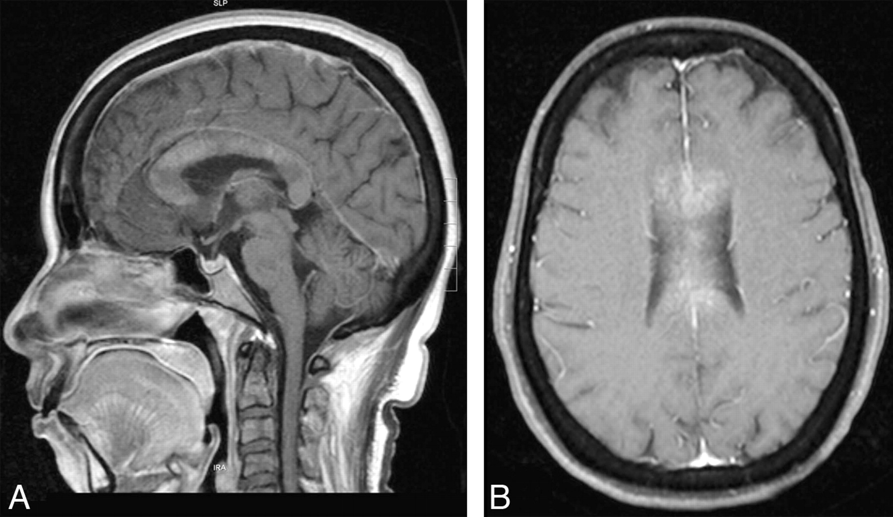
Case 1. Sagittal (A) and axial (B) T1-weighted postcontrast images show enhancement of the callosum.

At presentation, the patient was alert and afebrile, with a healing skin ulcer. Findings of a neurologic examination were unremarkable. She had no history of seizures or antiepileptic treatment before admission. Metabolic and hematologic parameters were normal. Additional imaging showed reduced choline and NAA on proton MR spectroscopy, restricted diffusion, and low perfusion on the perfusion imaging, which raised doubt concerning the diagnosis. Biopsy was deferred. She was treated with a short course of steroids. MR imaging repeated 6 weeks later showed near-complete resolution of the lesion with gliosis in the genu and thinning of the CC (Fig 3A). She had no neurologic deficits on follow-up.

Fig 3.
  • Download figure
  • Open in new tab
  • Download powerpoint
Fig 3.

A, Sagittal T2-weighted image performed after 6 weeks shows complete resolution of the pancallosal edema. There is evidence of thinning of the callosum with hyperintensity (arrow), suggesting callosal gliosis. B, Sagittal T2-weighted image of the patient obtained after 2 weeks shows complete resolution of the pancallosal hyperintensity.

Case 2

A 27-year-old man presented to us with fever associated with headache and erythematous rash over his body. On day 5 of illness, he had a transient alteration in the sensorium with mild left-arm weakness, which spontaneously subsided in 30 minutes. On day 6 of his illness, he had 2 similar attacks without any preceding convulsive movements. Clinical examination revealed a drowsy patient with subtle left-arm weakness.

Hematologic and biochemical parameters and CSF and electroencephalogram findings were normal. His neuroimaging showed a pancallosal lesion extending to the centrum semiovale symmetrically with restricted diffusion (Fig 1E−H) without contrast enhancement. Serology for dengue was positive. He was observed in a ward without any specific medications. A repeat MR imaging after 2 weeks showed complete resolution of the lesions (Fig 3B). The patient was asymptomatic on follow-up.

Discussion

Prevalence of CC lesions has been reported at 3% in a large series of 450 patients undergoing MR imaging.1 While the CC can be involved in vascular, traumatic, demyelinating, neoplastic, infective, and degenerative illnesses,2 transient involvement has been reported with encephalitis of different etiologies,3,5 antiepileptic drug use and a peri-ictal state. Among infectious etiologies, influenza has been most often associated with transient splenial involvement.6,7

Tada et al8 proposed the terminology “MERS” to explain a clinical syndrome sharing common features of mild encephalopathy, seizures, and good outcome on follow-up. However, in all these reports, the involvement was restricted to the splenium, with some patients having adjacent white matter involvement as well.

Takanashi9 described another entity called “AESD” in children. Neuroimaging showed reduced diffusion in the frontal or frontoparietal subcortical white matter during days 3–9, which reversed on follow-up. Okumura et al4 described 2 patterns of lesions in AESD, a diffuse pattern with poor outcome and a central sparing pattern, which was clinically mild. None of their patients had CC involvement.

Both of our patients had similar clinical presentations, with a background of a febrile illness. The presentation with encephalopathy and minimal focal signs within the first week of illness and symmetric involvement of the CC makes postinfectious demyelination less likely. This clinical pattern has been described in most of the previously reported cases of MERS.3,6–8

MR imaging showed pancallosal involvement with restricted diffusion in both of our patients. The first patient had a bulky CC, with patchy contrast enhancement, which was initially mistaken for glioma. The most consistently reported pathophysiologic mechanisms for CC involvement with restricted diffusion are transient intramyelinic edema and inflammatory infiltrates.10 Some of the recent reports of MERS have shown persistence of lesion/gliotic changes on follow-up, as in our first patient.5,11

Imaging characteristics of neurodengue have rarely been reported, to our knowledge. In a large series of neurodengue,12 no characteristic imaging pattern was noted. A similar observation was made by Wasay et al13 in their series of 6 patients. Their reported findings were cerebral edema and cortical and cord hyperintensities. Even though the findings of callosal hyperintensity with restricted diffusion have never been reported with neurodengue, we presume that the imaging finding may be related to AFE syndrome rather than the virus per se. More imaging studies on neurodengue would be required to clarify the observation.

We report these 2 cases to highlight a few points. Transient pancallosal involvement with restricted diffusion is a novel observation in AFE of any etiology. It may represent an extended spectrum of MERS because the presentation and outcome were similar. Encephalopathy associated with dengue has been linked to a poor outcome as opposed to our patient who had mild symptoms, which resolved spontaneously. Last, the appearance of the lesion can be mistaken for a glioma because of the associated swelling and contrast enhancement. However, the clinical setting and additional MR imaging sequences, including diffusion and perfusion, can differentiate the 2, thereby avoiding invasive biopsy.

References

  1. 1.↵
    1. McLeod NA,
    2. William JP,
    3. Machen B,
    4. et al
    . Normal and abnormal morphology of corpus callosum. Neurology 1987; 37: 1240–42
    Abstract/FREE Full Text
  2. 2.↵
    1. Uchino Y,
    2. Takase K,
    3. Nomiyama R,
    4. et al
    . Acquired lesions of the corpus callosum: MR imaging. Eur Radiol 2006; 16: 905–14
    CrossRefPubMed
  3. 3.↵
    1. Takanashi J,
    2. Barkovich AJ,
    3. Shiihara T,
    4. et al
    . Widening spectrum of a reversible splenial lesion with transiently reduced diffusion. AJNR Am J Neuroradiol 2006; 27: 836–38
    Abstract/FREE Full Text
  4. 4.↵
    1. Okumura A,
    2. Kidokoro H,
    3. Tsuji T,
    4. et al
    . Differences of clinical manifestations according to the patterns of brain lesions in acute encephalopathy with reduced diffusion in the bilateral hemispheres. AJNR Am J Neuroradiol 2009; 30: 825–30
    Abstract/FREE Full Text
  5. 5.↵
    1. Maeda M,
    2. Tsukahara H,
    3. Terada S,
    4. et al
    . Reversible splenial lesions with restricted diffusion in a wide spectrum of diseases and conditions. J Neuroradiol 2006; 33: 229–36
    PubMed
  6. 6.↵
    1. Takanashi J,
    2. Barkovich AJ,
    3. Yamaguchi K,
    4. et al
    . Influenza-associated encephalitis/encephalopathy with a reversible lesion in the splenium of the corpus callosum: a case report and literature review. AJNR Am J Neuroradiol 2004; 25: 798–802
    Abstract/FREE Full Text
  7. 7.↵
    1. Bulakbasi N,
    2. Kocaoglu M,
    3. Tayfun C,
    4. et al
    . Transient splenial lesion of the corpus callosum in clinically mild influenza-associated encephalitis/encephalopathy. AJNR Am J Neuroradiol 2006; 27: 1983–86
    Abstract/FREE Full Text
  8. 8.↵
    1. Tada H,
    2. Takanashi J,
    3. Barkovich AJ
    . Clinically mild encephalitis/encephalopathy with a reversible splenial lesion. Neurology 2004; 63: 1854–58
    Abstract/FREE Full Text
  9. 9.↵
    1. Takanashi J
    . Two newly proposed infectious encephalitis/encephalopathy syndromes. Brain Dev 2009; 31: 521–28
    CrossRefPubMed
  10. 10.↵
    1. Kobuchi N,
    2. Tsukahara H,
    3. Kawamura Y,
    4. et al
    . Reversible diffusion-weighted MR findings of Salmonella enteritidis-associated encephalopathy. Eur Neurol 2003; 49: 182–84
    CrossRefPubMed
  11. 11.↵
    1. Doherty MJ,
    2. Jayadev S,
    3. Watson NF,
    4. et al
    . Clinical implications of splenium magnetic resonance imaging signal changes. Arch Neurol 2005; 62: 433–37
    CrossRefPubMed
  12. 12.↵
    1. Kalita J,
    2. Misra UK
    . EEG in dengue virus infection with neurological manifestations: a clinical and CT/MRI correlation. Clinical Neurophysiol 2006; 117: 2252–56. Epub 2006 Aug 22
    CrossRef
  13. 13.↵
    1. Wasay M,
    2. Channa R,
    3. Jumani M,
    4. et al
    . Encephalitis and myelitis associated with dengue viral infection: clinical and neuroimaging features. Clin Neurol Neurosurg 2008; 110: 635–40. Epub 2008 May 7
    CrossRefPubMed
  • Received July 20, 2010.
  • Accepted after revision July 31, 2010.
  • © 2011 by American Journal of Neuroradiology
View Abstract
PreviousNext
Back to top

In this issue

American Journal of Neuroradiology: 32 (9)
American Journal of Neuroradiology
Vol. 32, Issue 9
1 Oct 2011
  • Table of Contents
  • Index by author
Advertisement
Print
Download PDF
Email Article

Thank you for your interest in spreading the word on American Journal of Neuroradiology.

NOTE: We only request your email address so that the person you are recommending the page to knows that you wanted them to see it, and that it is not junk mail. We do not capture any email address.

Enter multiple addresses on separate lines or separate them with commas.
Reversible Pancallosal Signal Changes in Febrile Encephalopathy: Report of 2 Cases
(Your Name) has sent you a message from American Journal of Neuroradiology
(Your Name) thought you would like to see the American Journal of Neuroradiology web site.
CAPTCHA
This question is for testing whether or not you are a human visitor and to prevent automated spam submissions.
Citation Tools
Reversible Pancallosal Signal Changes in Febrile Encephalopathy: Report of 2 Cases
S.E. Sreedharan, J. Chellenton, M.P. Kate, C. Kesavadas
American Journal of Neuroradiology Oct 2011, 32 (9) E172-E174; DOI: 10.3174/ajnr.A2318

Citation Manager Formats

  • BibTeX
  • Bookends
  • EasyBib
  • EndNote (tagged)
  • EndNote 8 (xml)
  • Medlars
  • Mendeley
  • Papers
  • RefWorks Tagged
  • Ref Manager
  • RIS
  • Zotero
Share
Reversible Pancallosal Signal Changes in Febrile Encephalopathy: Report of 2 Cases
S.E. Sreedharan, J. Chellenton, M.P. Kate, C. Kesavadas
American Journal of Neuroradiology Oct 2011, 32 (9) E172-E174; DOI: 10.3174/ajnr.A2318
Reddit logo Twitter logo Facebook logo Mendeley logo
  • Tweet Widget
  • Facebook Like
  • Google Plus One
Purchase

Jump to section

  • Article
    • Abstract
    • Abbreviations
    • Case Reports
    • Discussion
    • References
  • Figures & Data
  • Info & Metrics
  • References
  • PDF

Related Articles

  • No related articles found.
  • PubMed
  • Google Scholar

Cited By...

  • No citing articles found.
  • Crossref
  • Google Scholar

This article has not yet been cited by articles in journals that are participating in Crossref Cited-by Linking.

More in this TOC Section

  • Atypical Diffusion-Restricted Lesion in 5-Fluorouracil Encephalopathy
  • Dural Infantile Hemangioma Masquerading as a Skull Vault Lesion
  • Multimodal Imaging of Spike Propagation: A Technical Case Report
Show more Case Reports

Similar Articles

Advertisement

News and Updates

  • Lucien Levy Best Research Article Award
  • Thanks to our 2022 Distinguished Reviewers
  • Press Releases

Resources

  • Evidence-Based Medicine Level Guide
  • How to Participate in a Tweet Chat
  • AJNR Podcast Archive
  • Ideas for Publicizing Your Research
  • Librarian Resources
  • Terms and Conditions

Opportunities

  • Share Your Art in Perspectives
  • Get Peer Review Credit from Publons
  • Moderate a Tweet Chat

American Society of Neuroradiology

  • Neurographics
  • ASNR Annual Meeting
  • Fellowship Portal
  • Position Statements

© 2023 by the American Society of Neuroradiology | Print ISSN: 0195-6108 Online ISSN: 1936-959X

Powered by HighWire