Advertisement

Main menu

  • Home
  • Content
    • Current Issue
    • Publication Preview--Ahead of Print
    • Past Issue Archive
    • Case of the Week Archive
    • Classic Case Archive
    • Case of the Month Archive
    • COVID-19 Content and Resources
  • About Us
    • About AJNR
    • Editorial Board
    • American Society of Neuroradiology
  • For Authors
    • Author Policies
    • Manuscript Submission Guidelines
  • Submit a Manuscript
  • Podcasts
    • Podcasts
    • Subscribe on iTunes
  • More
    • Subscribers
    • Permissions
    • Advertisers
    • Alerts
    • Feedback

User menu

  • Subscribe
  • Alerts
  • Log in

Search

  • Advanced search
American Journal of Neuroradiology

American Journal of Neuroradiology

  • Subscribe
  • Alerts
  • Log in

Advanced Search

  • Home
  • Content
    • Current Issue
    • Publication Preview--Ahead of Print
    • Past Issue Archive
    • Case of the Week Archive
    • Classic Case Archive
    • Case of the Month Archive
    • COVID-19 Content and Resources
  • About Us
    • About AJNR
    • Editorial Board
    • American Society of Neuroradiology
  • For Authors
    • Author Policies
    • Manuscript Submission Guidelines
  • Submit a Manuscript
  • Podcasts
    • Podcasts
    • Subscribe on iTunes
  • More
    • Subscribers
    • Permissions
    • Advertisers
    • Alerts
    • Feedback
  • Follow AJNR on Twitter
  • Visit AJNR on Facebook
  • Follow AJNR on Instagram
  • Join AJNR on LinkedIn
  • RSS Feeds
LetterLetter

Infarction of the Corpus Callosum: A Manifestation of Giant Cell Arteritis

J. Lempel, S. Bernstein and P. Kalina
American Journal of Neuroradiology October 2011, 32 (9) E182-E183; DOI: https://doi.org/10.3174/ajnr.A2763
J. Lempel
  • Find this author on Google Scholar
  • Find this author on PubMed
  • Search for this author on this site
S. Bernstein
  • Find this author on Google Scholar
  • Find this author on PubMed
  • Search for this author on this site
P. Kalina
  • Find this author on Google Scholar
  • Find this author on PubMed
  • Search for this author on this site
  • Article
  • Figures & Data
  • Info & Metrics
  • References
  • PDF
Loading

Cerebral vasculitis is a known cause of ischemic and hemorrhagic strokes and has been described as one of the rare but important causes of corpus callosum infarction. Biopsy-proved giant cell arteritis causing callosal infarction is an exceedingly rare finding because a tissue specimen is usually not obtained and conclusions are drawn on the basis of clinical and radiologic findings alone. We present a case of callosal infarction, which evolved and eventually affected large portions of both cerebral hemispheres.

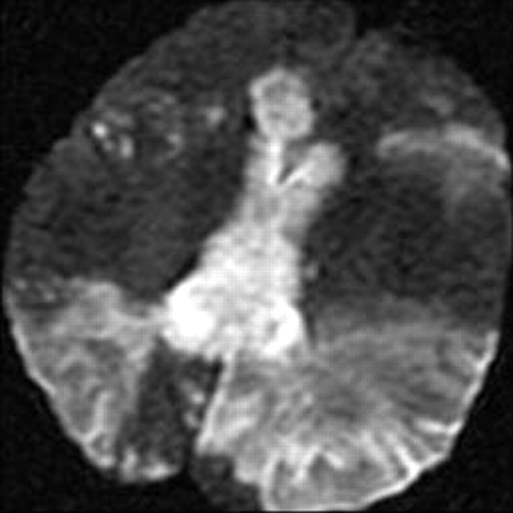
A 63-year-old woman presented to our hospital with left-sided numbness and neglect, cognitive changes, and apraxia. One month earlier, she was found to have a C-reactive protein level of 8.0 mg/dL (normal <0.5 mg/dL) and 75% stenosis in both femoral arteries. These results prompted a temporal artery biopsy, yielding a diagnosis of giant cell arteritis. On admission, head CT demonstrated low attenuation in the splenium of the corpus callosum (Fig 1). Considerations included infarction, tumor, demyelination, or infection. Subsequent CT angiography (CTA) revealed segmental narrowing and beading involving the petrous segment of both internal carotid arteries (Fig 2) and along the splenial or posterior pericallosal arteries within the posterior circulation. The condition of the patient continued to decline during the following week, culminating in a coma. An MR image obtained 12 days after the initial presentation revealed involvement of the entire corpus callosum, both cerebral hemispheres, and the right cerebellum with restricted diffusion indicating infarction secondary to intracranial vasculitis (Fig 3). In addition, hemorrhage was noted in the cortical gyri (Fig 4).

Fig 1.
  • Download figure
  • Open in new tab
  • Download powerpoint
Fig 1.

Noncontrast head CT scan demonstrates low attenuation in the splenium of the corpus callosum (arrow).

Fig 2.
  • Download figure
  • Open in new tab
  • Download powerpoint
Fig 2.

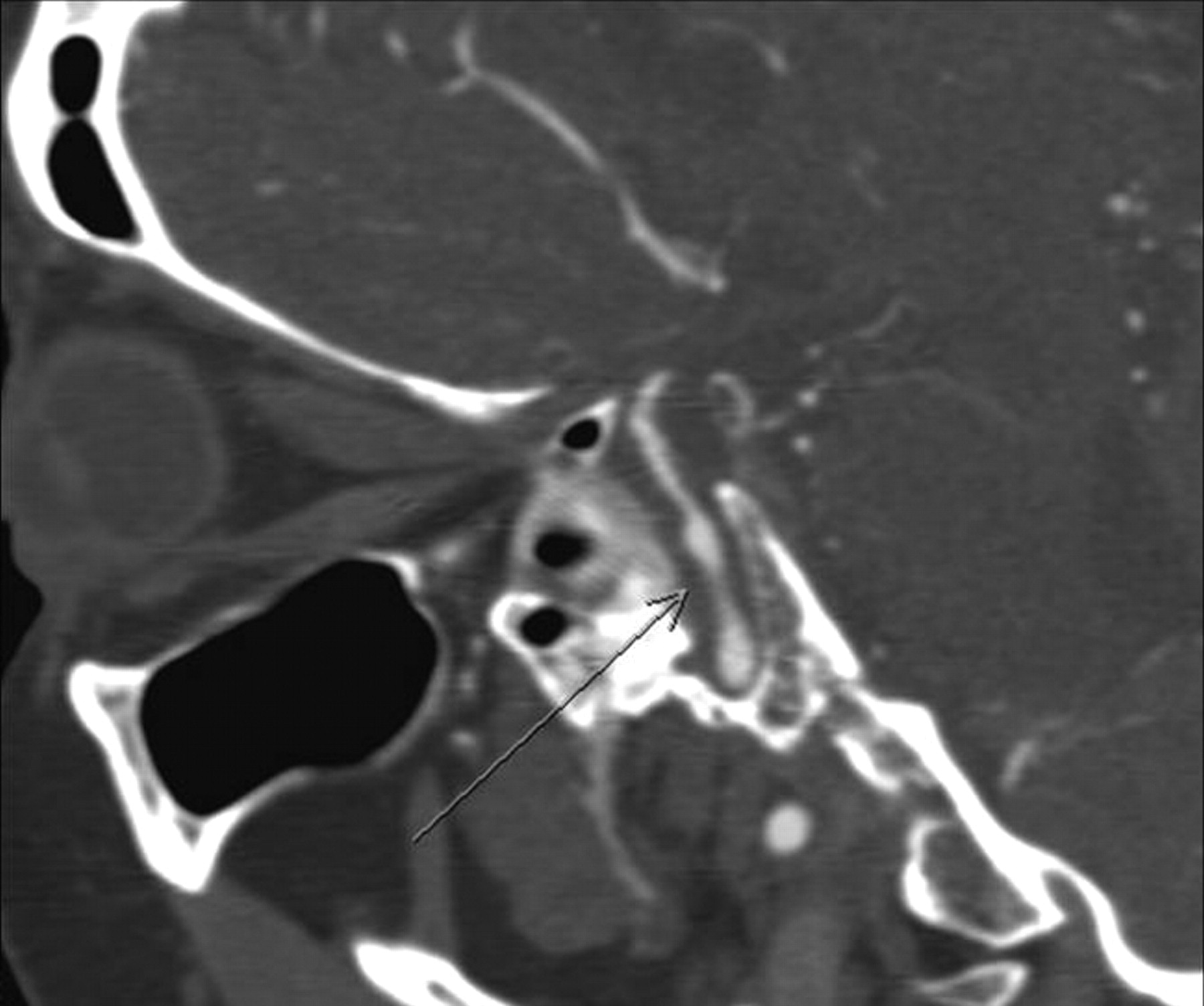
Oblique sagittal reformatted maximum intensity projection image from a CTA image depicts beading and narrowing of the left internal carotid artery (arrow).

Fig 3.
  • Download figure
  • Open in new tab
  • Download powerpoint
Fig 3.

Diffusion-weighted image shows hyperintensity in the corpus callosum and within both cerebral hemispheres.

Fig 4.
  • Download figure
  • Open in new tab
  • Download powerpoint
Fig 4.

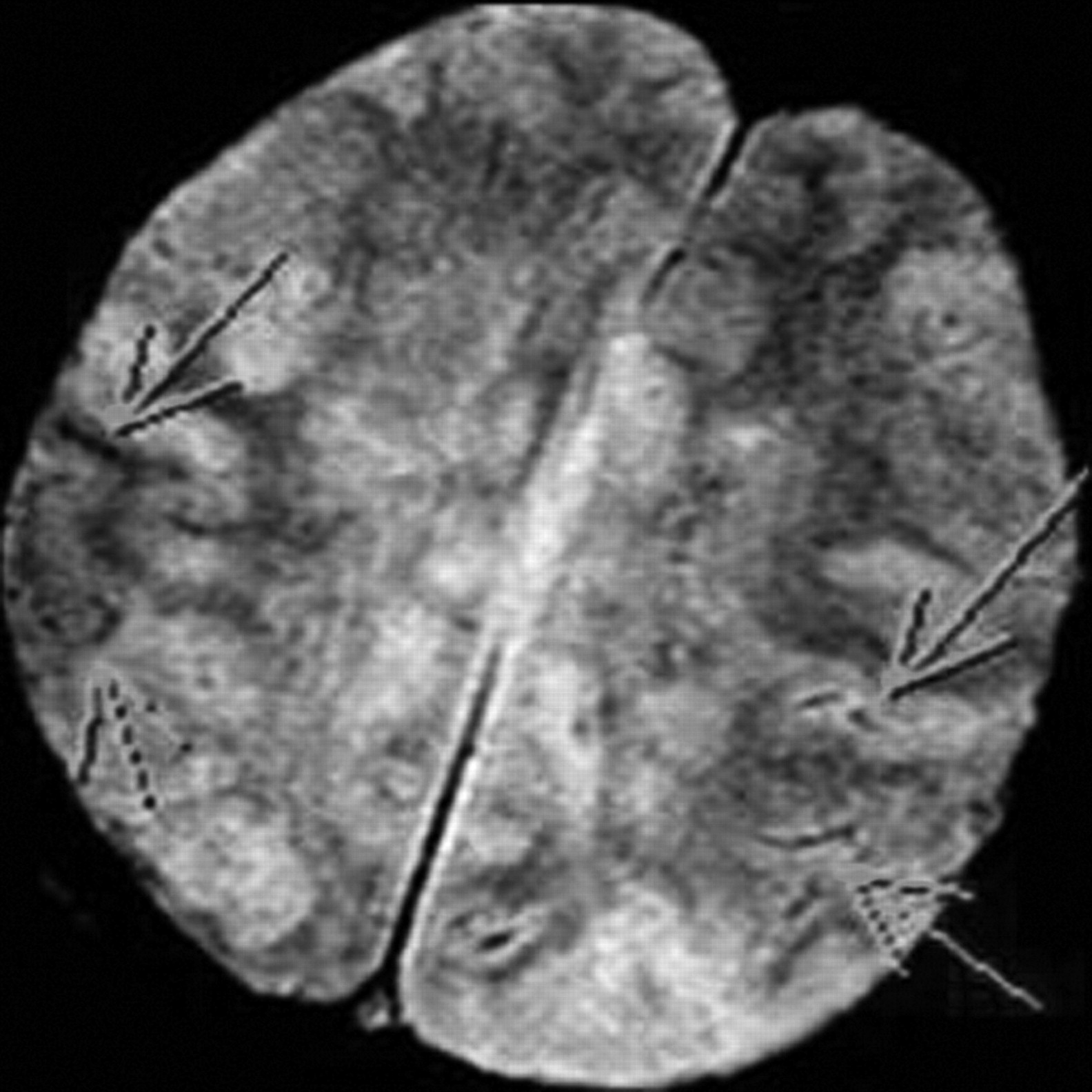
T2-weighted gradient recalled-echo image shows curvilinear areas of decreased signal intensity within the left parietal lobe, indicating susceptibility artifacts resulting from hemorrhage (arrows).

Lesions of the corpus callosum can present a diagnostic dilemma, both for the radiologist and the clinician. Clinically, they are associated with neuropsychiatric symptoms, mainly interhemispheric disconnection syndromes. Patients may experience gait disorders, apraxia, agraphia, and tactile anomia.1 While the corpus callosum is affected by varying disease processes, ischemia tends to spare this unique white matter structure. One accepted theory that explains relative callosal immunity to infarction relates to its rich blood supply delivered from the anterior and posterior circulations. In a study by Chrysikopoulos et al,2 the authors described the splenium as being most commonly involved in thromboembolic causes of callosal infarction, very likely due to the predilection for posterior cerebral artery versus anterior cerebral artery territory ischemia. In our patient, the lesion began in the splenium and progressed rostrally to involve the body and genu and eventually a large portion of both cerebral hemispheres. The global involvement in our case was secondary to diffuse vasculitis rather than more focal thromboembolic disease.

Johnson et al3 described a case of biopsy-proved cerebral vasculitis that was seen as bilateral parieto-occipital “masses” that crossed the splenium of the corpus callosum. Greenan et al4 described a series of 7 clinically suspected cases of cerebral vasculitis in which only 1 was biopsy-proved arteritis, but that single case did not involve the corpus callosum. Other cases in that series did cause infarcts in the deep white matter, including the corpus callosum and capsular tracts, but their diagnosis of vasculitis was based on clinical and radiographic findings alone. We have thus presented an unusual case of corpus callosum infarction with a known causal entity of vasculitis.

References

  1. 1.↵
    1. Giroud M,
    2. Dumas R
    . Clinical and topographical range of callosal infarction: a clinical and radiological correlation study. J Neurol Neurosurg Psychiatry 1995; 59: 238–42
    Abstract/FREE Full Text
  2. 2.↵
    1. Chrysikopoulos H,
    2. Andreou J,
    3. Roussakis A,
    4. et al
    . Infarction of the corpus callosum: computed tomography and magnetic resonance imaging. Eur J Radiol 1997; 25: 2–8
    CrossRefPubMed
  3. 3.↵
    1. Johnson M,
    2. Maciunas R,
    3. Dutt P,
    4. et al
    . Granulomatous angiitis masquerading as a mass lesion. Surg Neurol 1989; 31: 49–53
    CrossRefPubMed
  4. 4.↵
    1. Greenan TJ,
    2. Grossman RI,
    3. Goldberg HI
    . Cerebral vasculitis: MR imaging and angiographic correlation. Radiology 1992; 182: 65–72
    PubMed
  • © 2011 by American Journal of Neuroradiology
PreviousNext
Back to top

In this issue

American Journal of Neuroradiology: 32 (9)
American Journal of Neuroradiology
Vol. 32, Issue 9
1 Oct 2011
  • Table of Contents
  • Index by author
Advertisement
Print
Download PDF
Email Article

Thank you for your interest in spreading the word on American Journal of Neuroradiology.

NOTE: We only request your email address so that the person you are recommending the page to knows that you wanted them to see it, and that it is not junk mail. We do not capture any email address.

Enter multiple addresses on separate lines or separate them with commas.
Infarction of the Corpus Callosum: A Manifestation of Giant Cell Arteritis
(Your Name) has sent you a message from American Journal of Neuroradiology
(Your Name) thought you would like to see the American Journal of Neuroradiology web site.
CAPTCHA
This question is for testing whether or not you are a human visitor and to prevent automated spam submissions.
Citation Tools
Infarction of the Corpus Callosum: A Manifestation of Giant Cell Arteritis
J. Lempel, S. Bernstein, P. Kalina
American Journal of Neuroradiology Oct 2011, 32 (9) E182-E183; DOI: 10.3174/ajnr.A2763

Citation Manager Formats

  • BibTeX
  • Bookends
  • EasyBib
  • EndNote (tagged)
  • EndNote 8 (xml)
  • Medlars
  • Mendeley
  • Papers
  • RefWorks Tagged
  • Ref Manager
  • RIS
  • Zotero
Share
Infarction of the Corpus Callosum: A Manifestation of Giant Cell Arteritis
J. Lempel, S. Bernstein, P. Kalina
American Journal of Neuroradiology Oct 2011, 32 (9) E182-E183; DOI: 10.3174/ajnr.A2763
del.icio.us logo Digg logo Reddit logo Twitter logo CiteULike logo Facebook logo Google logo Mendeley logo
  • Tweet Widget
  • Facebook Like
  • Google Plus One
Purchase

Jump to section

  • Article
    • References
  • Figures & Data
  • Info & Metrics
  • References
  • PDF

Related Articles

  • No related articles found.
  • PubMed
  • Google Scholar

Cited By...

  • Severe Intracranial Involvement in Giant Cell Arteritis: 5 Cases and Literature Review
  • The corpus callosum in the diagnosis of multiple sclerosis and other CNS demyelinating and inflammatory diseases
  • Crossref (5)
  • Google Scholar

This article has been cited by the following articles in journals that are participating in Crossref Cited-by Linking.

  • Severe Intracranial Involvement in Giant Cell Arteritis: 5 Cases and Literature Review
    Roaa S. Alsolaimani, Sankalp V. Bhavsar, Nader A. Khalidi, Christian Pagnoux, Jennifer L. Mandzia, KengYeow Tay, Lillian J. Barra
    The Journal of Rheumatology 2016 43 3
  • Management of primary and secondary central nervous system vasculitis
    Carlo Salvarani, Nicolò Pipitone, Gene G. Hunder
    Current Opinion in Rheumatology 2016 28 1
  • Clinical Features, Etiology, and 6-Month Prognosis of Isolated Corpus Callosum Infarction
    Zhiyong Zhang, Xiufeng Meng, Wei Liu, Zunjing Liu
    BioMed Research International 2019 2019
  • Diffuse corpus callosum infarction — Rare vascular entity with differing etiology
    Rohan Mahale, Anish Mehta, Kiran Buddaraju, Aju Abraham John, Mahendra Javali, Rangasetty Srinivasa
    Journal of the Neurological Sciences 2016 360
  • A case with unusual stroke and fulminant outcome in a Hispanic male
    Harry E. Davis II
    American Journal of Case Reports 2013 14

More in this TOC Section

  • Medicare Coverage of Amyloid PET: Implications for Clinical Practice
  • Fair Performance of CT in Diagnosing Unilateral Vocal Fold Paralysis
  • Reply:
Show more LETTERS

Similar Articles

Advertisement

News and Updates

  • Lucien Levy Best Research Article Award
  • Thanks to our 2022 Distinguished Reviewers

Resources

  • Evidence-Based Medicine Level Guide
  • AJNR Podcast Archive
  • Librarian Resources
  • Terms and Conditions

Opportunities

  • Get Peer Review Credit from Publons

American Society of Neuroradiology

  • Neurographics
  • ASNR Annual Meeting
  • Fellowship Portal

© 2023 by the American Society of Neuroradiology | Print ISSN: 0195-6108 Online ISSN: 1936-959X

Powered by HighWire