Advertisement

Main menu

  • Home
  • Content
    • Current Issue
    • Publication Preview--Ahead of Print
    • Past Issue Archive
    • Case of the Week Archive
    • Classic Case Archive
    • Case of the Month Archive
    • COVID-19 Content and Resources
  • For Authors
  • About Us
    • About AJNR
    • Editors
    • American Society of Neuroradiology
  • Submit a Manuscript
  • Podcasts
    • Subscribe on iTunes
    • Subscribe on Stitcher
  • More
    • Subscribers
    • Permissions
    • Advertisers
    • Alerts
    • Feedback
  • Other Publications
    • ajnr

User menu

  • Subscribe
  • Alerts
  • Log in

Search

  • Advanced search
American Journal of Neuroradiology

American Journal of Neuroradiology

  • Subscribe
  • Alerts
  • Log in

Advanced Search

  • Home
  • Content
    • Current Issue
    • Publication Preview--Ahead of Print
    • Past Issue Archive
    • Case of the Week Archive
    • Classic Case Archive
    • Case of the Month Archive
    • COVID-19 Content and Resources
  • For Authors
  • About Us
    • About AJNR
    • Editors
    • American Society of Neuroradiology
  • Submit a Manuscript
  • Podcasts
    • Subscribe on iTunes
    • Subscribe on Stitcher
  • More
    • Subscribers
    • Permissions
    • Advertisers
    • Alerts
    • Feedback
  • Follow AJNR on Twitter
  • Visit AJNR on Facebook
  • Follow AJNR on Instagram
  • Join AJNR on LinkedIn
  • RSS Feeds
LetterLETTER

Comments on an Article by Ginat and Schatz

Y.J. Park and T.H. Park
American Journal of Neuroradiology October 2012, 33 (9) E122-E123; DOI: https://doi.org/10.3174/ajnr.A3331
Y.J. Park
aDepartment of Radiology Kangnam Severance Hospital Yonsei University School of Medicine Seoul, Korea
  • Find this author on Google Scholar
  • Find this author on PubMed
  • Search for this author on this site
T.H. Park
bDepartment of Plastic and Reconstructive Surgery Kangbuk Samsung Hospital Sungkyunkwan University School of Medicine Seoul, Korea
  • Find this author on Google Scholar
  • Find this author on PubMed
  • Search for this author on this site
  • Article
  • Figures & Data
  • Info & Metrics
  • References
  • PDF
Loading

We read with great interest the article by Ginat and Schatz entitled “Imaging Features of Midface Injectable Fillers and Associated Complications.”1 The authors reviewed several midfacial foreign-body granulomas caused by injection with various medical fillers. They addressed an important area of investigation.

Soft-tissue filler injections have become very popular. Foreign-body granuloma is one of the delayed complications of filler injection that is challenging to treat and consequently results in an embarrassing appearance.2 Considering the popularity of noninvasive filling procedures, we totally agree with the Ginat and Schatz that radiologists should be familiar with the imaging features of commonly used medical injectables and avoid confounding these with true pathology.1

Above all, accurate diagnosis based on clinical suspicion is most important for definite treatment.3 In addition, due to cost-effectiveness, surgeons do not routinely perform imaging once a clinical diagnosis is obtained. Although an imaging study can help many clinicians to determine the injected materials, results of imaging do not affect the treatment approach whatever the materials are. We do not even perform pathologic studies in every case to identify the injected materials because they reveal only mild-to-moderate inflammatory reactions without providing definite information regarding the materials, while incurring additional cost to the patients.4

We would like to make several points: As a referral center for this entity in our country, we have treated >200 patients during 10 years (Fig 1). Although Ginat and Schatz showed several CT, MR imaging, and PET-CT examinations with patients injected with various fillers, we think radiologic findings can vary according to the patient's specific status—that is, radiologic findings according to various injected fillers that were presented by Ginat and Schatz cannot be applied in all cases. Thus, surgeons do not routinely perform radiologic examinations in all cases to determine the injected materials.

Fig 1.
  • Download figure
  • Open in new tab
  • Download powerpoint
Fig 1.

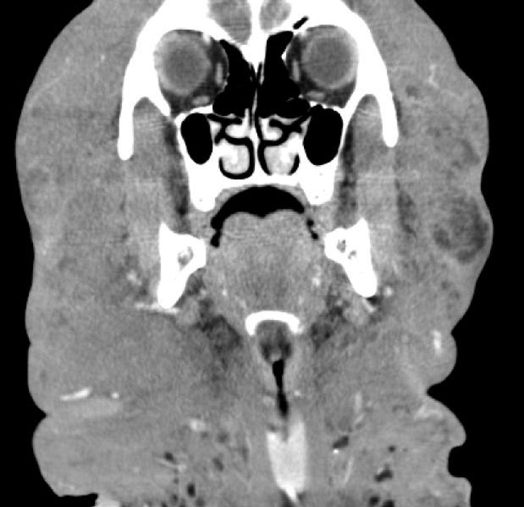
Diffuse subcutaneous soft tissue enlargement with a honeycomb appearance and increased vascularity in the entire head and neck, which were compatible with lymphedema due to foreign-body injection.

Ginat and Schatz suggested scarring as one of the late complications after filler injections. Although scarring can be a complication of all surgical procedures, none of the authors of medical literature reported that scar formation can be one of the complications of filler injections. Rather, as we hypothesized in our previous study, filler injection causes soft-tissue expansion and “creeping effect,” leading to less scar formation.2

Moreover, Ginat and Schatz argued that imaging can be helpful for delineating the extent of the excess material. However, plastic surgeons do not perform imaging studies to delineate the extent of overcorrection. Rather, some surgeons intentionally overcorrect the deformity to get higher patient satisfaction. In addition, acute complications such as initial signs of tissue necrosis, which are caused by excessive filler material injection, should be immediately reversed by using hyaluronidase injection or needle aspiration without hesitation. Surely, follow-up imaging can be of help to evaluate the success of the intervention.

In addition, Ginat and Schatz argued that chronic inflammation and lymphatic obstruction can lead to scar formation, referencing 2 previous articles.5,6 To our knowledge, previous studies by Rapaport et al5 and Mastruserio et al6 do not mention scar formation as a complication of the filling procedure at all. We also think that scar formation should be changed to “granuloma formation.”

Attempts to rejuvenate the aging hands have recently gained popularity; with development of dermal fillers, patients have various options.7 Recently, calcium hydroxyapatite (CaHA) fillers have been considered suitable for resurfacing the aging hand.8⇓⇓⇓⇓–13 In 2010, Bidic et al14 examined the anatomic superstructure of the dorsal hand soft tissues, which is relevant to hand rejuvenation, by using duplex sonography. According to our long-term follow-up, CaHA fillers occasionally result in hard palpable bonelike granulomas (Fig 2). According to previous studies, CaHA filler–related foreign-body granulomas cause increased FDG uptake in PET-CT imaging studies.15⇓–17 The mechanism of increased FDG uptake is likely associated with glycolysis in cellular elements, which are recruited at the site of injection.15 The attenuation of CaHA filler–related foreign-body granulomas is less than that of either cortical or medullary bone.16 We believe that radiologic studies of complications of hand rejuvenation can also be of interest to many clinicians.

Fig 2.
  • Download figure
  • Open in new tab
  • Download powerpoint
Fig 2.

Gross specimen of a CaHA filler–related foreign body granuloma.

Overall, the present study by Ginat and Schatz focused on foreign-body granuloma and its radiologic findings. Radiologic study cannot detect most early and late complications except some extensive skin necrosis and foreign-body granulomas. However, to the best of our knowledge, the article by Ginat and Schatz is the first attempt to introduce the imaging features of injected filler materials. We believe this issue has merit for many plastic and dermatologic surgeons as well as radiologists, and further study with more patients should be performed to validate the present study.

References

  1. 1.↵
    1. Ginat DT,
    2. Schatz CJ
    . Imaging features of midface injectable fillers and associated complications. AJNR Am J Neuroradiol 2012 Jul 26. [Epub ahead of print]
  2. 2.↵
    1. Park TH,
    2. Seo SW,
    3. Kim JK,
    4. et al
    . The efficacy of perilesional surgical approach for foreign body granuloma. Plast Reconstr Surg 2011; 127: 121e– 23e
    CrossRefPubMed
  3. 3.↵
    1. Park TH,
    2. Seo SW,
    3. Kim JK,
    4. et al
    . Clinical experience with polymethylmethacrylate microsphere filler complications. Aesthetic Plast Surg 2012; 36: 421– 26
    CrossRefPubMed
  4. 4.↵
    1. Park TH,
    2. Seo SW,
    3. Kim JK,
    4. et al
    . Clinical outcome in a series of 173 cases of foreign body granuloma: improved outcomes with a novel surgical technique. J Plast Reconstr Aesthet Surg 2012; 65: 29– 34
    CrossRefPubMed
  5. 5.↵
    1. Rapaport MJ,
    2. Vinnik C,
    3. Zarem H
    . Injectable silicone: cause of facial nodules, cellulitis, ulceration, and migration. Aesthetic Plast Surg 1996; 20: 267– 76
    CrossRefPubMed
  6. 6.↵
    1. Mastruserio DN,
    2. Pesqueira MJ,
    3. Cobb MW
    . Severe granulomatous reaction and facial ulceration occurring after subcutaneous silicone injection. J Am Acad Dermatol 1996; 34: 849– 52
    CrossRefPubMed
  7. 7.↵
    1. Park TH,
    2. Yeo KK,
    3. Seo SW,
    4. et al
    . Clinical experience with complications of hand rejuvenation. J Plast Reconstr Aesthet Surg 2012 Jul 2. [Epub ahead of print]
  8. 8.↵
    1. Busso M,
    2. Applebaum D
    . Hand augmentation with Radiesse (calcium hydroxylapatite). Dermatol Ther 2007; 20: 385– 87
    CrossRefPubMed
  9. 9.↵
    1. Bank DE
    . A novel approach to treatment of the aging hand with Radiesse. J Drugs Dermatol 2009; 8: 1122– 26
    PubMed
  10. 10.↵
    1. Edelson KL
    . Hand recontouring with calcium hydroxylapatite (Radiesse). J Cosmet Dermatol 2009; 8: 44– 51
    CrossRefPubMed
  11. 11.↵
    1. Marmur ES,
    2. Al Quran H,
    3. De Sa Earp AP,
    4. et al
    . A five-patient satisfaction pilot study of calcium hydroxylapatite injection for treatment of aging hands. Dermatol Surg 2009; 35: 1978– 84
    CrossRefPubMed
  12. 12.↵
    1. Gargasz SS,
    2. Carbone MC
    . Hand rejuvenation using Radiesse. Plast Reconstr Surg 2010; 125: 259e– 60e
    CrossRefPubMed
  13. 13.↵
    1. Sadick NS
    . A 52-week study of safety and efficacy of calcium hydroxylapatite for rejuvenation of the aging hand. J Drugs Dermatol 2011; 10: 47– 51
    PubMed
  14. 14.↵
    1. Bidic SM,
    2. Hatef DA,
    3. Rohrich RJ
    . Dorsal hand anatomy relevant to volumetric rejuvenation. Plast Reconstr Surg 2010; 126: 163– 68
    PubMed
  15. 15.↵
    1. Feeney JN,
    2. Fox JJ,
    3. Akhurst T
    . Radiological impact of the use of calcium hydroxylapatite dermal fillers. Clin Radiol 2009; 64: 897– 902
    CrossRefPubMed
  16. 16.↵
    1. Vazquez J,
    2. Rosenthal DI
    . Bilateral, symmetrical soft tissue calcifications in the face. Skeletal Radiol 2010; 39: 387– 89
    CrossRefPubMed
  17. 17.↵
    1. Carruthers A,
    2. Liebeskind M,
    3. Carruthers J,
    4. et al
    . Radiographic and computed tomographic studies of calcium hydroxylapatite for treatment of HIV-associated facial lipoatrophy and correction of nasolabial folds. Dermatol Surg 2008; 34 ( suppl 1): S78– 84
    CrossRefPubMed
  • © 2012 by American Journal of Neuroradiology
PreviousNext
Back to top

In this issue

American Journal of Neuroradiology: 33 (9)
American Journal of Neuroradiology
Vol. 33, Issue 9
1 Oct 2012
  • Table of Contents
  • Index by author
Advertisement
Print
Download PDF
Email Article

Thank you for your interest in spreading the word on American Journal of Neuroradiology.

NOTE: We only request your email address so that the person you are recommending the page to knows that you wanted them to see it, and that it is not junk mail. We do not capture any email address.

Enter multiple addresses on separate lines or separate them with commas.
Comments on an Article by Ginat and Schatz
(Your Name) has sent you a message from American Journal of Neuroradiology
(Your Name) thought you would like to see the American Journal of Neuroradiology web site.
CAPTCHA
This question is for testing whether or not you are a human visitor and to prevent automated spam submissions.
Citation Tools
Comments on an Article by Ginat and Schatz
Y.J. Park, T.H. Park
American Journal of Neuroradiology Oct 2012, 33 (9) E122-E123; DOI: 10.3174/ajnr.A3331

Citation Manager Formats

  • BibTeX
  • Bookends
  • EasyBib
  • EndNote (tagged)
  • EndNote 8 (xml)
  • Medlars
  • Mendeley
  • Papers
  • RefWorks Tagged
  • Ref Manager
  • RIS
  • Zotero
Share
Comments on an Article by Ginat and Schatz
Y.J. Park, T.H. Park
American Journal of Neuroradiology Oct 2012, 33 (9) E122-E123; DOI: 10.3174/ajnr.A3331
Reddit logo Twitter logo Facebook logo Mendeley logo
  • Tweet Widget
  • Facebook Like
  • Google Plus One
Purchase

Jump to section

  • Article
    • References
  • Figures & Data
  • Info & Metrics
  • References
  • PDF

Related Articles

  • No related articles found.
  • PubMed
  • Google Scholar

Cited By...

  • No citing articles found.
  • Crossref
  • Google Scholar

This article has not yet been cited by articles in journals that are participating in Crossref Cited-by Linking.

More in this TOC Section

  • Fair Performance of CT in Diagnosing Unilateral Vocal Fold Paralysis
  • Reply:
  • Regarding “Altered Blood Flow in the Ophthalmic and Internal Carotid Arteries in Patients with Age-Related Macular Degeneration Measured Using Noncontrast MR Angiography at 7T”
Show more LETTERS

Similar Articles

Advertisement

News and Updates

  • Lucien Levy Best Research Article Award
  • Thanks to our 2022 Distinguished Reviewers
  • Press Releases

Resources

  • Evidence-Based Medicine Level Guide
  • How to Participate in a Tweet Chat
  • AJNR Podcast Archive
  • Ideas for Publicizing Your Research
  • Librarian Resources
  • Terms and Conditions

Opportunities

  • Share Your Art in Perspectives
  • Get Peer Review Credit from Publons
  • Moderate a Tweet Chat

American Society of Neuroradiology

  • Neurographics
  • ASNR Annual Meeting
  • Fellowship Portal
  • Position Statements

© 2023 by the American Society of Neuroradiology | Print ISSN: 0195-6108 Online ISSN: 1936-959X

Powered by HighWire