
Abstract
BACKGROUND AND PURPOSE: Odontoid lateral mass interval asymmetry can be within the normal spectrum or the result of traumatic atlantoaxial injury. We sought to set radiographic guidelines for further investigation of odontoid lateral mass interval asymmetry in cervical spine CT studies of pediatric trauma patients.
MATERIALS AND METHODS: Fourteen children with C1–2 ligamentous injury or atlantoaxial rotational fixation/subluxation were retrospectively identified. We identified an additional 56 children fulfilling the following inclusion criteria: 1) They underwent C-spine CT to exclude traumatic injury, and 2) C-spine clearance and follow-up. Those were matched for age, sex, and severity of traumatic insult with the injured group. Clinical data were collected, and we measured the following parameters: anterior atlantodental interval; odontoid lateral mass interval; and the rotation of the head, C1, and C2.
RESULTS: A significant difference (P < .001) was found between the groups in cervical tenderness and torticollis. There was a significant difference in the atlantodental interval value (3.3 ± 0.8 mm in injured and 2.2 ± 0.5 mm in noninjured). The directionality of head, C1, and C2 rotation was significantly (P < .05) more toward the same direction in the noninjured group. We found significant linear correlation between head rotation and ipsilateral odontoid lateral mass interval asymmetry only in the noninjured at C1–2. With multivariant analysis, the presence of cervical tenderness and an abnormal atlantodental interval were the most significant variables.
CONCLUSIONS: Odontoid lateral mass interspace asymmetry in the absence of cervical tenderness and with a normal atlantodental interval is likely in the normal range and need not be further investigated.
ABBREVIATIONS:
- ADI
- atlantodental interval
- OLMI
- odontoid lateral mass interval
Cervical spine injury in children is rare relative to adults, with a reported incidence of 1%–2%.1,2 In children 8–10 years of age and younger, the upper cervical spine is more vulnerable to injury due to anatomic and developmental considerations.1,3,4 From a clinical perspective, exclusion of cervical spine injury in young children might be challenging because clinical decision tools are not as accurate as in adults.5 Although radiographs are advocated as the first line of screening for cervical spine injury in children with Glasgow Coma Scale >8 to reduce radiation, multidetector CT is often performed.
Odontoid lateral mass interval (OLMI) asymmetry was reported present in healthy adult and pediatric populations whether traumatized or not.6⇓⇓⇓⇓⇓–12 It is thought to arise from anatomic variation, head rotation, and muscle spasm. Nevertheless OLMI asymmetry might also imply ligamentous injury or atlantoaxial rotational fixation/subluxation at the C1–C2 level which, if present, could potentially lead to a catastrophic sequela.4,13,14 Therefore, when facing a CT study of a trauma victim with pure OLMI asymmetry and no fracture, radiologists and clinicians may find such injury difficult to exclude.
The differential diagnostic possibilities of OLMI asymmetry without fracture in trauma settings include C1–2 ligamentous injury and atlantoaxial rotational fixation/subluxation. Consequently, further work-up in such cases includes dynamic CT with head rotation,4 which leads to increased radiation exposure, or MR imaging with the added risk of child sedation and increasing costs. To our knowledge, no prior study has compared injured and noninjured populations with OLMI asymmetry to define which children could be cleared and which need additional work-up.
In this study, we sought to find radiologic and clinical parameters that could differentiate injured and noninjured patients with OLMI asymmetry in the scenario of acute trauma.
Materials and Methods
The study was approved by the institutional review board with a waiver of informed consent.
Patients
For this retrospective study, we included children 2–18 years of age. Children younger than 2 years of age were excluded to avoid inaccurate measurements due to insufficient vertebral ossification.
We reviewed our clinical data base during a 4-year time frame and identified 14 children diagnosed with C1–2 ligamentous injury without fracture or with atlantoaxial rotational fixation/subluxation. Patients with insufficient clinical data, no CT scans, and congenital malformations or diseases affecting the upper cervical spine were excluded. All patients underwent either MR imaging or dynamic CT to confirm the diagnosis. These 14 patients constitute the injured group.
During the same time frame, 2730 cervical spine CTs were performed to rule out traumatic injury. We reviewed the CT results and medical records of those patients and identified 381 children fulfilling the following inclusion criteria: 1) They underwent cervical spine CT to rule out traumatic injury, and 2) had eventual cervical spine clearance based on CT results, physical examination, and documented uneventful clinical follow-up. From this group, we selected 56 consecutive children matched for age, sex, and severity of traumatic insult with the injured group; these children constituted a control, noninjured group.
For each individual, relevant clinical data were collected from the patient's electronic records.
CT Technique and Measurements
CT scans were performed by using 16- to 64-section machines (GE Healthcare; Milwaukee, Wisconsin) with section widths ranging from 1 to 2.5 mm. All CT studies included had CT data appropriate for performance of MPR.
Measurements were performed on a PACS workstation. Axial, coronal, and sagittal reformats were constructed on the PACS to allow maximal symmetry. All measurements were performed by a single investigator (A.E.) to eliminate the possibility of interobserver variability.
Each individual right and left OLMI interspace was measured on the axial and coronal planes at the midlateral mass level (Fig 1). OLMI asymmetry was calculated in the coronal and axial planes by subtracting the value of the right interspace from that of the left one. The values were noted in absolute and real numbers to determine the degree and directionality of the asymmetry.
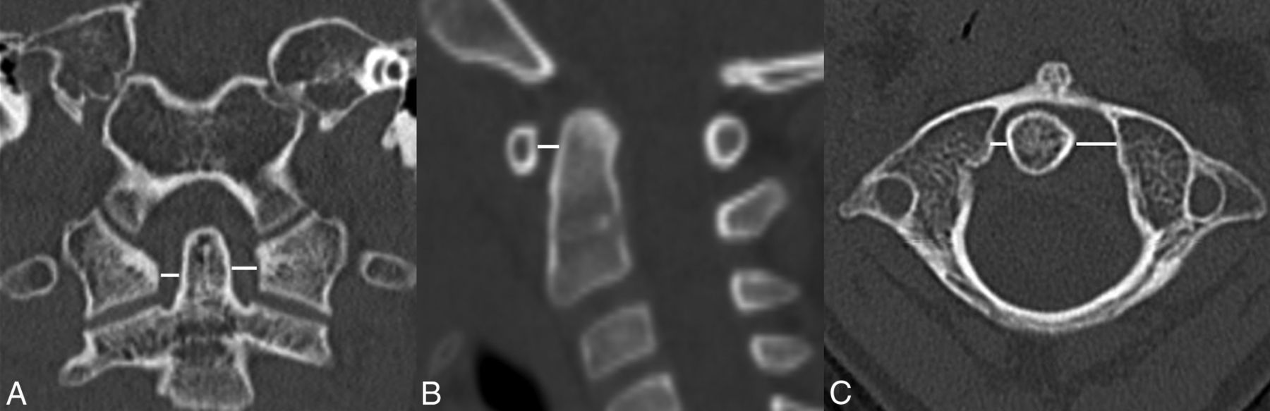
Coronal, sagittal, and axial CT sections at the craniocervical junction that show OLMI measurement (white line) at the midlateral mass level (A and C) and ADI measurement (B).
For each individual, the atlantodental interval (ADI) was also measured on the midsagittal plane (Fig 1).
Head rotation was measured in degrees by using the angle tool of the PACS toolbox. The rotation of the head, C1, and C2 relative to the CT table was measured by drawing an angle in which 1 ray was a line along the midline axis of each structure (head/C1/C2) and the second ray was parallel to the CT table, pointing toward the right side of the screen (Fig 2). The rotation of each structure relative to the other was calculated by subtracting the angle of the lower structure from that of the upper one (eg, rotation of C2 relative to C1 equals the C1 angle minus the C2 angle). Therefore, rightward rotation was given a negative value, and left head rotation had a positive value. Those measurements were noted in absolute and real values to evaluate the degree and directionality of the rotation.

Axial CT image at the C1 level shows measurement of C1 rotation.
Statistical Analysis
The data were analyzed with SPSS, Version 17 (IBM, Armonk, New York). The differences between the 2 groups in quantitative variables were analyzed by Mann-Whitney U and t tests. The differences between the 2 groups in the categoric variables were tested by Fisher exact and Pearson χ2 tests. The Pearson correlation was used to find the relation between the degree and directionality of head rotation and OLMI asymmetry. Multivariate analysis by logistic regression was used to study the parameters that determined whether the patient was in the noninjured or the injured group. P ≤ .05 was considered significant.
Results
The demographics and clinical data of the injured and noninjured groups are summarized in the Table. There was no statistically significant difference between the groups in age, sex, and percentage of patients with loss of consciousness. Cervical tenderness was present in 78.6% of patients in the injured group compared with 8.7% of the noninjured group. Torticollis was present in 61.5% of the injured patients and absent in the noninjured group. Those differences were statistically significant (P < .001) (Table).
Demographics and clinical characteristics of the study groups
OLMI asymmetry was 3.8 ± 2.2 mm (mean) in the injured group and 1.4 ± 0.7 mm in the noninjured group; this difference was statistically significant (P < .001). There was no significant difference between measurements in the coronal or axial planes, and the values represent the average of the 2 measurements. ADI was significantly different between the groups: −3.3 ± 0.8 mm in the injured group and 2.2 ± 0.5 mm in the noninjured group (P < .001).
During head rotation, the directionality of the head, C1, and C2 rotation was significantly (P < .05) more toward the same direction in the noninjured group. In the 2 groups, there was a trend toward OLMI increase during head rotation. We found a significant linear correlation between head rotation and OLMI asymmetry only in the noninjured group at the C1–2 level. During head rotation to the right, there was a significant increase in OLMI asymmetry toward the right (the right OLMI was larger) (P < .05) and vice versa on head rotation to the left. No such correlation was found in other levels and in the injured group.
On multivariate analysis, cervical tenderness and an abnormal ADI were the most significant variables that differentiated the injured and noninjured groups (P < .001).
Discussion
The dilemma concerning whether additional work-up is needed for a child with OLMI asymmetry is encountered almost on a daily basis in busy trauma centers. Accumulating data show that OLMI asymmetry can be a normal variant in adults6⇓⇓–9,11,12 and children,10 or it can be related to head positioning. Nevertheless, OLMI asymmetry can also imply ligamentous cervical spine injury at the craniocervical junction, which can be considerably disabling and even lethal.4,13,14 In this study, we compared injured and noninjured children who presented to the emergency department similarly to enable us to find parameters that will differentiate between the groups. Such comparison has not been performed before in the literature, to the best of our knowledge.
In our study, we compared OLMI asymmetry and head rotation in pediatric patients with known traumatic injury without fracture at the C1–2 level with matched pediatric trauma patients without cervical spine injury. We found significant differences between injured and noninjured patients in the following parameters: Cervical tenderness and torticollis were significantly more common in the injured patients; OLMI asymmetry and ADI were significantly larger in the injured group. Analysis of head rotation parameters showed that rotation of the head, C1, and C2 was significantly more toward the same direction in the noninjured group and there was linear correlation between the direction of head rotation and OLMI asymmetry at the C1–2 level only in noninjured patients.
The mean OLMI asymmetry in the noninjured patients was 1.4 ± 0.7 mm, which is similar to results obtained in prior studies in children,10 therefore confirming the validity of our data. The asymmetry in the injured group was 3.8 ± 2.2 mm, which overlaps with the noninjured group. In a study by Wolansky et al,8 an asymmetry >3 mm was found to be abnormal in adults. On the other hand, Billmann et al12 did not find a significant correlation between OLMI asymmetry and traumatic injury in adults. It seems that on the basis of our results and prior studies, a division of normal and abnormal based on OLMI asymmetry alone cannot be applied.
ADI was found to be one of the significant parameters in multivariate analysis that differentiated injured from noinjured children. The maximal interval in the noninjured group was 2.7 mm, and the minimal interval in the injured group was 2.5 mm, giving almost complete separation between the groups. Bertozzi et al15 studied cervical spine parameters at the craniocervical junction in noninjured children and found a similar maximal ADI of 2.6 mm.
The analysis of head rotation parameters gives additional tools to differentiate the groups. In the noninjured group, we found increasing OLMI distance on head rotation ipsilateral to the rotation side. Those normal relations were also shown by Sutherland et al16 in postmortem examinations. Pang and Li,17 in a study of head rotation in children, found that most of the rotation occurs at the C1–2 level and has a predictable behavior. This behavior is disturbed when the child has an atlantoaxial rotatory fixation.18 Possibly the ligamentous disruption in the injured group in our study prevented normal motion. Additional studies of head rotation parameters in patients with ligamentous injury at the C1–2 level may confirm this finding.
One of the limitations in this study is excluding ligamentous injury in the noninjured patients without MR imaging. However, other studies also used normal CT findings and no evidence of cervical spine injury on emergency department discharge as tools for confirming “no injury.”19 MR imaging is an expensive tool, and screening every patient with cervical spine trauma by using MR imaging is not justified. We additionally confirmed clearance of cervical spine injury on a clinic follow-up visit in our noninjured group. Another limitation of our study is its retrospective nature. Finally, the relatively small number of injured patients in our study is another limitation and probably reflects low prevalence of C1–C2 injury in children, at least at our institution. Further multicenter prospective studies that use the parameters we suggest for injury exclusion are needed.
Conclusions
OLMI asymmetry in the absence of cervical tenderness and with normal ADI (<2.6 mm) is likely due to head positioning and should not be further investigated unless high clinical suspicion exists.
Footnotes
Disclosures: David M. Yousem—UNRELATED: Expert Testimony: medicolegal cases, self-employed; Payment for Lectures (including service on Speakers Bureaus): American College of Radiology Education Center Course Director*; Royalties: 3 books with Elsevier; Payment for Development of Educational Presentations: CMEInfo.com for Continuing Medical Education courses.* Izlem Izbudak—UNRELATED: Grants/Grants Pending: Siemens,* Comments: MRI DTI research grant. *Money paid to the institution.
Paper previously presented at: Annual Meeting of the American Society of Neuroradiology, June 4–9, 2011; Seattle, Washington.
References
- Received March 22, 2015.
- Accepted after revision May 18, 2015.
- © 2016 by American Journal of Neuroradiology


Jump to section
Related Articles
Cited By...
- No citing articles found.