Advertisement

Main menu

  • Home
  • Content
    • Current Issue
    • Accepted Manuscripts
    • Article Preview
    • Past Issue Archive
    • Video Articles
    • AJNR Case Collection
    • Case of the Week Archive
    • Case of the Month Archive
    • Classic Case Archive
  • Special Collections
    • AJNR Awards
    • Low-Field MRI
    • Alzheimer Disease
    • ASNR Foundation Special Collection
    • Photon-Counting CT
    • View All
  • Multimedia
    • AJNR Podcasts
    • AJNR SCANtastic
    • Trainee Corner
    • MRI Safety Corner
    • Imaging Protocols
  • For Authors
    • Submit a Manuscript
    • Submit a Video Article
    • Submit an eLetter to the Editor/Response
    • Manuscript Submission Guidelines
    • Statistical Tips
    • Fast Publishing of Accepted Manuscripts
    • Graphical Abstract Preparation
    • Imaging Protocol Submission
    • Author Policies
  • About Us
    • About AJNR
    • Editorial Board
    • Editorial Board Alumni
  • More
    • Become a Reviewer/Academy of Reviewers
    • Subscribers
    • Permissions
    • Alerts
    • Feedback
    • Advertisers
    • ASNR Home

User menu

  • Alerts
  • Log in

Search

  • Advanced search
American Journal of Neuroradiology

American Journal of Neuroradiology

ASHNR American Society of Functional Neuroradiology ASHNR American Society of Pediatric Neuroradiology ASSR
  • Alerts
  • Log in

Advanced Search

  • Home
  • Content
    • Current Issue
    • Accepted Manuscripts
    • Article Preview
    • Past Issue Archive
    • Video Articles
    • AJNR Case Collection
    • Case of the Week Archive
    • Case of the Month Archive
    • Classic Case Archive
  • Special Collections
    • AJNR Awards
    • Low-Field MRI
    • Alzheimer Disease
    • ASNR Foundation Special Collection
    • Photon-Counting CT
    • View All
  • Multimedia
    • AJNR Podcasts
    • AJNR SCANtastic
    • Trainee Corner
    • MRI Safety Corner
    • Imaging Protocols
  • For Authors
    • Submit a Manuscript
    • Submit a Video Article
    • Submit an eLetter to the Editor/Response
    • Manuscript Submission Guidelines
    • Statistical Tips
    • Fast Publishing of Accepted Manuscripts
    • Graphical Abstract Preparation
    • Imaging Protocol Submission
    • Author Policies
  • About Us
    • About AJNR
    • Editorial Board
    • Editorial Board Alumni
  • More
    • Become a Reviewer/Academy of Reviewers
    • Subscribers
    • Permissions
    • Alerts
    • Feedback
    • Advertisers
    • ASNR Home
  • Follow AJNR on Twitter
  • Visit AJNR on Facebook
  • Follow AJNR on Instagram
  • Join AJNR on LinkedIn
  • RSS Feeds

AJNR Awards, New Junior Editors, and more. Read the latest AJNR updates

Brief ReportSpine Imaging and Spine Image-Guided Interventions
Open Access

A Novel Patient-Positioning Device for Dynamic CT Myelography

Andrew L. Callen, Rich Wojcik and Michael Bojanowski
American Journal of Neuroradiology October 2023, DOI: https://doi.org/10.3174/ajnr.A8023
Andrew L. Callen
aFrom the Department of Radiology (A.L.C.), Neuroradiology Section, University of Colorado Anschutz Medical Campus, Aurora, Colorado
  • Find this author on Google Scholar
  • Find this author on PubMed
  • Search for this author on this site
  • ORCID record for Andrew L. Callen
Rich Wojcik
bK1C Consulting LLC (R.W., M.B.), Denver, Colorado
  • Find this author on Google Scholar
  • Find this author on PubMed
  • Search for this author on this site
  • ORCID record for Rich Wojcik
Michael Bojanowski
bK1C Consulting LLC (R.W., M.B.), Denver, Colorado
  • Find this author on Google Scholar
  • Find this author on PubMed
  • Search for this author on this site
  • Article
  • Figures & Data
  • Supplemental
  • Info & Metrics
  • Responses
  • References
  • PDF
Loading

Abstract

SUMMARY: We describe a novel patient-positioning device for dynamic CT myelography. Dynamic CT myelography requires angling the patient’s spine to distribute dense contrast along the dependent thecal sac. The proposed device is constructed of a low-density reinforced polymer frame and can be raised or lowered to various heights with a hand-operated mechanism, allowing precise adjustment of the spinal angle and control of the contrast bolus, increasing the safety, reproducibility, and sensitivity of dynamic CT myelography.

ABBREVIATION:

dCTM
dynamic CT myelography

Dynamic CT myelography (dCTM) is an effective technique for the localization of CSF leaks and the detection of CSF venous fistulas.1 Unlike conventional CT myelography, in which the goal is the even diffusion of contrast material throughout the subarachnoid space, in dCTM, the goal is to distribute dense, unmixed contrast along the dependent thecal sac.2 When one looks for a CSF venous fistula or a ruptured meningeal diverticulum, the patient is in a decubitus position so that dense contrast fills the nerve root sleeves and meningeal diverticula. When a ventral dural defect is suspected, the patient is positioned prone with the hips elevated to localize the site of subarachnoid contrast extravasation into the ventral epidural space.3 Unlike digital subtraction myelography, which can use a tilt table to facilitate gravity-dependent egress of contrast from the lumbar puncture site toward the craniocervical junction, patient positioning in CT typically relies on the use of either a foam wedge or an inflatable mattress device positioned under the patient’s hips to angle the spine.4 A foam wedge can lead to a predictable, constant movement of contrast, but positioning a patient on top of the wedge from the beginning of the procedure precludes the accurate measurement of spinal opening pressure, and the prone position can often require additional pillows or other material to elevate the hips sufficiently to overcome a patient’s lumbar lordosis (Fig 1).Additionally, beginning the patient in the angled position can often result in decreased tension of the thecal sac, causing difficulty achieving a subarachnoid puncture. An inflatable mattress allows the patient to begin the examination in the horizontal position, followed by temporary inflation and deflation before scanning. However, the time required for inflation can be variable, and patient positioning can be unstable, often requiring assistants to secure the patient while the device is in the inflated position. In the case of type 1 or 2 CSF leaks, misjudging the timing of the contrast bolus or delays due to needing to reposition a patient after contrast injection can lead to poorer visualization of a dural defect and decreased diagnostic accuracy (Fig 2). To overcome the drawbacks of each commonly used technique, we designed a unique patient-positioning device to accurately and safely manipulate the spinal angle during dCTM, to increase the safety, reproducibility, and sensitivity of the examination.

FIG 1.
  • Download figure
  • Open in new tab
  • Download powerpoint
FIG 1.

Spine angulation using a foam wedge versus using the elevation device in suspected ventral dural defects. A, A patient with a suspected ventral dural defect, positioned on top of a foam wedge for dCTM. CT image obtained after contrast injection shows pooling of contrast at the patient’s lumbar lordosis (arrows) due to insufficient hip elevation. B, A separate patient with a suspected ventral dural defect on top of the novel positioning device. CT image obtained after contrast injection demonstrates egress of contrast from the puncture site to the craniocervical junction. Smaller FOV image demonstrates extravasation of contrast from the subarachnoid space into the ventral epidural space at C7–T1 (arrow), consistent with a ventral dural defect.

FIG 2.
  • Download figure
  • Open in new tab
  • Download powerpoint
FIG 2.

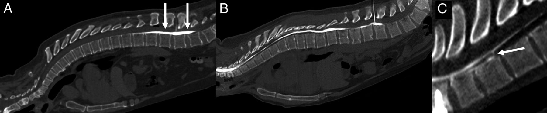
A ventral dural defect at T2–T3. A, Image obtained immediately after contrast injection demonstrates clear extravasation of contrast (arrow) from the subarachnoid space into the ventral epidural space, precisely localizing this patient’s dural defect. B, A second image obtained 38 seconds after the first demonstrates rapid diffusion of contrast throughout the ventral epidural fluid collection (arrows), obscuring the precise site of the dural defect.

Device Description

The device consists of a reinforced polymer frame covered with heavy duty canvas fabric and supportive foam cushioning, secured to the CT table at the end distal to the gantry by straps and 3D-printed plastic inserts. The inserts are specific to the channel along the length of the patient’s bed, which can vary among manufacturers. The device was initially designed for a Somatom Alpha CT table (Siemens). This CT table uses a channel size of 0.219 inches (5.6 mm) in diameter with a 0.129-inch (3.3-mm) slot opening. If the channel diameter and opening on the patient bed are within ±0.010 inches (± 0.25 mm) of the dimensions above, then the straps will be compatible with other CT tables. If the channel diameters vary more than these dimensions, new inserts may be 3D-printed at minimal cost. The device is bisected by a pivot point. At the fixed end, there is a hand-operated winch, which connects to the other end of the device by a polyester strap that runs beneath the frame (Fig 3). Underneath the mobile end proximal to the gantry, there are polyurethane wheels that allow translation along the Z-axis of the bore, depending on the length of the strap, which is controlled by the hand winch. The portion of the device that is included in the FOV is constructed of reinforced polymer and engineered polymers to minimize streak artifacts and the patient dose (Fig 4). The only metallic components present are along the most distal and proximal ends of the device, which remain outside of the FOV.

FIG 3.
  • Download figure
  • Open in new tab
  • Download powerpoint
FIG 3.

Device schematic.

FIG 4.
  • Download figure
  • Open in new tab
  • Download powerpoint
FIG 4.

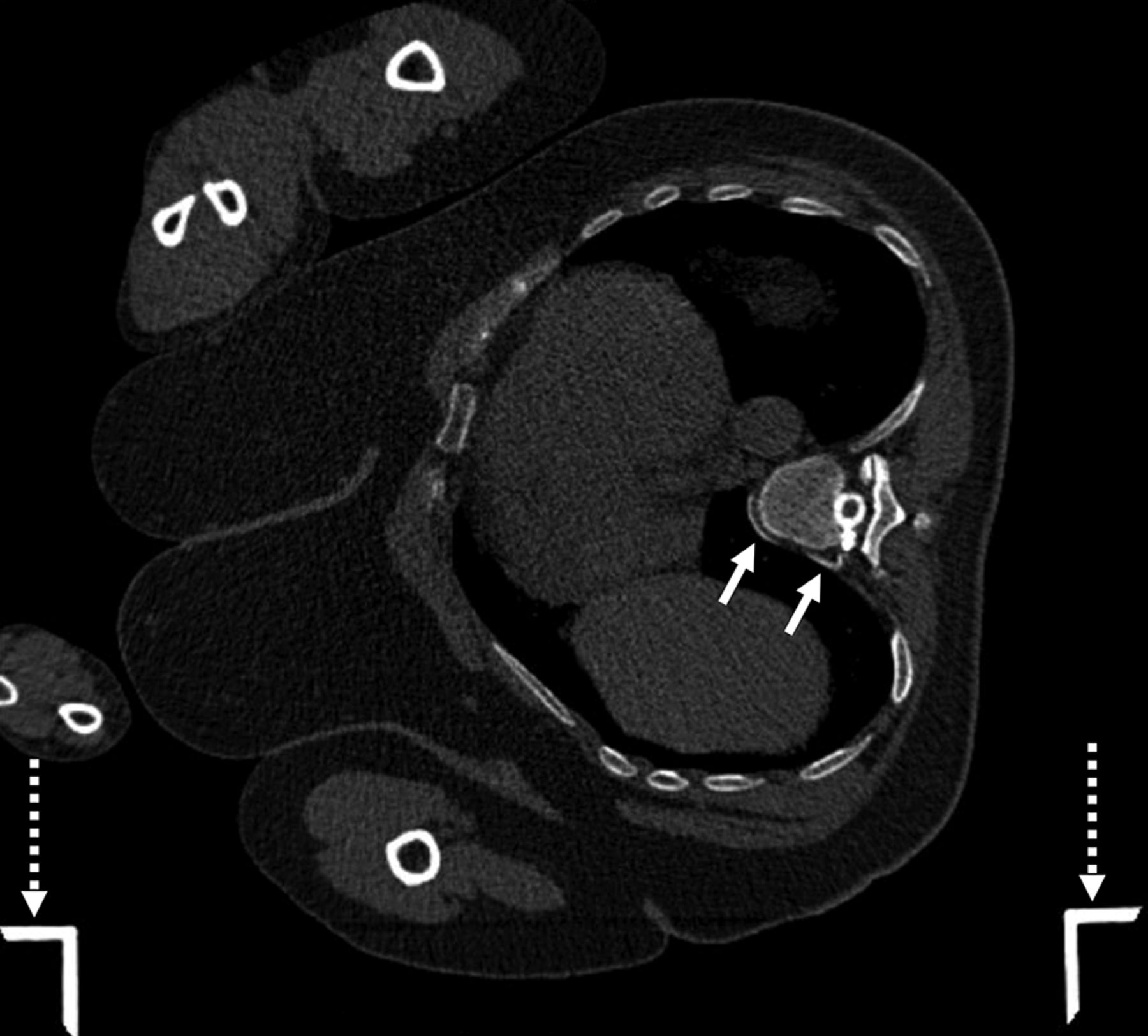
Localization of a CSF venous fistula using the positioning device. Axial CT image with a wide FOV demonstrates a patient in the right lateral decubitus position on top of the positioning device during dCTM with a CSF venous fistula (solid arrows) arising from a right T8–T9 meningeal diverticulum. The device frame (dashed arrows) generates no streak artifacts.

The overall device measures 91 inches long × 21 inches wide × 9 inches tall (231 × 54 × 23 cm), with the bed portion measuring 77 inches long × 20 inches wide (196 × 51 cm). It can support a patient weighing up to 300 pounds (136 kg) and up to 76 inches (193 cm) in height. Initially designed for the Somatom Alpha CT machine, the fixation straps can accommodate multiple CT machine types as long as the CT table uses a patient bed that is 17–19 inches wide with a minimum length of 91 inches and a curvature height of 2 inches from the edges to the bottom of the bed.

Device Use

The device can be unpacked and secured to the CT table in <5 minutes, best achieved with 2 operators. The patient can be positioned either decubitus or prone and is secured to the device with 2 fabric straps (Fig 5). At the mobile end proximal to the gantry, there are 2 fixed handles that can be held by the patient in the prone position. The patient can begin the examination in the horizontal or elevated position, depending on the preference of the operator. The height of the device can be adjusted depending on the size of the patient and is limited only by the width of his or her hips to allow the maximal elevation possible without obstructing movement into the gantry. Patient elevation takes approximately 8–12 seconds (Supplementary Online Video). Because the patient’s elevation is controlled and predictable, the needle may be left in the patient during height adjustment to allow additional contrast administration during the examination if necessary. If the patient is too large to fit in the gantry in the elevated position, the device can be elevated and subsequently lowered with the patient outside the gantry before scanning. Because the device is secured to the CT table, the patient may rotate to the contralateral decubitus position while remaining on the device if desired. Additionally, the patient may remain on the device during patching or other CT-guided procedures. Angling the patient, if desired, may allow needle trajectories that would otherwise require tilting of the CT gantry.

FIG 5.
  • Download figure
  • Open in new tab
  • Download powerpoint
FIG 5.

Example of the device in use in decubitus (A, Flat. B, Elevated) and prone (C, Flat, D, Elevated) patient positions.

CONCLUSIONS

We describe a dCTM patient-positioning device that offers several practical advantages over current commonly used methods for angling the spine on the CT table. By providing accurate and safe manipulation of the spinal angle, the device has the potential to increase diagnostic accuracy and procedural reproducibility and can accommodate patients of varying sizes. Patients can be positioned decubitus or prone and can start the examination in a horizontal or angled position. Because elevation is smooth and controlled, height can be adjusted while leaving the needle in place if needed. We believe that the incorporation of this device in clinical practice will improve patient outcomes and streamline the dCTM procedure for radiologists. Further studies are warranted to validate its effectiveness, assess patient and provider satisfaction as well as explore potential applications for manipulating the spinal angle in other CT-guided procedures. To date, we have performed 32 dCTM examinations using the device, all of which have been executed without compromised image quality. A patent is pending for the device, and it is our hope that it will be commercially available soon.

Acknowledgments

We acknowledge Ashesh A. Thaker, MD, Vincent M. Timpone, MD, and Josh Grip for input early in the design process.

Footnotes

  • Grant support from the Considine Family Foundation was used for the development of this device.

  • Disclosure forms provided by the authors are available with the full text and PDF of this article at www.ajnr.org.

Indicates open access to non-subscribers at www.ajnr.org

References

  1. 1.↵
    1. Kranz PG,
    2. Gray L,
    3. Amrhein TJ
    . Decubitus CT myelography for detecting subtle CSF leaks in spontaneous intracranial hypotension. AJNR Am J Neuroradiol 2019;40:754–56 doi:10.3174/ajnr.A5995 pmid:30819772
    Abstract/FREE Full Text
  2. 2.↵
    1. Mamlouk MD,
    2. Ochi RP,
    3. Jun P, et al
    . Decubitus CT myelography for CSF-venous fistulas: a procedural approach. AJNR Am J Neuroradiol 2021;42:32–36 doi:10.3174/ajnr.A6844 pmid:33122215
    Abstract/FREE Full Text
  3. 3.↵
    1. Thielen KR,
    2. Sillery JC,
    3. Morris JM, et al
    . Ultrafast dynamic computed tomography myelography for the precise identification of high-flow cerebrospinal fluid leaks caused by spiculated spinal osteophytes. J Neurosurg Spine 2015;22:324–31 doi:10.3171/2014.10.SPINE14209 pmid:25555057
    CrossRefPubMed
  4. 4.↵
    1. Callen AL,
    2. Timpone VM,
    3. Schwertner A, et al
    . Algorithmic multimodality approach to diagnosis and treatment of spinal CSF leak and venous fistula in patients with spontaneous intracranial hypotension. AJR Am J Roentgenol 2022;219:292–301 doi:10.2214/AJR.22.27485
    CrossRefPubMed
  • Received August 2, 2023.
  • Accepted after revision September 3, 2023.
  • © 2023 by American Journal of Neuroradiology
PreviousNext
Back to top
Advertisement
Print
Download PDF
Email Article

Thank you for your interest in spreading the word on American Journal of Neuroradiology.

NOTE: We only request your email address so that the person you are recommending the page to knows that you wanted them to see it, and that it is not junk mail. We do not capture any email address.

Enter multiple addresses on separate lines or separate them with commas.
A Novel Patient-Positioning Device for Dynamic CT Myelography
(Your Name) has sent you a message from American Journal of Neuroradiology
(Your Name) thought you would like to see the American Journal of Neuroradiology web site.
CAPTCHA
This question is for testing whether or not you are a human visitor and to prevent automated spam submissions.
Cite this article
Andrew L. Callen, Rich Wojcik, Michael Bojanowski
A Novel Patient-Positioning Device for Dynamic CT Myelography
American Journal of Neuroradiology Oct 2023, DOI: 10.3174/ajnr.A8023

Citation Manager Formats

  • BibTeX
  • Bookends
  • EasyBib
  • EndNote (tagged)
  • EndNote 8 (xml)
  • Medlars
  • Mendeley
  • Papers
  • RefWorks Tagged
  • Ref Manager
  • RIS
  • Zotero
0 Responses
Respond to this article
Share
Bookmark this article
A Novel Patient-Positioning Device for Dynamic CT Myelography
Andrew L. Callen, Rich Wojcik, Michael Bojanowski
American Journal of Neuroradiology Oct 2023, DOI: 10.3174/ajnr.A8023
del.icio.us logo Twitter logo Facebook logo Mendeley logo
  • Tweet Widget
  • Facebook Like
  • Google Plus One
Purchase

Jump to section

  • Article
    • Abstract
    • ABBREVIATION:
    • CONCLUSIONS
    • Acknowledgments
    • Footnotes
    • References
  • Figures & Data
  • Supplemental
  • Info & Metrics
  • Responses
  • References
  • PDF

Related Articles

  • PubMed
  • Google Scholar

Cited By...

  • Assessing the Diagnostic Value of Brain White Matter Hyperintensities and Clinical Symptoms in Predicting the Detection of CSF-Venous Fistula in Patients with Suspected Spontaneous Intracranial Hypotension
  • Spinal CSF Leaks: The Neuroradiologist Transforming Care
  • Myelographic Techniques for the Localization of CSF-Venous Fistulas: Updates in 2024
  • Crossref (3)
  • Google Scholar

This article has been cited by the following articles in journals that are participating in Crossref Cited-by Linking.

  • Myelographic Techniques for the Localization of CSF-Venous Fistulas: Updates in 2024
    Ajay A. Madhavan, Waleed Brinjikji, Jeremy K. Cutsforth-Gregory, Timothy J. Amrhein, Peter G. Kranz, John C. Benson, Felix E. Diehn, Ben A. Johnson-Tesch, Greta B. Liebo, Vance T. Lehman, Ian T. Mark, Pearse P. Morris, Michael P. Oien, Darya P. Shlapak, Jared T. Verdoorn
    American Journal of Neuroradiology 2024 45 10
  • Spinal CSF Leaks: The Neuroradiologist Transforming Care
    Mark D. Mamlouk, Andrew L. Callen, Ajay A. Madhavan, Niklas Lützen, Lalani Carlton Jones, Ian T. Mark, Waleed Brinjikji, John C. Benson, Jared T. Verdoorn, D.K. Kim, Timothy J. Amrhein, Linda Gray, William P. Dillon, Marcel M. Maya, Thien J. Huynh, Vinil N. Shah, Tomas Dobrocky, Eike I. Piechowiak, Joseph Levi Chazen, Michael D. Malinzak, Jessica L. Houk, Peter G. Kranz
    American Journal of Neuroradiology 2024 45 11
  • Assessing the Diagnostic Value of Brain White Matter Hyperintensities and Clinical Symptoms in Predicting the Detection of CSF-Venous Fistula in Patients with Suspected Spontaneous Intracranial Hypotension
    Samantha L. Pisani Petrucci, Nadya Andonov, Peter Lennarson, Marius Birlea, Chantal O’Brien, Danielle Wilhour, Abigail Anderson, Jeffrey L. Bennett, Andrew L. Callen
    American Journal of Neuroradiology 2025 46 5

More in this TOC Section

  • Optimization of Photon Counting CT Myelography
  • Characteristics of SIH Type I Culprit Lesions
  • Management Outcomes For VO Spine Biopsy
Show more SPINE IMAGING AND SPINE IMAGE-GUIDED INTERVENTIONS

Similar Articles

Advertisement

Indexed Content

  • Current Issue
  • Accepted Manuscripts
  • Article Preview
  • Past Issues
  • Editorials
  • Editor's Choice
  • Fellows' Journal Club
  • Letters to the Editor
  • Video Articles

Cases

  • Case Collection
  • Archive - Case of the Week
  • Archive - Case of the Month
  • Archive - Classic Case

More from AJNR

  • Trainee Corner
  • Imaging Protocols
  • MRI Safety Corner
  • Book Reviews

Multimedia

  • AJNR Podcasts
  • AJNR Scantastics

Resources

  • Turnaround Time
  • Submit a Manuscript
  • Submit a Video Article
  • Submit an eLetter to the Editor/Response
  • Manuscript Submission Guidelines
  • Statistical Tips
  • Fast Publishing of Accepted Manuscripts
  • Graphical Abstract Preparation
  • Imaging Protocol Submission
  • Evidence-Based Medicine Level Guide
  • Publishing Checklists
  • Author Policies
  • Become a Reviewer/Academy of Reviewers
  • News and Updates

About Us

  • About AJNR
  • Editorial Board
  • Editorial Board Alumni
  • Alerts
  • Permissions
  • Not an AJNR Subscriber? Join Now
  • Advertise with Us
  • Librarian Resources
  • Feedback
  • Terms and Conditions
  • AJNR Editorial Board Alumni

American Society of Neuroradiology

  • Not an ASNR Member? Join Now

© 2025 by the American Society of Neuroradiology All rights, including for text and data mining, AI training, and similar technologies, are reserved.
Print ISSN: 0195-6108 Online ISSN: 1936-959X

Powered by HighWire