Advertisement

Main menu

  • Home
  • Content
    • Current Issue
    • Accepted Manuscripts
    • Article Preview
    • Past Issue Archive
    • Video Articles
    • AJNR Case Collection
    • Case of the Week Archive
    • Case of the Month Archive
    • Classic Case Archive
  • Special Collections
    • AJNR Awards
    • ASNR Foundation Special Collection
    • Most Impactful AJNR Articles
    • Photon-Counting CT
    • Spinal CSF Leak Articles (Jan 2020-June 2024)
  • Multimedia
    • AJNR Podcasts
    • AJNR SCANtastic
    • Trainee Corner
    • MRI Safety Corner
    • Imaging Protocols
  • For Authors
    • Submit a Manuscript
    • Submit a Video Article
    • Submit an eLetter to the Editor/Response
    • Manuscript Submission Guidelines
    • Statistical Tips
    • Fast Publishing of Accepted Manuscripts
    • Graphical Abstract Preparation
    • Imaging Protocol Submission
    • Author Policies
  • About Us
    • About AJNR
    • Editorial Board
    • Editorial Board Alumni
  • More
    • Become a Reviewer/Academy of Reviewers
    • Subscribers
    • Permissions
    • Alerts
    • Feedback
    • Advertisers
    • ASNR Home

User menu

  • Alerts
  • Log in

Search

  • Advanced search
American Journal of Neuroradiology

American Journal of Neuroradiology

ASHNR American Society of Functional Neuroradiology ASHNR American Society of Pediatric Neuroradiology ASSR
  • Alerts
  • Log in

Advanced Search

  • Home
  • Content
    • Current Issue
    • Accepted Manuscripts
    • Article Preview
    • Past Issue Archive
    • Video Articles
    • AJNR Case Collection
    • Case of the Week Archive
    • Case of the Month Archive
    • Classic Case Archive
  • Special Collections
    • AJNR Awards
    • ASNR Foundation Special Collection
    • Most Impactful AJNR Articles
    • Photon-Counting CT
    • Spinal CSF Leak Articles (Jan 2020-June 2024)
  • Multimedia
    • AJNR Podcasts
    • AJNR SCANtastic
    • Trainee Corner
    • MRI Safety Corner
    • Imaging Protocols
  • For Authors
    • Submit a Manuscript
    • Submit a Video Article
    • Submit an eLetter to the Editor/Response
    • Manuscript Submission Guidelines
    • Statistical Tips
    • Fast Publishing of Accepted Manuscripts
    • Graphical Abstract Preparation
    • Imaging Protocol Submission
    • Author Policies
  • About Us
    • About AJNR
    • Editorial Board
    • Editorial Board Alumni
  • More
    • Become a Reviewer/Academy of Reviewers
    • Subscribers
    • Permissions
    • Alerts
    • Feedback
    • Advertisers
    • ASNR Home
  • Follow AJNR on Twitter
  • Visit AJNR on Facebook
  • Follow AJNR on Instagram
  • Join AJNR on LinkedIn
  • RSS Feeds

AJNR is seeking candidates for the AJNR Podcast Editor. Read the position description.

OtherHEAD AND NECK

Bilateral Aberrant Internal Carotid Arteries with Bilateral Persistent Stapedial Arteries and Bilateral Duplicated Internal Carotid Arteries

John D. Roll, Martin A. Urban, Theodore C. Larson, Philippe Gailloud, Pradeep Jacob and H. Ric Harnsberger
American Journal of Neuroradiology April 2003, 24 (4) 762-765;
John D. Roll
  • Find this author on Google Scholar
  • Find this author on PubMed
  • Search for this author on this site
Martin A. Urban
  • Find this author on Google Scholar
  • Find this author on PubMed
  • Search for this author on this site
Theodore C. Larson
  • Find this author on Google Scholar
  • Find this author on PubMed
  • Search for this author on this site
Philippe Gailloud
  • Find this author on Google Scholar
  • Find this author on PubMed
  • Search for this author on this site
Pradeep Jacob
  • Find this author on Google Scholar
  • Find this author on PubMed
  • Search for this author on this site
H. Ric Harnsberger
  • Find this author on Google Scholar
  • Find this author on PubMed
  • Search for this author on this site
  • Article
  • Figures & Data
  • Info & Metrics
  • Responses
  • References
  • PDF
Loading

Abstract

Summary: A 5-year-old boy was evaluated for a left retrotympanic mass found at otoscopy. Subsequent petrous bone CT and MR angiographic examinations demonstrated bilateral aberrant internal carotid, bilateral stapedial artery persistence, and bilateral duplicated internal carotid arteries. Imaging findings and their clinical relevance are discussed. A second case of unilateral aberrant internal carotid artery with a persistent stapedial artery is included for comparison.

An aberrant internal carotid artery (ICA) in the middle ear is a rare congenital finding. If the diagnosis is not made before middle ear surgery is performed, hemorrhage, stroke, or death may occur as a result of injury to the vessel.

A persistent stapedial artery (PSA) is a more benign congenital diagnosis; however, symptoms of hearing loss and tinnitus may be present. Tinnitus was noted to resolve in one case after lysis and cauterization of the PSA (1). Stapes surgery is complicated by the presence of a PSA because the artery passes through the obturator foramen of the stapes. Stapes surgery may result in injury to the PSA; however, Govaerts et al (2) are not aware of cases in which damage to the PSA has led to unfavorable results.

A duplicated ICA is a rare congenital variant of little consequence other than possible associated findings such as an aberrant ICA. If neck or tonsil surgery is contemplated, knowledge of the presence of duplicated ICAs may be important.

Case Reports

Case 1

A 5-year-old boy presented to his pediatrician for a follow-up visit after treatment for otitis media of the left ear. A retrotympanic mass was noted during otoscopy of the left ear. Subsequent CT of the petrous bones demonstrated bilateral aberrant ICAs (Fig 1A), and an absent foramen spinosum bilaterally (Fig 2A). Bilateral PSAs were identified in the middle ear and facial nerve canal (Fig 3A). The exocranial orifice of the carotid canal was noted to be present but hypoplastic (Fig 4A,5).

Fig 1.
  • Download figure
  • Open in new tab
  • Download powerpoint
Fig 1.

Aberrant ICAs (arrows) in the middle ear.

A, Case 1. CT scan demonstrates bilateral aberrant ICAs.

B, Case 2. CT scan demonstrates left aberrant ICA.

Fig 2.
  • Download figure
  • Open in new tab
  • Download powerpoint
Fig 2.

Absent foramen spinosum.

A, Case 1. CT scan demonstrates bilateral absence of the foramen spinosum. A foramen of Vesalius is noted on this image on the right (arrow) and was present on the left on additional images (not shown).

B, Case 2. CT scan demonstrates a right foramen spinosum on the right (arrow) and absence on the left.

Fig 3.
  • Download figure
  • Open in new tab
  • Download powerpoint
Fig 3.

Thickened contents of the horizontal facial canal.

A, Case 1. CT scan shows bilateral thickening from PSAs.

B, Case 2. CT scan shows left-sided thickening.

Fig 4.
  • Download figure
  • Open in new tab
  • Download powerpoint
Fig 4.

Carotid canals and inferior tympanic canals.

A, Case 1. CT scan shows bilateral hypoplastic carotid canals (horizontal arrows) and enlarged bilateral inferior tympanic canals (vertical arrows) through which the aberrant ICA (inferior tympanic artery portion) passes.

B, Case 2. CT scan shows a normal right carotid canal (horizontal arrow on the patient’s right), a hypoplastic carotid canal (horizontal arrow on the patient’s left), and the inferior tympanic canal (vertical arrow) with the aberrant ICA.

Fig 5.
  • Download figure
  • Open in new tab
  • Download powerpoint
Fig 5.

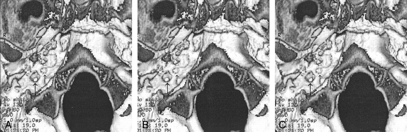
Case 1. 3D CT scans of the of the exocranial foramen with stereoscopic views. Images A and B are for cross-eyed viewing, and images B and C are for parallel-eye viewing. The right inferior tympanic canal (left arrow) shows a bony constriction. The hypoplastic right carotid canal is shown with the right arrow.

The patient had no symptoms related to these anatomic findings. Audiograms showed mild low-frequency hearing loss involving the left ear and normal hearing in the right ear. This has remained stable over the past 5 years.

When the patient was aged 10 years, his parents noticed a left-ear bruit when they placed their ear next to the left ear of the patient. With exercise, this increased to a palpable thrill. A subtle right-ear bruit was audible with a stethoscope. However, the patient only reported hearing a mild bruit immediately after exertion. Repeat CT demonstrated no apparent change. MR angiography (MRA) demonstrated the known bilateral aberrant ICAs with the PSAs and confirmed flow in the native hypoplastic carotid canals (Fig 6). Also noted was the origin of right occipital artery from the larger of the duplicated right cervical ICA.

Fig 6.
  • Download figure
  • Open in new tab
  • Download powerpoint
Fig 6.

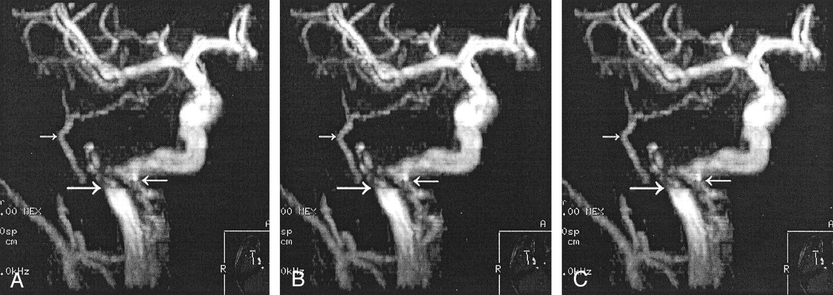
Case 1. Stereoscopic MRA images of the right carotid. Images A and B are for cross-eyed viewing, and images B and C are for parallel-eyed viewing. The top left arrow shows the PSA supplying the middle meningeal distribution. The right arrow shows the duplicated ICA (carotid branch of the ascending pharyngeal artery). The bottom left arrow points to the turbulent blood flow at the inferior tympanic canal in the aberrant ICA. Views of the left ICA showed similar findings (not shown).

Case 2

An otolaryngologist evaluated a 10-year-old girl for otitis media. At otoscopy, a left retrotympanic pulsatile mass was noted. Subsequent CT scans of the petrous bone demonstrated a left aberrant ICA (Fig 1B), an absent left foramen spinosum (Fig 2B), a left PSA (Fig 3B), and a hypoplastic native exocranial carotid canal on the left side (Fig 4B). No further imaging studies were performed. Audiograms revealed a mild low-frequency hearing loss involving the left ear and normal hearing in the right ear. No clinical symptoms were referable to these anatomic findings.

Discussion

Many authors (1–6) have discussed the embryologic development of the cervical and cranial vasculature. Now, an aberrant ICA is generally accepted to be a collateral pathway that occurs as a result of agenesis of the first embryonic segment of the ICA. The inferior tympanic branch of the ascending pharyngeal artery anastomoses with the caroticotympanic artery (hyoid artery remnant) of the ICA. The aberrant ICA variant may therefore be more accurately referred to as “the inferior tympano-caroticotympanic variant,” as suggested by Lasjaunias (3). The inferior tympanic artery passes through the inferior tympanic canal (Jacobsen canal) at the skull base, with a resultant characteristic narrowing of the vessel.

Characteristic angiographic findings of aberrant ICA include lateral extension of the ICA well beyond the vestibular line of Lapayowker (7).

CT is considered one of the most reliable ways to diagnose an aberrant ICA (8–10). Findings include the following: A reduced-caliber aberrant ICA enters the middle ear through the enlarged inferior tympanic canal within the caroticojugular spine. The vessel takes a sharp turn anteriorly and crosses the promontory to continue on to the horizontal portion of the petrous carotid. Because the aberrant ICA is present in the middle ear, the normal bone covering the artery is absent. The vertical portion of the carotid canal may be absent or hypoplastic.

Dephasing of the spins due to turbulence, with loss of the signal intensity on the three-dimensional (3D) time-of-flight MRA image in case 1, is evident beyond the expected location of the inferior tympanic canal; this effect accounted for the patient’s bruits. MRA findings of aberrant ICA have been described (11); these include a lateral and superior location of the carotid genu in the middle ear on the anteroposterior projection and the posterior and superior location of this structure on the lateral projection.

The stapedial artery is persistent when the embryonic stapedial artery does not regress completely. This condition usually results in the stapedial artery supplying the middle meningeal artery territory. Congenital absence of the foramen spinosum is present in 3.2% of the population (12); therefore, additional findings demonstrating the course of the PSA are needed to confirm its presence. These findings include a small canaliculus exiting the carotid canal (in cases in which aberrant ICA is not present), the presence of a linear soft-tissue attenuation crossing the middle ear over the promontory, an enlarged facial nerve canal, or a separate and parallel canal (13).

In a case presented by Lasjaunias (3), courtesy of R. Willinsky, the duplicated ICAs had an appearance strikingly similar to that of case 1. The anatomic explanation of the two channels carrying the ICA flow is that one channel is the inferior tympanic artery branch of the ascending pharyngeal artery, and the other channel is the carotid branch of the superior pharyngeal branch of the pharyngeal trunk of the ascending pharyngeal artery. In case 1, we propose the same anatomic explanation for the bilateral duplicated channels of the ICAs. We hypothesize that the stenosis at the inferior tympanic canal bilaterally results in the need for an additional collateral supply from the carotid branch of the ascending pharyngeal artery.

The surgical incidence of PSA is quoted as 1:5,000 to 1:10,000 (0.02–0.01%) with stapes surgery (2). This incidence is different from that from a histopathologic study in which five PSAs were found in 1,045 temporal bones, for an incidence of 0.48% (14).

The incidence of aberrant ICA and the incidence of aberrant ICA with PSA is unknown; however, several case reports exist (1,6,15,16). Retrospective and prospective evaluation of cases of aberrant ICA may be helpful to determine the true incidence of this association. The incidence of a duplicated ICA is also unknown.

Six previous reports of bilateral aberrant ICAs (17–22) and 13 previous cases of bilateral PSAs (2,6,23) are known. No cases of bilateral duplicated ICAs or cases of bilateral aberrant carotid arteries with bilateral PSAs have been previously reported.

Conclusion

Recognizing the findings of an aberrant ICA is important to avoid potentially severe surgical complications. The identification of PSAs is also helpful in the evaluation of tinnitus and hearing loss.

We present what we believe to be the first reported case of bilateral aberrant ICAs with bilateral PSAs and bilateral duplicated ICAs.

Footnotes

  • Presented at the 40th Annual Meeting of the American Society of Neuroradiology, Vancouver, B.C., May 10–17, 2002.

References

  1. ↵
    Silbergleit R, Quint DJ, Mehta BA, Patel SC, Metes JJ, Noujaim SE. The persistent stapedial artery. AJNR Am J Neuroradiol 2000;21:572–577
    Abstract/FREE Full Text
  2. ↵
    Govaerts PJ, Marquet TF, Cremers CWRJ, Offeciers FE. Persistent stapedial artery: does it prevent successful surgery? Ann Otol Rhinol Laryngol 1993;102:724–728
    Abstract/FREE Full Text
  3. ↵
    Lasjaunias P, Berenstein A, Ter Brugge KG. Surgical Neuroangiography, Clinical Vascular Anatomy and Variations. 2nd ed. New York: Springer-Verlag;2001
  4. Padget DH. The development of the cranial arteries in the human embryo. Contrib Embroyl 1948;32:205–261
  5. Congdod ED. Transformation of the aortic-arch system during the development of the human embryo. Contrib Embryol 1922;14:47–110
  6. ↵
    Steffen TH. Vascular anomalies of the middle ear. Laryngoscope 1968;78:171–197
    CrossRefPubMed
  7. ↵
    Lapayowker MS, Liebman EP, Ronis ML, Safer JN. Presentation of the internal carotid artery as a tumor of the middle ear. Radiology 1971;98:293–297
    CrossRefPubMed
  8. ↵
    Swartz JD, Buzarnic ML, Naidich TP, Lowry LD, Doan HT. Aberrant internal carotid artery lying within the middle ear. Neuroradiology 1985;27:322–326
    CrossRefPubMed
  9. Lo WWM, Solti-Bohman LG, McElveen JT. Aberrant Carotid artery: radiologic diagnosis with emphasis on high-resolution computed tomography. Radiographics 1985;5:985–993
    PubMed
  10. ↵
    McElveen JT, Lo WWM, El Gabri TH, Nigri P. Aberrant internal carotid artery: classic findings on computed tomography. Otolaryngol Head Neck Surg 1986;94:616–621
    PubMed
  11. ↵
    Davis WL, Harnsberger HR. MR Angiography of an aberrant internal carotid artery. AJNR Am J Neuroradiol 1991;12:1225
    FREE Full Text
  12. ↵
    Ginsberg LE, Pruett SW, Chen MYM, Elster AD. Skull-based foramina of the middle cranial fossa: reassessment of normal variation with high-resolution CT. AJNR Am J Neuroradiol 1994;15:283–291
    Abstract/FREE Full Text
  13. ↵
    Thiers FA, Sukai O, Poe DS, Curtin HD. Persistent stapedial artery: CT findings. AJNR Am J Neuroradiol 2000;21:1551–1554
    Abstract/FREE Full Text
  14. ↵
    Moreano EH, Puparella MM, Zelterman D, Goycoolea MV. Prevalence of facial canal dehiscence and of persistent stapedial artery in the human middle ear: a report of 1000 temporal bones. Laryngoscope 1994;104:309–320
    PubMed
  15. ↵
    Guinto FC, Garrabrant EC, Radcliffe WB. Radiology of the persistent stapedial artery. Radiology 1972;105:365–369
    CrossRefPubMed
  16. ↵
    Lasjaunias P, Moret J, Manelfe C, Théron J, Hasso T, Seeger J Arterial anomalies at the base of the skull. Neuroradiology 1977;13:267–272
    PubMed
  17. ↵
    Goldman NC, Singleton GT, Holly EH. Aberrant internal carotid artery. Arch Otolaryngol 1971;94:269–273
    CrossRefPubMed
  18. Saito H, Chikamory Y, Yanagihara N. Aberrant carotid artery in the middle ear. Arch Oto Rhino Laryngol 1975;209:83–87
  19. Campbell G, Renner G, Estrem SA. Bilateral aberrant internal carotid arteries. Otolaryng Head Neck Surg 1992;107:124–128
    PubMed
  20. Glasscock ME, Seshul M, Seshul MB. Bilateral aberrant internal carotid artery case presentation. Arch Otolaryngol Head Neck Surg 1993;119:335–339
    CrossRefPubMed
  21. Ashikaga R, Araki Y, Ishida O. Bilateral aberrant internal carotid arteries. Neuroradiology 1995;37:655–657
    CrossRefPubMed
  22. ↵
    Benton C, Bellet PS. Imaging of congenital anomalies of the temporal bone. Neuroimaging Clin N Am 2000;10:35–53
    PubMed
  23. ↵
    Marion M, Hinojosa R, Kahn AA. Persistence of the stapedial artery: a histopathologic study. Otolaryngol Head Neck Surg 1985;93:298–312
    PubMed
  • Received July 10, 2002.
  • Accepted after revision July 18, 2002.
  • Copyright © American Society of Neuroradiology
View Abstract
PreviousNext
Back to top

In this issue

American Journal of Neuroradiology: 24 (4)
American Journal of Neuroradiology
Vol. 24, Issue 4
1 Apr 2003
  • Table of Contents
  • Index by author
Advertisement
Print
Download PDF
Email Article

Thank you for your interest in spreading the word on American Journal of Neuroradiology.

NOTE: We only request your email address so that the person you are recommending the page to knows that you wanted them to see it, and that it is not junk mail. We do not capture any email address.

Enter multiple addresses on separate lines or separate them with commas.
Bilateral Aberrant Internal Carotid Arteries with Bilateral Persistent Stapedial Arteries and Bilateral Duplicated Internal Carotid Arteries
(Your Name) has sent you a message from American Journal of Neuroradiology
(Your Name) thought you would like to see the American Journal of Neuroradiology web site.
CAPTCHA
This question is for testing whether or not you are a human visitor and to prevent automated spam submissions.
Cite this article
John D. Roll, Martin A. Urban, Theodore C. Larson, Philippe Gailloud, Pradeep Jacob, H. Ric Harnsberger
Bilateral Aberrant Internal Carotid Arteries with Bilateral Persistent Stapedial Arteries and Bilateral Duplicated Internal Carotid Arteries
American Journal of Neuroradiology Apr 2003, 24 (4) 762-765;

Citation Manager Formats

  • BibTeX
  • Bookends
  • EasyBib
  • EndNote (tagged)
  • EndNote 8 (xml)
  • Medlars
  • Mendeley
  • Papers
  • RefWorks Tagged
  • Ref Manager
  • RIS
  • Zotero
0 Responses
Respond to this article
Share
Bookmark this article
Bilateral Aberrant Internal Carotid Arteries with Bilateral Persistent Stapedial Arteries and Bilateral Duplicated Internal Carotid Arteries
John D. Roll, Martin A. Urban, Theodore C. Larson, Philippe Gailloud, Pradeep Jacob, H. Ric Harnsberger
American Journal of Neuroradiology Apr 2003, 24 (4) 762-765;
del.icio.us logo Twitter logo Facebook logo Mendeley logo
  • Tweet Widget
  • Facebook Like
  • Google Plus One

Jump to section

  • Article
    • Abstract
    • Case Reports
    • Discussion
    • Conclusion
    • Footnotes
    • References
  • Figures & Data
  • Info & Metrics
  • Responses
  • References
  • PDF

Related Articles

  • No related articles found.
  • PubMed
  • Google Scholar

Cited By...

  • Aberrant internal carotid artery in the middle ear: the duplication variant
  • Dangerous Extracranial-Intracranial Anastomoses and Supply to the Cranial Nerves: Vessels the Neurointerventionalist Needs to Know
  • Crossref
  • Google Scholar

This article has not yet been cited by articles in journals that are participating in Crossref Cited-by Linking.

More in this TOC Section

  • Correlation of Apparent Diffusion Coefficient at 3T with Prognostic Parameters of Retinoblastoma
  • Parathyroid Lesions: Characterization with Dual-Phase Arterial and Venous Enhanced CT of the Neck
  • MR Evaluation of Sinonasal Angiomatous Polyp
Show more HEAD AND NECK

Similar Articles

Advertisement

Indexed Content

  • Current Issue
  • Accepted Manuscripts
  • Article Preview
  • Past Issues
  • Editorials
  • Editor's Choice
  • Fellows' Journal Club
  • Letters to the Editor
  • Video Articles

Cases

  • Case Collection
  • Archive - Case of the Week
  • Archive - Case of the Month
  • Archive - Classic Case

Special Collections

  • AJNR Awards
  • ASNR Foundation Special Collection
  • Most Impactful AJNR Articles
  • Photon-Counting CT
  • Spinal CSF Leak Articles (Jan 2020-June 2024)

More from AJNR

  • Trainee Corner
  • Imaging Protocols
  • MRI Safety Corner

Multimedia

  • AJNR Podcasts
  • AJNR Scantastics

Resources

  • Turnaround Time
  • Submit a Manuscript
  • Submit a Video Article
  • Submit an eLetter to the Editor/Response
  • Manuscript Submission Guidelines
  • Statistical Tips
  • Fast Publishing of Accepted Manuscripts
  • Graphical Abstract Preparation
  • Imaging Protocol Submission
  • Evidence-Based Medicine Level Guide
  • Publishing Checklists
  • Author Policies
  • Become a Reviewer/Academy of Reviewers
  • News and Updates

About Us

  • About AJNR
  • Editorial Board
  • Editorial Board Alumni
  • Alerts
  • Permissions
  • Not an AJNR Subscriber? Join Now
  • Advertise with Us
  • Librarian Resources
  • Feedback
  • Terms and Conditions
  • AJNR Editorial Board Alumni

American Society of Neuroradiology

  • Not an ASNR Member? Join Now

© 2025 by the American Society of Neuroradiology All rights, including for text and data mining, AI training, and similar technologies, are reserved.
Print ISSN: 0195-6108 Online ISSN: 1936-959X

Powered by HighWire