Advertisement

Main menu

  • Home
  • Content
    • Current Issue
    • Accepted Manuscripts
    • Article Preview
    • Past Issue Archive
    • Video Articles
    • AJNR Case Collection
    • Case of the Week Archive
    • Case of the Month Archive
    • Classic Case Archive
  • Special Collections
    • AJNR Awards
    • ASNR Foundation Special Collection
    • Most Impactful AJNR Articles
    • Photon-Counting CT
    • Spinal CSF Leak Articles (Jan 2020-June 2024)
  • Multimedia
    • AJNR Podcasts
    • AJNR SCANtastic
    • Trainee Corner
    • MRI Safety Corner
    • Imaging Protocols
  • For Authors
    • Submit a Manuscript
    • Submit a Video Article
    • Submit an eLetter to the Editor/Response
    • Manuscript Submission Guidelines
    • Statistical Tips
    • Fast Publishing of Accepted Manuscripts
    • Graphical Abstract Preparation
    • Imaging Protocol Submission
    • Author Policies
  • About Us
    • About AJNR
    • Editorial Board
    • Editorial Board Alumni
  • More
    • Become a Reviewer/Academy of Reviewers
    • Subscribers
    • Permissions
    • Alerts
    • Feedback
    • Advertisers
    • ASNR Home

User menu

  • Alerts
  • Log in

Search

  • Advanced search
American Journal of Neuroradiology

American Journal of Neuroradiology

ASHNR American Society of Functional Neuroradiology ASHNR American Society of Pediatric Neuroradiology ASSR
  • Alerts
  • Log in

Advanced Search

  • Home
  • Content
    • Current Issue
    • Accepted Manuscripts
    • Article Preview
    • Past Issue Archive
    • Video Articles
    • AJNR Case Collection
    • Case of the Week Archive
    • Case of the Month Archive
    • Classic Case Archive
  • Special Collections
    • AJNR Awards
    • ASNR Foundation Special Collection
    • Most Impactful AJNR Articles
    • Photon-Counting CT
    • Spinal CSF Leak Articles (Jan 2020-June 2024)
  • Multimedia
    • AJNR Podcasts
    • AJNR SCANtastic
    • Trainee Corner
    • MRI Safety Corner
    • Imaging Protocols
  • For Authors
    • Submit a Manuscript
    • Submit a Video Article
    • Submit an eLetter to the Editor/Response
    • Manuscript Submission Guidelines
    • Statistical Tips
    • Fast Publishing of Accepted Manuscripts
    • Graphical Abstract Preparation
    • Imaging Protocol Submission
    • Author Policies
  • About Us
    • About AJNR
    • Editorial Board
    • Editorial Board Alumni
  • More
    • Become a Reviewer/Academy of Reviewers
    • Subscribers
    • Permissions
    • Alerts
    • Feedback
    • Advertisers
    • ASNR Home
  • Follow AJNR on Twitter
  • Visit AJNR on Facebook
  • Follow AJNR on Instagram
  • Join AJNR on LinkedIn
  • RSS Feeds

AJNR is seeking candidates for the AJNR Podcast Editor. Read the position description.

LetterLetter

Prenatal Diagnosis of a Vein of Galen Aneurysmal Malformation with Fetal MR Imaging Study

Anna Messori, Gabriele Polonara and Ugo Salvolini
American Journal of Neuroradiology October 2003, 24 (9) 1923-1925;
Anna Messori
  • Find this author on Google Scholar
  • Find this author on PubMed
  • Search for this author on this site
Gabriele Polonara
  • Find this author on Google Scholar
  • Find this author on PubMed
  • Search for this author on this site
Ugo Salvolini
  • Find this author on Google Scholar
  • Find this author on PubMed
  • Search for this author on this site
  • Article
  • Figures & Data
  • Info & Metrics
  • Responses
  • References
  • PDF
Loading

We read with interest the article by Jones et al (1), in which the authors describe their experience in diagnosing and treating 13 children with vein of Galen aneurysmal malformation (VGAM) during a 14-year period. Their experience confirms that children with VGAM presenting during the neonatal period have a generally much worse prognosis than do those presenting later in childhood. Only two of the eight patients who presented during the neonatal period achieved normal or near-normal outcomes; five died, and one experienced significant impairment. Neonatal presentation with congestive heart failure is frequent with these malformations; if they are diagnosed prenatally, early delivery by means of cesarean section may be proposed to enable more effective cardiac evaluation and management. However, the authors also emphasize that only four cases among the 13 were diagnosed prenatally, and despite the increased use of routine prenatal sonography in recent years, they identified no trend toward a greater number of prenatal diagnoses. We would like to point out that fetal MR imaging may play a role for prenatal diagnosis of VGAM and to emphasize its value for appropriate management of these cases.

In 1995, we had experience with one such case. A 34-year-old gravida 2, para 0 woman was referred at 38 weeks and 5 days of gestation for evaluation of a routine sonography-detected anechoic intracranial lesion. Doppler ultrasonography revealed turbulent blood flow within the lesion, 20 mm in diameter, situated in the midline, and a possible diagnosis of VGAM was suggested (Fig 1). Intrauterine MR imaging was performed without curarization or sedation on a 1.0-T superconducting unit (Magnetom Impact; Siemens, Erlangen, Germany). It confirmed the diagnosis of VGAM and showed otherwise normal brain parenchyma, without hydrocephalus (Fig 2). Fetal echocardiography showed slight cardiomegaly and slightly distended superior cava, brachiocephalic, and internal jugular veins. After 2 days, a male infant weighing 2860 g was delivered by cesarean section. Apgar scores were 9 at 1 min and 9 at 5 min; echocardiography showed no signs of congestive heart failure. Postnatal MR imaging and MR angiography were performed with the patient under narcosis at 35 days (not shown) and at 75 days of life (Figs 3 and 4); the child’s clinical condition remained good, and his development was normal. At 6 months of life, he underwent digital angiography and graded catheter embolization, which were performed during a 4-month period. For these procedures, the child was referred to another institution; therefore, no information is provided herein. The result was occlusion of 80% of the VGAM. At 7-year follow-up, the patient’s physical and psychomotor development was normal.

Although ultrasonography is the imaging technique of choice for assessment of fetal CNS abnormalities, intrauterine MR imaging has several advantages over it (2) and provides additional information that can aid in the management of these cases. Its use for revealing VGAM has rarely been reported (3–5). In fetuses with ultrasonographic findings consistent with VGAM, complementary MR imaging enables correct diagnosis, allows for better evaluation of the lesion, and aids in decision making, which also depends on an accurate assessment of the condition of the brain parenchyma. Elective early cesarean delivery can enable optimal care of the baby’s cardiac function, minimizing the risks related to the sudden diversion of flow through the malformation at birth. Importantly, monitoring with postnatal MR imaging and MR angiography can also help in understanding the right time to perform treatment. Prompt postdelivery embolization may not be the best management option. In their article, Jones et al note that embolization during the first few weeks of life is unlikely to result in good outcomes and that endovascular treatment should be delayed, if possible, for several months. Thanks to the absence of congestive heart failure, the child whose case we reported had treatment delayed for 6 months, and he eventually achieved a good outcome. His case also confirms that graded embolization procedures may provide the best results in these cases and that incomplete obliteration of the lesion may be ideal (1, 6).

References

  1. ↵
    Jones BV, Ball WS, Tomsick TA, Millard J, Crone KR. Vein of Galen aneurysmal malformation: diagnosis and treatment of 13 children with extended clinical follow-up. AJNR Am J Neuroradiol 2002;23:1717–1724
    Abstract/FREE Full Text
  2. ↵
    Yuh WT, Nguyen HD, Fisher DJ, et al. MR of fetal central nervous system abnormalities. AJNR Am J Neuroradiol 1994;15:459–464
    Abstract/FREE Full Text
  3. ↵
    Yamashita Y, Abe T, Ohara N, et al. Successful treatment of neonatal aneurysmal dilatation of the vein of Galen: the role of prenatal diagnosis and trans-arterial embolization. Neuroradiology 1992;34:457–459
    CrossRefPubMed
  4. Campi A, Scotti G, Filippi M, Gerevini S, Strigimi F, Lasjaunias P. Antenatal diagnosis of vein of Galen aneurysmal malformation: MR study of fetal brain and postnatal follow-up. Neuroradiology 1996;38:87–90
    CrossRefPubMed
  5. ↵
    Kurihara N, Tokieda K, Ikeda K, et al. Prenatal MR findings in a case of aneurysm of the vein of Galen. Pediatr Radiol 2001;31:160–162
    CrossRefPubMed
  6. ↵
    Campi A, Rodesch G, Scotti G, Lasjaunias P. Aneurysmal malformation of the vein of Galen in three patients: clinical and radiological follow-up. Neuroradiology 1998;40:816–821
    PubMed

Reply:

We appreciate the interest in our article expressed by Messori et al. In their letter, they provide an interesting example of a case of VGAM that included prenatal MR imaging as a major component of the diagnostic evaluation. Their experience in the treatment parallels ours, as they describe.

Our comment regarding the absence of a trend toward prenatal discovery of VGAM in our patients was essentially an anecdotal observation. Clearly, we conducted no epidemiologic investigation regarding the prevalence of prenatal sonography in our referral base, nor did we actively pursue information regarding the presence or timing of prenatal ultrasonography in our 13 cases. It may be a logical hypothesis that prenatal diagnosis of VGAM is on the rise, but that is a question to be answered by a different type of study.

The use of fetal MR imaging in the evaluation of VGAM is an interesting and ever more important topic. Fetal MR imaging is being used more and more frequently to further assess abnormalities identified on ultrasonograms. This is the role described by Messori et al; the lesion had been correctly identified by ultrasonography, but the extent and effect on the surrounding brain and cardiac structures was more confidently assessed by MR imaging. Because the child was essentially at term, the issue of exigent delivery is not in debate. However, Messori et al note that the use of cesarean section allowed greater control of the perinatal environment. It remains to be seen whether a cesarean delivery is physiologically less stressful for an infant with VGAM, but this is not an unreasonable presumption.

The greater issue presented by the case is the usefulness or necessity of prenatal MR imaging of the fetus with VGAM. It is of little doubt that fetal MR imaging is rapidly becoming a new standard of care in the assessment of complex fetal abnormalities. VGAM certainly belongs in that category. Considering the useful information obtainable with fetal MR imaging regarding viability of the brain and degree of cardiac compromise, fetal MR imaging should be considered a reasonable diagnostic tool when this diagnosis is made based on prenatal ultrasonography. Is it essential in the management of these cases? No, but it may allow treatment decisions to be made in a more controlled clinical environment. We would contend that such examinations be performed at the center where the child will eventually be treated to allow a single treatment team to contribute to the decision process from the start.

We thank Messori et al for bringing to the forefront this aspect of the evaluation and treatment of VGAM and congratulate them on their successful management of this difficult problem.

Fig 1.
  • Download figure
  • Open in new tab
  • Download powerpoint
Fig 1.

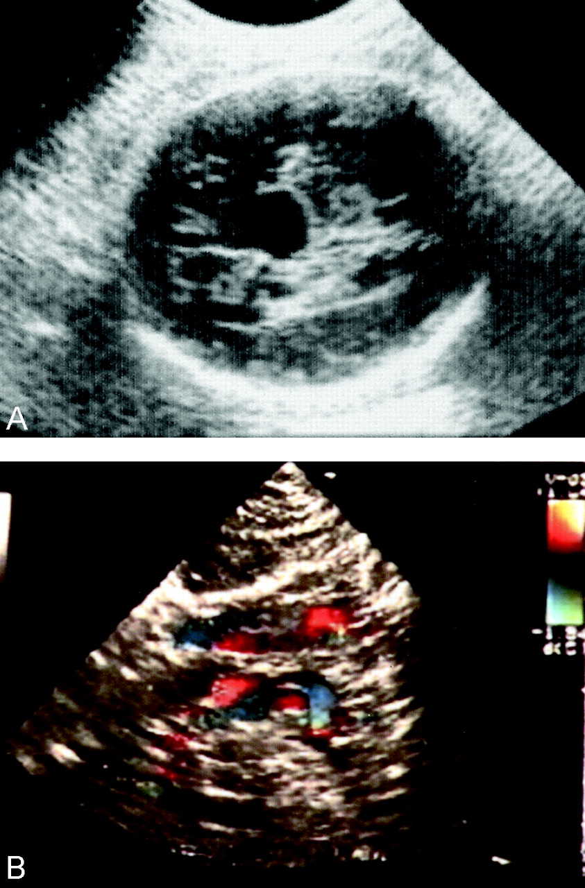
Ultrasonogram and color Doppler sonogram from the case of a 34-year-old gravida 2, para 0 woman who was referred at 38 weeks and 5 days of gestation.

A, Routine ultrasonogram shows an intracranial echo-free area.

B, Color Doppler sonogram shows turbulent blood flow within the lesion (courtesy of Dr. G. Ciotti, Department of Obstetrics, Children’s Hospital “G. Salesi,” Ancona, Italy).

Fig 2.
  • Download figure
  • Open in new tab
  • Download powerpoint
Fig 2.

Fetal MR images obtained at 38 weeks and 5 days of gestation (body coil; fast low angle shot 2D; 44/10 [TR/TE]; flip angle, 40 degrees; section thickness, 5 mm; field of view, 380 mm; matrix, 192 × 256).

A, Midsagittal view of the fetal brain shows a distended midline vascular structure; the brain parenchyma appears otherwise normal, and there is no ventricular dilation.

B, Axial view.

Fig 3.
  • Download figure
  • Open in new tab
  • Download powerpoint
Fig 3.

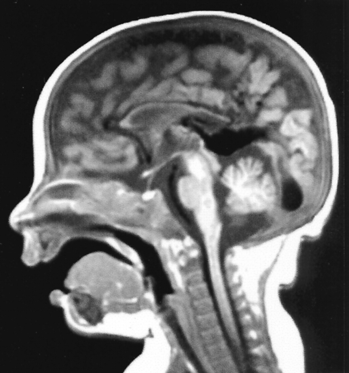
Postnatal midsagittal T1-weighted spin-echo MR image obtained at 75 days of life (500/15; section thickness, 5 mm; field of view, 230 mm; matrix, 220 × 256) shows an interhemispheric vascular malformation at the level of the posterior third and splenium of the corpus callosum, flowing into a markedly distended vein of Galen and into the straight sinus.

Fig 4.
  • Download figure
  • Open in new tab
  • Download powerpoint
Fig 4.

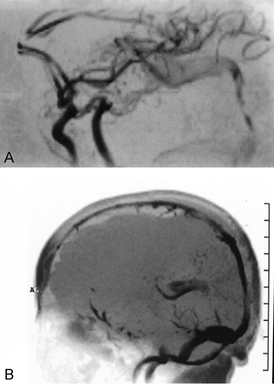
Postnatal MR angiograms obtained at 75 days of life.

A, Arterial flow (fast imaging with steady-state free precession 3D, magnetization transfer-tilted optimized nonsaturating excitation, 44/10).

B, Venous flow (fast low angle shot 2D, 40/10). Although turbulent flow through the vein of Galen makes it impossible to directly show it, the images indirectly show the malformation via its arterial supply and the venous flow into the straight sinus.

  • Copyright © American Society of Neuroradiology
PreviousNext
Back to top

In this issue

American Journal of Neuroradiology: 24 (9)
American Journal of Neuroradiology
Vol. 24, Issue 9
1 Oct 2003
  • Table of Contents
  • Index by author
Advertisement
Print
Download PDF
Email Article

Thank you for your interest in spreading the word on American Journal of Neuroradiology.

NOTE: We only request your email address so that the person you are recommending the page to knows that you wanted them to see it, and that it is not junk mail. We do not capture any email address.

Enter multiple addresses on separate lines or separate them with commas.
Prenatal Diagnosis of a Vein of Galen Aneurysmal Malformation with Fetal MR Imaging Study
(Your Name) has sent you a message from American Journal of Neuroradiology
(Your Name) thought you would like to see the American Journal of Neuroradiology web site.
CAPTCHA
This question is for testing whether or not you are a human visitor and to prevent automated spam submissions.
Cite this article
Anna Messori, Gabriele Polonara, Ugo Salvolini
Prenatal Diagnosis of a Vein of Galen Aneurysmal Malformation with Fetal MR Imaging Study
American Journal of Neuroradiology Oct 2003, 24 (9) 1923-1925;

Citation Manager Formats

  • BibTeX
  • Bookends
  • EasyBib
  • EndNote (tagged)
  • EndNote 8 (xml)
  • Medlars
  • Mendeley
  • Papers
  • RefWorks Tagged
  • Ref Manager
  • RIS
  • Zotero
0 Responses
Respond to this article
Share
Bookmark this article
Prenatal Diagnosis of a Vein of Galen Aneurysmal Malformation with Fetal MR Imaging Study
Anna Messori, Gabriele Polonara, Ugo Salvolini
American Journal of Neuroradiology Oct 2003, 24 (9) 1923-1925;
del.icio.us logo Twitter logo Facebook logo Mendeley logo
  • Tweet Widget
  • Facebook Like
  • Google Plus One

Jump to section

  • Article
    • References
  • Figures & Data
  • Info & Metrics
  • Responses
  • References
  • PDF

Related Articles

  • No related articles found.
  • PubMed
  • Google Scholar

Cited By...

  • No citing articles found.
  • Crossref
  • Google Scholar

This article has not yet been cited by articles in journals that are participating in Crossref Cited-by Linking.

More in this TOC Section

  • Brain AVM’s Nidus: What if We Hadn’t Understood Anything?
  • Letter to the Editor regarding “Automated Volumetric Software in Dementia: Help or Hindrance to the Neuroradiologist?”
  • Reply:
Show more Letters

Similar Articles

Advertisement

Indexed Content

  • Current Issue
  • Accepted Manuscripts
  • Article Preview
  • Past Issues
  • Editorials
  • Editor's Choice
  • Fellows' Journal Club
  • Letters to the Editor
  • Video Articles

Cases

  • Case Collection
  • Archive - Case of the Week
  • Archive - Case of the Month
  • Archive - Classic Case

Special Collections

  • AJNR Awards
  • ASNR Foundation Special Collection
  • Most Impactful AJNR Articles
  • Photon-Counting CT
  • Spinal CSF Leak Articles (Jan 2020-June 2024)

More from AJNR

  • Trainee Corner
  • Imaging Protocols
  • MRI Safety Corner

Multimedia

  • AJNR Podcasts
  • AJNR Scantastics

Resources

  • Turnaround Time
  • Submit a Manuscript
  • Submit a Video Article
  • Submit an eLetter to the Editor/Response
  • Manuscript Submission Guidelines
  • Statistical Tips
  • Fast Publishing of Accepted Manuscripts
  • Graphical Abstract Preparation
  • Imaging Protocol Submission
  • Evidence-Based Medicine Level Guide
  • Publishing Checklists
  • Author Policies
  • Become a Reviewer/Academy of Reviewers
  • News and Updates

About Us

  • About AJNR
  • Editorial Board
  • Editorial Board Alumni
  • Alerts
  • Permissions
  • Not an AJNR Subscriber? Join Now
  • Advertise with Us
  • Librarian Resources
  • Feedback
  • Terms and Conditions
  • AJNR Editorial Board Alumni

American Society of Neuroradiology

  • Not an ASNR Member? Join Now

© 2025 by the American Society of Neuroradiology All rights, including for text and data mining, AI training, and similar technologies, are reserved.
Print ISSN: 0195-6108 Online ISSN: 1936-959X

Powered by HighWire