Advertisement

Main menu

  • Home
  • Content
    • Current Issue
    • Accepted Manuscripts
    • Article Preview
    • Past Issue Archive
    • Video Articles
    • AJNR Case Collection
    • Case of the Week Archive
    • Case of the Month Archive
    • Classic Case Archive
  • Special Collections
    • AJNR Awards
    • ASNR Foundation Special Collection
    • Most Impactful AJNR Articles
    • Photon-Counting CT
    • Spinal CSF Leak Articles (Jan 2020-June 2024)
  • Multimedia
    • AJNR Podcasts
    • AJNR SCANtastic
    • Trainee Corner
    • MRI Safety Corner
    • Imaging Protocols
  • For Authors
    • Submit a Manuscript
    • Submit a Video Article
    • Submit an eLetter to the Editor/Response
    • Manuscript Submission Guidelines
    • Statistical Tips
    • Fast Publishing of Accepted Manuscripts
    • Graphical Abstract Preparation
    • Imaging Protocol Submission
    • Author Policies
  • About Us
    • About AJNR
    • Editorial Board
    • Editorial Board Alumni
  • More
    • Become a Reviewer/Academy of Reviewers
    • Subscribers
    • Permissions
    • Alerts
    • Feedback
    • Advertisers
    • ASNR Home

User menu

  • Alerts
  • Log in

Search

  • Advanced search
American Journal of Neuroradiology

American Journal of Neuroradiology

ASHNR American Society of Functional Neuroradiology ASHNR American Society of Pediatric Neuroradiology ASSR
  • Alerts
  • Log in

Advanced Search

  • Home
  • Content
    • Current Issue
    • Accepted Manuscripts
    • Article Preview
    • Past Issue Archive
    • Video Articles
    • AJNR Case Collection
    • Case of the Week Archive
    • Case of the Month Archive
    • Classic Case Archive
  • Special Collections
    • AJNR Awards
    • ASNR Foundation Special Collection
    • Most Impactful AJNR Articles
    • Photon-Counting CT
    • Spinal CSF Leak Articles (Jan 2020-June 2024)
  • Multimedia
    • AJNR Podcasts
    • AJNR SCANtastic
    • Trainee Corner
    • MRI Safety Corner
    • Imaging Protocols
  • For Authors
    • Submit a Manuscript
    • Submit a Video Article
    • Submit an eLetter to the Editor/Response
    • Manuscript Submission Guidelines
    • Statistical Tips
    • Fast Publishing of Accepted Manuscripts
    • Graphical Abstract Preparation
    • Imaging Protocol Submission
    • Author Policies
  • About Us
    • About AJNR
    • Editorial Board
    • Editorial Board Alumni
  • More
    • Become a Reviewer/Academy of Reviewers
    • Subscribers
    • Permissions
    • Alerts
    • Feedback
    • Advertisers
    • ASNR Home
  • Follow AJNR on Twitter
  • Visit AJNR on Facebook
  • Follow AJNR on Instagram
  • Join AJNR on LinkedIn
  • RSS Feeds

AJNR is seeking candidates for the AJNR Podcast Editor. Read the position description.

OtherBRAIN

Noninvasive Detection of a Ruptured Aneurysm at a Basilar Artery Fenestration with Submillimeter Multisection CT Angiography

Filippo Cademartiri, Dragan Stojanov, Diederik W.J. Dippel, Aad van der Lugt and Herve Tanghe
American Journal of Neuroradiology November 2003, 24 (10) 2009-2010;
Filippo Cademartiri
  • Find this author on Google Scholar
  • Find this author on PubMed
  • Search for this author on this site
Dragan Stojanov
  • Find this author on Google Scholar
  • Find this author on PubMed
  • Search for this author on this site
Diederik W.J. Dippel
  • Find this author on Google Scholar
  • Find this author on PubMed
  • Search for this author on this site
Aad van der Lugt
  • Find this author on Google Scholar
  • Find this author on PubMed
  • Search for this author on this site
Herve Tanghe
  • Find this author on Google Scholar
  • Find this author on PubMed
  • Search for this author on this site
  • Article
  • Figures & Data
  • Supplemental
  • Info & Metrics
  • Responses
  • References
  • PDF
Loading

Abstract

Summary: The criterion standard for the detection of intracranial aneurysms is digital subtraction angiography. MR imaging and CT provide good accuracy in the evaluation of brain arteries and aneurysms. We herein report a case of a ruptured aneurysm at a basilar artery fenestration. The diagnosis was assessed with 16-row multisection CT angiography and was confirmed by using digital subtraction angiography. The patient was successfully treated with coil placement.

A fenestration or a segmental duplication of the basilar artery, previously reported exclusively as an anatomic variation, owes its clinical interest to the possible association with aneurysms localized at the junctions of the fenestrated segments (1). The fenestration is frequently located in the proximal basilar trunk, close to the vertebrobasilar junction (2). Saccular aneurysms associated with fenestration, especially in the middle or distal portion of the basilar artery, are infrequent (3). An aneurysm at the fenestrated basilar artery usually arises at the proximal end of the fenestration, with few exceptions (4).

Basilar artery fenestration, with or without cerebral aneurysm, has been diagnosed by using digital subtraction angiography and MR angiography (5). The capability of CT angiography to diagnose this malformation has not been reported. We herein describe the first case of saccular aneurysm at the proximal end of the fenestration located in the middle portion of the basilar artery diagnosed with the new generation of 16-row multisection CT scanners.

Case Report

A 37-year-old woman was admitted to the hospital with the following symptoms: coma, meningeal syndrome, and stiff neck. Unenhanced CT was performed and showed signs of subarachnoid hemorrhage.

Subsequently, multisection CT angiography (Sensation 16; Siemens, Forchheim, Germany) was performed and depicted an intracranial aneurysm. Scanning was performed with the following parameters: detector rows, 16; collimation, 0.75 mm; feed/rotation, 12.0 mm; and gantry rotation time, 500 ms. A volume of 80 mL of contrast medium was injected at 4.0 mL/s through an antecubital vein with an automatic power injector. The scanning time was 3.1 s. The effective reconstructed section width was 0.75 mm, with a reconstruction index of 0.5 mm. The reconstructed data sets were sent to a workstation for postprocessing to create multiplanar reconstructions, maximum intensity projections, and 3D volume renderings (Fig 1A–C; Videos 1–3 [To view, see supplemental data at www.ajnr.org]).

Fig 1.
  • Download figure
  • Open in new tab
  • Download powerpoint
Fig 1.

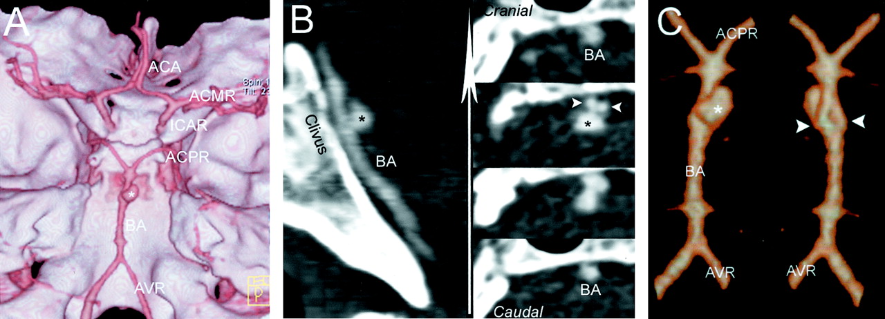
Scans from 16-row multisection CT and digital subtraction angiograms obtained before and after coiling.

A, 3D volume rendering of the multisection acquisition shows a panoramic view of the skull base with the position of the aneurysm (asterisk) at the middle segment of basilar artery.

B, Multiplanar reconstructions, obtained at the level of the clivus, show the position and configuration of the aneurysm and of the underlying basilar fenestration in the sagittal plane (left) and on sequential planes orthogonal to the clivus (right) through the basilar artery.

C, Segmented 3D volume rendering at the level of the basilar artery shows the aneurysm (craniocaudal view on left) and the fenestration (caudocranial view on right).

Multisection CT angiography showed both the saccular aneurysm and the fenestration with their reciprocal relationships. The proximal junction, lumen duplication, and distal junction were visualized. The neck of the aneurysm was depicted at the proximal end of the basilar fenestration (Fig 1B; Video 2). The direction of the dome of the aneurysm was posterior (orthogonal to the axis of the basilar artery).

Subsequently, digital subtraction angiography (Angiostar; Siemens, Erlangen, Germany) was performed for confirmation and treatment planning and revealed a fenestration of the middle segment of the basilar artery with a saccular aneurysm. Because of the surgical difficulties anticipated in approaching the aneurysm, the patient was successfully treated with endovascular embolization using GDCs (Boston Scientific, Target Therapeutics, Natick, MA).

Discussion

The basilar artery is formed by fusion of the plexiform primitive longitudinal neural arteries in a craniocaudal direction by approximately the 5th fetal week (6). If these embryonic precursors fail to fuse completely, duplication or fenestration of the basilar artery results.

The word fenestration refers to localized duplication of a vessel. Fenestrated basilar arteries are found in 1.33% to 6% of anatomic dissections (7). Their angiographic prevalence has been described as ranging from 0.04% to 0.6% (7, 8). In one study, basilar artery fenestration was detected in 1.7% of cases by using MR angiography.

An association of the fenestration with an aneurysm is not different from the typical association of the bifurcation in the circle of Willis with saccular aneurysms. The incidence of aneurysms in association with basilar fenestration is 7% (5). Medial defects, a common feature in both brain arteries and fenestrations, may predispose the arterial fenestration to aneurysm formation (9).

No reports in the literature describe the CT angiographic detection of a saccular aneurysm associated with basilar artery fenestration. In this case report, an association between a ruptured saccular aneurysm and a fenestration of the middle portion of the basilar artery is reported. An aneurysm was suspected after the clinical presentation of the patient and the detection of subarachnoid hemorrhage by unenhanced CT. Multisection CT angiography allowed us to make the diagnosis and showed the ruptured aneurysm and the congenital anatomic variant of the fenestration of the basilar artery. Digital subtraction angiography confirmed the findings of 16-row multisection CT.

One of the disadvantages reported with previous generations of CT scanners was the low scanning speed that resulted in visualization of cerebral veins together with cerebral arteries. This is because of the fast arteriovenous circulation time in the brain. Another reported disadvantage of CT angiography of cerebral arteries is the lack of spatial resolution needed to resolve small structures (eg, small aneurysms), especially those located at bone edges.

The new generation of spiral multisection CT scanners with 16 rows of detectors and 500-ms gantry rotation time allows fast data acquisition with high spatial resolution, reducing those pitfalls. Proper reconstructions and postprocessing allow better visualization of the vessels and their normal and abnormal morphologic features.

A limitation of this technique is blurring of the scan related to patient motion during scanning, even though the scanning time is very fast (3.1 s). Nevertheless, the limited amount of contrast material needed and the real-time display of the scans allows prompt scanning repetition. The advantages of multisection CT over digital subtraction angiography are related to its feasibility in any condition, its lower cost, its reduced invasiveness, and its high diagnostic accuracy.

References

  1. ↵
    De Caro R, Serafini MT, Galli S, Parenti A, Guidolin D, Munari PF. Anatomy of segmental duplication in the human basilar artery: possible site of aneurysm formation. Clin Neuropathol 1995;14:303–309
    PubMed
  2. ↵
    Imaizumi T, Saito K, Kobayashi T, Sakamoto Y, Komeichi T. Saccular aneurysm associated with fenestration of the distal segment of basilar artery [in Japanese]. No Shinkei Geka 1996;24:639–642
    PubMed
  3. ↵
    Nagashima H, Okudera H, Orz Y, Nakagawa F. Endovascular treatment of basilar trunk aneurysm associated with fenestration of the basilar artery. Neurosurg Rev 1999;22:219–221
    CrossRefPubMed
  4. ↵
    Fujimura M, Sugawara T, Higuchi H, Oku T, Seki H. A ruptured aneurysm at the distal end of the basilar artery fenestration associated with multiple fenestrations of the vertebrobasilar system: case report. Surg Neurol 1997;47:469–472
    PubMed
  5. ↵
    Uchino A, Kato A, Takase Y, Kudo S. Basilar artery fenestrations detected by MR angiography. Radiat Med 2001;19:71–74
    PubMed
  6. ↵
    Padget DH. The development of the cranial arteries in the human embryo. Contrib Embryol 1948;32:205–261
  7. ↵
    Sanders WP, Sorek PA, Mehta BA. Fenestration of intracranial arteries with special attention to associated aneurysms and other anomalies. AJNR Am J Neuroradiol 1993;14:675–680
    Abstract/FREE Full Text
  8. ↵
    Osborn GA. Diagnostic Cerebral Angiography. Philadelphia, Lippincott Williams & Wilkins;1999;190–193
  9. ↵
    Finlay HM, Canham PB. The layered fabric of cerebral artery fenestrations. Stroke 1994;25:1799–1806
    Abstract/FREE Full Text
  • Received February 26, 2003.
  • Accepted after revision April 13, 2003.
  • Copyright © American Society of Neuroradiology
View Abstract
PreviousNext
Back to top

In this issue

American Journal of Neuroradiology: 24 (10)
American Journal of Neuroradiology
Vol. 24, Issue 10
1 Nov 2003
  • Table of Contents
  • Index by author
Advertisement
Print
Download PDF
Email Article

Thank you for your interest in spreading the word on American Journal of Neuroradiology.

NOTE: We only request your email address so that the person you are recommending the page to knows that you wanted them to see it, and that it is not junk mail. We do not capture any email address.

Enter multiple addresses on separate lines or separate them with commas.
Noninvasive Detection of a Ruptured Aneurysm at a Basilar Artery Fenestration with Submillimeter Multisection CT Angiography
(Your Name) has sent you a message from American Journal of Neuroradiology
(Your Name) thought you would like to see the American Journal of Neuroradiology web site.
CAPTCHA
This question is for testing whether or not you are a human visitor and to prevent automated spam submissions.
Cite this article
Filippo Cademartiri, Dragan Stojanov, Diederik W.J. Dippel, Aad van der Lugt, Herve Tanghe
Noninvasive Detection of a Ruptured Aneurysm at a Basilar Artery Fenestration with Submillimeter Multisection CT Angiography
American Journal of Neuroradiology Nov 2003, 24 (10) 2009-2010;

Citation Manager Formats

  • BibTeX
  • Bookends
  • EasyBib
  • EndNote (tagged)
  • EndNote 8 (xml)
  • Medlars
  • Mendeley
  • Papers
  • RefWorks Tagged
  • Ref Manager
  • RIS
  • Zotero
0 Responses
Respond to this article
Share
Bookmark this article
Noninvasive Detection of a Ruptured Aneurysm at a Basilar Artery Fenestration with Submillimeter Multisection CT Angiography
Filippo Cademartiri, Dragan Stojanov, Diederik W.J. Dippel, Aad van der Lugt, Herve Tanghe
American Journal of Neuroradiology Nov 2003, 24 (10) 2009-2010;
del.icio.us logo Twitter logo Facebook logo Mendeley logo
  • Tweet Widget
  • Facebook Like
  • Google Plus One

Jump to section

  • Article
    • Abstract
    • Case Report
    • Discussion
    • References
  • Figures & Data
  • Supplemental
  • Info & Metrics
  • Responses
  • References
  • PDF

Related Articles

  • PubMed
  • Google Scholar

Cited By...

  • Management of basilar fenestration aneurysms: a systematic review with an illustrative case report
  • Crossref
  • Google Scholar

This article has not yet been cited by articles in journals that are participating in Crossref Cited-by Linking.

More in this TOC Section

  • Optimal MRI Sequence for Identifying Occlusion Location in Acute Stroke: Which Value of Time-Resolved Contrast-Enhanced MRA?
  • Evaluating the Effects of White Matter Multiple Sclerosis Lesions on the Volume Estimation of 6 Brain Tissue Segmentation Methods
  • Quiet PROPELLER MRI Techniques Match the Quality of Conventional PROPELLER Brain Imaging Techniques
Show more BRAIN

Similar Articles

Advertisement

Indexed Content

  • Current Issue
  • Accepted Manuscripts
  • Article Preview
  • Past Issues
  • Editorials
  • Editor's Choice
  • Fellows' Journal Club
  • Letters to the Editor
  • Video Articles

Cases

  • Case Collection
  • Archive - Case of the Week
  • Archive - Case of the Month
  • Archive - Classic Case

Special Collections

  • AJNR Awards
  • ASNR Foundation Special Collection
  • Most Impactful AJNR Articles
  • Photon-Counting CT
  • Spinal CSF Leak Articles (Jan 2020-June 2024)

More from AJNR

  • Trainee Corner
  • Imaging Protocols
  • MRI Safety Corner

Multimedia

  • AJNR Podcasts
  • AJNR Scantastics

Resources

  • Turnaround Time
  • Submit a Manuscript
  • Submit a Video Article
  • Submit an eLetter to the Editor/Response
  • Manuscript Submission Guidelines
  • Statistical Tips
  • Fast Publishing of Accepted Manuscripts
  • Graphical Abstract Preparation
  • Imaging Protocol Submission
  • Evidence-Based Medicine Level Guide
  • Publishing Checklists
  • Author Policies
  • Become a Reviewer/Academy of Reviewers
  • News and Updates

About Us

  • About AJNR
  • Editorial Board
  • Editorial Board Alumni
  • Alerts
  • Permissions
  • Not an AJNR Subscriber? Join Now
  • Advertise with Us
  • Librarian Resources
  • Feedback
  • Terms and Conditions
  • AJNR Editorial Board Alumni

American Society of Neuroradiology

  • Not an ASNR Member? Join Now

© 2025 by the American Society of Neuroradiology All rights, including for text and data mining, AI training, and similar technologies, are reserved.
Print ISSN: 0195-6108 Online ISSN: 1936-959X

Powered by HighWire