Advertisement

Main menu

  • Home
  • Content
    • Current Issue
    • Accepted Manuscripts
    • Article Preview
    • Past Issue Archive
    • Video Articles
    • AJNR Case Collection
    • Case of the Week Archive
    • Case of the Month Archive
    • Classic Case Archive
  • Special Collections
    • AJNR Awards
    • ASNR Foundation Special Collection
    • Most Impactful AJNR Articles
    • Photon-Counting CT
    • Spinal CSF Leak Articles (Jan 2020-June 2024)
  • Multimedia
    • AJNR Podcasts
    • AJNR SCANtastic
    • Trainee Corner
    • MRI Safety Corner
    • Imaging Protocols
  • For Authors
    • Submit a Manuscript
    • Submit a Video Article
    • Submit an eLetter to the Editor/Response
    • Manuscript Submission Guidelines
    • Statistical Tips
    • Fast Publishing of Accepted Manuscripts
    • Graphical Abstract Preparation
    • Imaging Protocol Submission
    • Author Policies
  • About Us
    • About AJNR
    • Editorial Board
    • Editorial Board Alumni
  • More
    • Become a Reviewer/Academy of Reviewers
    • Subscribers
    • Permissions
    • Alerts
    • Feedback
    • Advertisers
    • ASNR Home

User menu

  • Alerts
  • Log in

Search

  • Advanced search
American Journal of Neuroradiology

American Journal of Neuroradiology

ASHNR American Society of Functional Neuroradiology ASHNR American Society of Pediatric Neuroradiology ASSR
  • Alerts
  • Log in

Advanced Search

  • Home
  • Content
    • Current Issue
    • Accepted Manuscripts
    • Article Preview
    • Past Issue Archive
    • Video Articles
    • AJNR Case Collection
    • Case of the Week Archive
    • Case of the Month Archive
    • Classic Case Archive
  • Special Collections
    • AJNR Awards
    • ASNR Foundation Special Collection
    • Most Impactful AJNR Articles
    • Photon-Counting CT
    • Spinal CSF Leak Articles (Jan 2020-June 2024)
  • Multimedia
    • AJNR Podcasts
    • AJNR SCANtastic
    • Trainee Corner
    • MRI Safety Corner
    • Imaging Protocols
  • For Authors
    • Submit a Manuscript
    • Submit a Video Article
    • Submit an eLetter to the Editor/Response
    • Manuscript Submission Guidelines
    • Statistical Tips
    • Fast Publishing of Accepted Manuscripts
    • Graphical Abstract Preparation
    • Imaging Protocol Submission
    • Author Policies
  • About Us
    • About AJNR
    • Editorial Board
    • Editorial Board Alumni
  • More
    • Become a Reviewer/Academy of Reviewers
    • Subscribers
    • Permissions
    • Alerts
    • Feedback
    • Advertisers
    • ASNR Home
  • Follow AJNR on Twitter
  • Visit AJNR on Facebook
  • Follow AJNR on Instagram
  • Join AJNR on LinkedIn
  • RSS Feeds

AJNR is seeking candidates for the AJNR Podcast Editor. Read the position description.

OtherSPINE

CT-Guided Percutaneous Biopsy of Thoracic and Lumbar Spine: A New Coaxial Technique

Daniel Yaffe, Ghal Greenberg, Joseph Leitner, Reuven Gipstein, Myra Shapiro and Gil N. Bachar
American Journal of Neuroradiology November 2003, 24 (10) 2111-2113;
Daniel Yaffe
  • Find this author on Google Scholar
  • Find this author on PubMed
  • Search for this author on this site
Ghal Greenberg
  • Find this author on Google Scholar
  • Find this author on PubMed
  • Search for this author on this site
Joseph Leitner
  • Find this author on Google Scholar
  • Find this author on PubMed
  • Search for this author on this site
Reuven Gipstein
  • Find this author on Google Scholar
  • Find this author on PubMed
  • Search for this author on this site
Myra Shapiro
  • Find this author on Google Scholar
  • Find this author on PubMed
  • Search for this author on this site
Gil N. Bachar
  • Find this author on Google Scholar
  • Find this author on PubMed
  • Search for this author on this site
  • Article
  • Figures & Data
  • Info & Metrics
  • Responses
  • References
  • PDF
Loading

Abstract

Summary: Herein we describe the technique of CT-guided lumbar or thoracic bone biopsy performed with a larger bore needle and coaxial system. The use of the external sheath cannula as a coaxial system led to an accurate diagnosis in all 19 patients who underwent the procedure. Bleeding at the biopsy site occurred in two patients and was controlled by insertion of Gelfoam. No other complications were encountered. We suggest that our procedure is more effective, reliable, safe, and rapid than the traditional technique.

Modern imaging techniques can depict small and even asymptomatic bone lesions. Open biopsy is a major surgical procedure associated with morbidity and complications. Percutaneous biopsy under fluoroscopic or CT (CT) guidance is a safe and almost painless, and is preferred for lesions that have a soft-tissue component or are located close to vital structures. The reported accuracy of CT-guided spinal bone biopsy is 67–97%, and the complication rate ranges from 0–26% (1–7).

CT-guided bone biopsy can be performed with different types of needles (7, 8). The Ackerman needle (Cook Medical, Bloomington, IN) has the advantage of a coaxial system and has been found to be safe for biopsy retrieval; however, our initial experience with the Ackermann needle was unsatisfactory because of the presence of a crush artifact in a few cases, which made the histologic diagnosis difficult. This prompted us to try a larger bore needle with a newly developed coaxial system. Our aim is to describe our biopsy technique, which led to an accurate diagnosis in 19 consecutive patients.

Description of the Technique

The study group consisted of 19 consecutive patients (seven men, 12 women) aged 25 to 83 years (mean ± SD, 64.1 ± 14.9 years) who underwent CT-guided lumbar (n = 12) or thoracic (n = 7) bone biopsy in our center. Lesions were located in the following regions: one in T7, one in T8, three in T10, two in T11, two in L2, three in L3, five in L4 and two in L5. Indications for biopsy were osteolytic lesions (n = 11), osteoblastic lesions (n = 3), pathologic fracture (n = 3), and sclerotic collapse vertebra (n = 2). Patients with lesions in the sacral bone or those who underwent fine needle aspiration were excluded.

The study was approved by the institutional ethics committee and all patients gave informed consent before undergoing the examination. Aspirin and other non-steroidal, anti-inflammatory medications were discontinued 7 days before the procedure. Immediately before the biopsy, complete blood count, activated partial thromboplastin time, and prothrombin time were measured.

Twelve biopsies were performed with the 8-gauge Jamshidi needle (Manan Medical Products, Inc., Northbrook, IL) and seven with the 8-gauge Waldemar spinal biopsy needle (Link, Hamburg, Germany) (Fig 1). All biopsies were done under CT guidance (Elscint 2400 Elite, Haifa, Israel or Picker International Inc., Cleveland, OH) with the patients in the prone position. Maximum attention was addressed to the patient’s comfort. The lesion was localized, and the point of entry on the skin was marked in accordance with the biopsy technique (4–7). Three approaches were used: posterolateral (n = 2) for the upper lumbar vertebrae; transcostotransversal (n = 6) for the thoracic vertebrae; and transpedicular (n = 11) for the L4-L5 vertebrae and the thoracic spine. At the beginning of the procedure, a local anesthetic (1% Esracaine) was injected subcutaneously with a 25-gauge needle. Thereafter, to anesthetize the tract up to the periosteum, an 18-gauge, 24-cm-long needle (Fig 2) was inserted. When the tip of the needle reached the periosteum, additional Esracaine was injected, and the hub of the needle was cut, transforming it into a guidewire (Fig 2). An external sheath measuring 5.2 mm in diameter was slid over the guidewire by using a rotary motion for deeper penetration, and the wire was removed. The biopsy needle was inserted through the cannula. Before the bone was entered, a scan was obtained to check the location and direction of the needle. The periostium was penetrated by moving the Jamshidi or the Waldemar needle in a clockwise-counterclockwise motion with the sharp pointed stylet. The biopsy needle was then advanced slowly through the cortex to reach the lesion (Fig 3).

Fig 1.
  • Download figure
  • Open in new tab
  • Download powerpoint
Fig 1.

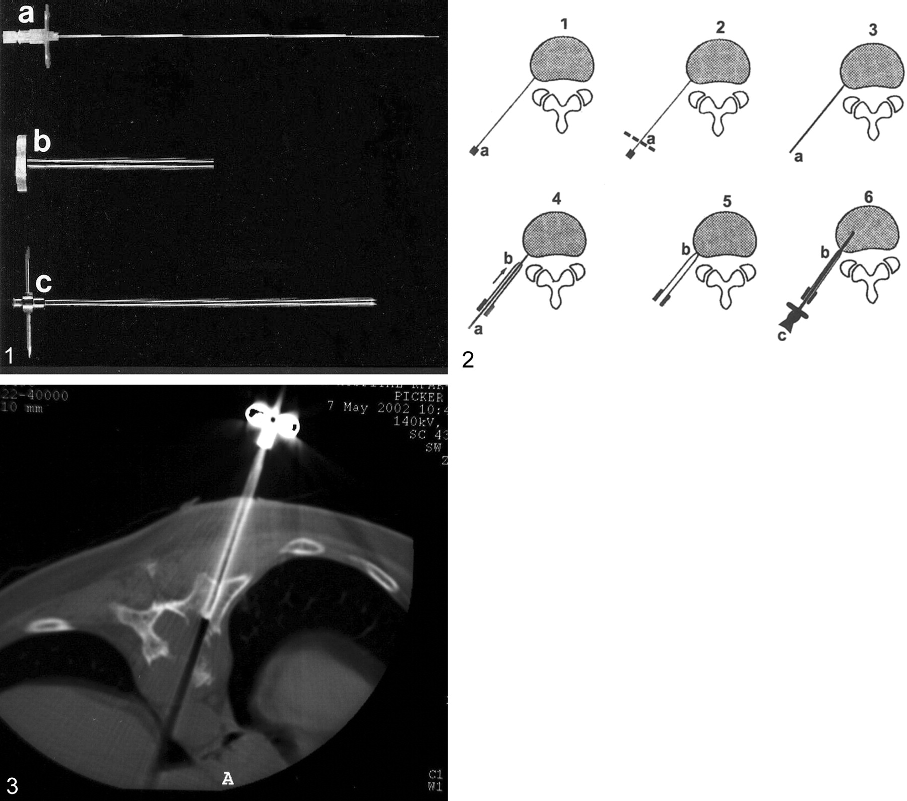
Equipment for coaxial vertebral biopsy.

A, An 18-gauge, 24-cm-long anesthetic needle.

B, external sheath cannula 5.2 mm in diameter.

C, Waldemar spine biopsy needle.

Fig 2.
  • Download figure
  • Open in new tab
  • Download powerpoint
Fig 2.

Percutaneous vertebral biopsy with the coaxial system. Parts shown in Figure 1 are labeled.

Fig 3.
  • Download figure
  • Open in new tab
  • Download powerpoint
Fig 3.

A 35-year-old patient with lytic lesion (plasmocytoma) involving the body of T10. Percutanous trans costal-transverse approach: the tip of the biopsy needle is visible at the margin of the mass.

An effort was made to obtain several specimens from the same tract, by using the external cannula in the coaxial system. The needle was retracted under continuous suction.

Biopsy material was obtained in all cases. None of the biopsy findings were falsely negative. The use of the external sheath cannula as a coaxial system led to an accurate diagnosis in all 19 cases (100%). The final diagnosis was osteoporotic fracture in two patients, multiple myeloma or plasmocytoma in five, metastasis in three, B cell lymphoma in three, and diskopathy in one. No malignancy was found on the basis of biopsy study in five patients, including three patients with a lytic lesion with a sclerotic border on CT scans and two with sclerotic collapse. In all five cases, clinical follow-up confirmed that the lesions were benign. Bleeding at the biopsy site occurred in two patients with a vascular lesion and was controlled by insertion of Gelfoam through the cannula. No other complications were encountered. Neurologic examinations showed no change after the biopsy procedure. The procedure was well tolerated by all patients and no patient complained of radicular pain either immediately after the procedure or during the next few postoperative days.

Discussion

Percutaneous biopsy performed with local anesthesia is a safe and highly accurate (1, 4, 5, 6, 8–11). Kornblum et al (1) reported a 71% accuracy rate in a series of 103 biopsies, and Murphy et al (9), who used a combination of aspiration and trephine technique, reported a 94% accuracy rate. Armstrong and Chalmers (10) reported a diagnostic yield of 68% in their aspiration biopsy series. DeSantos et al (11) achieved a 93% accuracy of biopsy findings of skeletal neoplasms. In a review of 120 bone biopsies Tehranzadeh et al (8) found an overall success rate of 72%. There were 70 (58%) true-positive results, 17 (14%) true-negative results, and 17 (14%) false-negative results; in 16 cases, the tissue was considered inadequate for diagnosis.

The major limitations of vertebral lesion biopsy are crushing and insufficent sample size. Intraprocedural trauma to the biopsy specimen was also the reason for our initial unsatisfactory results with the Ackermann needle. Although it was possible to identify malignant cells, tumor morphology was unrecognizable. Therefore, in the present series, we applied the larger bore (8-gauge) Jamshidi or Waldemar needle, which has a core cut of 3 mm compared with that of 1.5 mm for the Ackermann. This needle was sufficent to avoid crushing injury to the tissue. We were able to collect a precisely cut bone specimen within the lumen of the needle. The needle could be used for both lytic and sclerotic lesions. The transformation of the long anesthesia needle into a guidewire shortened the procedure and prevented the need for several insertion trials. To obtain two or three biopsies from a single lesion, we used a cannula of 5.2-mm diameter, so several biopsies could be retrieved via a single tract. This procedure proved to be practical and safe with both the Jamshidi and Waldemar needle sets. Of the 19 vertebral biopsies performed with this method, 11 (58%) results were true-positive, and eight (42%) were true-negative at a clinical follow-up of 12–60 months. The overall success rate was 100%.

Conclusion

Percutaneous vertebral biopsy is associated with a 0–26% rate of complications (1–9); the most frequently reported are pulmonary, neurologic, and infectious disorders. However, none of our patients had either immediate or long-term complications of the procedure. Two patients (10.5%) had excessive bleeding which was easily controlled by insertion of Gelfoam through the coaxial system. Bleeding is not considered a specific complication of this technique. The coaxial technique for CT-guided percutaneous biopsy of the spine has three advantages. First, intact specimens of adequate size can be obtained in most cases. Second, the use of the coaxial system enabled us to retrieve two or three biopsies through a single tract for better diagnosis. Third, the cannula can also be used to control bleeding in vascular lesions. We suggest that our system is more effective, reliable, safe, and rapid than the traditional technique.

References

  1. ↵
    Kornblum BK, Wesolowski DP, Fischgrund JS, et al. Computed tomography-guided biopsy of the spine: a review of 103 patients. Spine 1998;23:81–85
    CrossRefPubMed
  2. Babu NV, Titus VTK, Chittaranjan S, et al. Computed tomographically guided biopsy of the spine. Spine 1994;21:2436–2442
  3. Hadjipavlou AG, Arya S, Crow WN, et al. Percutaneous transpedicular biopsy of the spine. J Intervent Radiol 1996;11:103–108
  4. ↵
    Ghelman B, Lospinuso MF, Levine DB, et al. Percutaneous computed-tomography-guided biopsy of the thoracic and lumbar spine. Spine 1991;16:736–739
    CrossRefPubMed
  5. ↵
    Pierot L, Boulin A. Percutaneous biopsy of the thoracic and lumbar spine: transpedicular approach under fluoroscopic guidance. AJNR Am J Neuroradiol 1999;20:23–25
    Abstract/FREE Full Text
  6. ↵
    Jelinek JS, Kransdorf MJ, Gray R, et al. Percutaneous transpedicular biopsy of vertebral body lesions. Spine 1996;21:2035–2040
    CrossRefPubMed
  7. ↵
    Kattapuram SV, Rosenthal D. Percutaneous biopsy of skeletal lesions. AJR Am J Roentgenol 1991;157:935–942
    PubMed
  8. ↵
    Tehranzadeh J, Freiberger RH, Ghelman B. Closed skeletal needle biopsy: review of 120 cases. AJR Am J Roentgenol 1983;140:113–115
    PubMed
  9. ↵
    Murphy WA, Destouet JM, Gilula LA. Percutaneous skeletal biopsy in 1981: a procedure for radiologists, results, review and recommendations. Radiology 1981;139:545–549
    PubMed
  10. ↵
    Armstrong P, Chalmers AH. Needle aspiration-biopsy of the spine in suspected disc space infection. Br J Radiol 1978;51:333–337
    Abstract/FREE Full Text
  11. ↵
    DeSantos LA, Murray JA, Ayala AG. The value of percutaneous needle biopsy in the management of primary bone tumors. Cancer 1979;43:735–744
    CrossRefPubMed
  • Received May 12, 2003.
  • Accepted after revision May 23, 2003.
  • Copyright © American Society of Neuroradiology
View Abstract
PreviousNext
Back to top

In this issue

American Journal of Neuroradiology: 24 (10)
American Journal of Neuroradiology
Vol. 24, Issue 10
1 Nov 2003
  • Table of Contents
  • Index by author
Advertisement
Print
Download PDF
Email Article

Thank you for your interest in spreading the word on American Journal of Neuroradiology.

NOTE: We only request your email address so that the person you are recommending the page to knows that you wanted them to see it, and that it is not junk mail. We do not capture any email address.

Enter multiple addresses on separate lines or separate them with commas.
CT-Guided Percutaneous Biopsy of Thoracic and Lumbar Spine: A New Coaxial Technique
(Your Name) has sent you a message from American Journal of Neuroradiology
(Your Name) thought you would like to see the American Journal of Neuroradiology web site.
CAPTCHA
This question is for testing whether or not you are a human visitor and to prevent automated spam submissions.
Cite this article
Daniel Yaffe, Ghal Greenberg, Joseph Leitner, Reuven Gipstein, Myra Shapiro, Gil N. Bachar
CT-Guided Percutaneous Biopsy of Thoracic and Lumbar Spine: A New Coaxial Technique
American Journal of Neuroradiology Nov 2003, 24 (10) 2111-2113;

Citation Manager Formats

  • BibTeX
  • Bookends
  • EasyBib
  • EndNote (tagged)
  • EndNote 8 (xml)
  • Medlars
  • Mendeley
  • Papers
  • RefWorks Tagged
  • Ref Manager
  • RIS
  • Zotero
0 Responses
Respond to this article
Share
Bookmark this article
CT-Guided Percutaneous Biopsy of Thoracic and Lumbar Spine: A New Coaxial Technique
Daniel Yaffe, Ghal Greenberg, Joseph Leitner, Reuven Gipstein, Myra Shapiro, Gil N. Bachar
American Journal of Neuroradiology Nov 2003, 24 (10) 2111-2113;
del.icio.us logo Twitter logo Facebook logo Mendeley logo
  • Tweet Widget
  • Facebook Like
  • Google Plus One

Jump to section

  • Article
    • Abstract
    • Description of the Technique
    • Discussion
    • Conclusion
    • References
  • Figures & Data
  • Info & Metrics
  • Responses
  • References
  • PDF

Related Articles

  • No related articles found.
  • PubMed
  • Google Scholar

Cited By...

  • Diagnostic yield, accuracy, and complication rate of CT-guided biopsy for spinal lesions: a systematic review and meta-analysis
  • Percutaneous Spine Biopsy: A Meta-Analysis
  • Crossref
  • Google Scholar

This article has not yet been cited by articles in journals that are participating in Crossref Cited-by Linking.

More in this TOC Section

  • MP2RAGE 7T in MS Lesions of the Cervical Spine
  • Bern Score Validity for SIH
  • Resisted Inspiration for CSF-Venous Fistula
Show more Spine

Similar Articles

Advertisement

Indexed Content

  • Current Issue
  • Accepted Manuscripts
  • Article Preview
  • Past Issues
  • Editorials
  • Editor's Choice
  • Fellows' Journal Club
  • Letters to the Editor
  • Video Articles

Cases

  • Case Collection
  • Archive - Case of the Week
  • Archive - Case of the Month
  • Archive - Classic Case

Special Collections

  • AJNR Awards
  • ASNR Foundation Special Collection
  • Most Impactful AJNR Articles
  • Photon-Counting CT
  • Spinal CSF Leak Articles (Jan 2020-June 2024)

More from AJNR

  • Trainee Corner
  • Imaging Protocols
  • MRI Safety Corner

Multimedia

  • AJNR Podcasts
  • AJNR Scantastics

Resources

  • Turnaround Time
  • Submit a Manuscript
  • Submit a Video Article
  • Submit an eLetter to the Editor/Response
  • Manuscript Submission Guidelines
  • Statistical Tips
  • Fast Publishing of Accepted Manuscripts
  • Graphical Abstract Preparation
  • Imaging Protocol Submission
  • Evidence-Based Medicine Level Guide
  • Publishing Checklists
  • Author Policies
  • Become a Reviewer/Academy of Reviewers
  • News and Updates

About Us

  • About AJNR
  • Editorial Board
  • Editorial Board Alumni
  • Alerts
  • Permissions
  • Not an AJNR Subscriber? Join Now
  • Advertise with Us
  • Librarian Resources
  • Feedback
  • Terms and Conditions
  • AJNR Editorial Board Alumni

American Society of Neuroradiology

  • Not an ASNR Member? Join Now

© 2025 by the American Society of Neuroradiology All rights, including for text and data mining, AI training, and similar technologies, are reserved.
Print ISSN: 0195-6108 Online ISSN: 1936-959X

Powered by HighWire