Advertisement

Main menu

  • Home
  • Content
    • Current Issue
    • Accepted Manuscripts
    • Article Preview
    • Past Issue Archive
    • Video Articles
    • AJNR Case Collection
    • Case of the Week Archive
    • Case of the Month Archive
    • Classic Case Archive
  • Special Collections
    • AJNR Awards
    • ASNR Foundation Special Collection
    • Most Impactful AJNR Articles
    • Photon-Counting CT
    • Spinal CSF Leak Articles (Jan 2020-June 2024)
  • Multimedia
    • AJNR Podcasts
    • AJNR SCANtastic
    • Trainee Corner
    • MRI Safety Corner
    • Imaging Protocols
  • For Authors
    • Submit a Manuscript
    • Submit a Video Article
    • Submit an eLetter to the Editor/Response
    • Manuscript Submission Guidelines
    • Statistical Tips
    • Fast Publishing of Accepted Manuscripts
    • Graphical Abstract Preparation
    • Imaging Protocol Submission
    • Author Policies
  • About Us
    • About AJNR
    • Editorial Board
    • Editorial Board Alumni
  • More
    • Become a Reviewer/Academy of Reviewers
    • Subscribers
    • Permissions
    • Alerts
    • Feedback
    • Advertisers
    • ASNR Home

User menu

  • Alerts
  • Log in

Search

  • Advanced search
American Journal of Neuroradiology

American Journal of Neuroradiology

ASHNR American Society of Functional Neuroradiology ASHNR American Society of Pediatric Neuroradiology ASSR
  • Alerts
  • Log in

Advanced Search

  • Home
  • Content
    • Current Issue
    • Accepted Manuscripts
    • Article Preview
    • Past Issue Archive
    • Video Articles
    • AJNR Case Collection
    • Case of the Week Archive
    • Case of the Month Archive
    • Classic Case Archive
  • Special Collections
    • AJNR Awards
    • ASNR Foundation Special Collection
    • Most Impactful AJNR Articles
    • Photon-Counting CT
    • Spinal CSF Leak Articles (Jan 2020-June 2024)
  • Multimedia
    • AJNR Podcasts
    • AJNR SCANtastic
    • Trainee Corner
    • MRI Safety Corner
    • Imaging Protocols
  • For Authors
    • Submit a Manuscript
    • Submit a Video Article
    • Submit an eLetter to the Editor/Response
    • Manuscript Submission Guidelines
    • Statistical Tips
    • Fast Publishing of Accepted Manuscripts
    • Graphical Abstract Preparation
    • Imaging Protocol Submission
    • Author Policies
  • About Us
    • About AJNR
    • Editorial Board
    • Editorial Board Alumni
  • More
    • Become a Reviewer/Academy of Reviewers
    • Subscribers
    • Permissions
    • Alerts
    • Feedback
    • Advertisers
    • ASNR Home
  • Follow AJNR on Twitter
  • Visit AJNR on Facebook
  • Follow AJNR on Instagram
  • Join AJNR on LinkedIn
  • RSS Feeds

AJNR is seeking candidates for the AJNR Podcast Editor. Read the position description.

Case ReportCase Report

Dural Infantile Hemangioma Masquerading as a Skull Vault Lesion

C. Philpott, A. Wray, D. MacGregor and L. Coleman
American Journal of Neuroradiology June 2012, 33 (6) E85-E87; DOI: https://doi.org/10.3174/ajnr.A2752
C. Philpott
  • Find this author on Google Scholar
  • Find this author on PubMed
  • Search for this author on this site
A. Wray
  • Find this author on Google Scholar
  • Find this author on PubMed
  • Search for this author on this site
D. MacGregor
  • Find this author on Google Scholar
  • Find this author on PubMed
  • Search for this author on this site
L. Coleman
  • Find this author on Google Scholar
  • Find this author on PubMed
  • Search for this author on this site
  • Article
  • Figures & Data
  • Info & Metrics
  • Responses
  • References
  • PDF
Loading

Abstract

SUMMARY: We describe a case of intracranial dural IH initially diagnosed as a primary skull vault lesion hemangioma due to associated focal hyperostosis. Histopathologic examination of the dural component confirmed IH. The case is discussed in the context of IH within the neural axis.

ABBREVIATIONS:

Glut-1
glucose transporter-1 by immunohistochemistry
IH
infantile hemangioma

IHs are the most common benign neoplasms of infancy. Although intracranial lesions are relatively rare, they occur predominantly in the basal cisterns. IH presenting in a dural location has never previously been described, to our knowledge.

Case Report

A 12-month-old girl, a monozygotic twin, presented with an enlarged but stable head circumference greater than the 95th percentile with no neurologic signs. Sonography revealed mild ventriculomegaly and enlargement of the extra-axial spaces, compatible with benign enlargement of these spaces. MR imaging was performed, revealing an unexpected finding of focal expansion of the cancellous diploe of the right parietal bone with diffuse thickening and enhancement of the underlying dura (Figs 1 and 2).The dural abnormality was adjacent to the bony lesion but extended well beyond the lesion, including the dura of the tentorium. Apart from mild distortion of the adjacent posterior parietal lobe, there was no abnormality of the underlying brain.

Fig 1.
  • Download figure
  • Open in new tab
  • Download powerpoint
Fig 1.

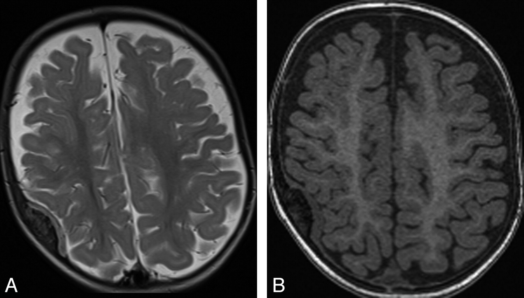
Axial T2 (A) and T1-weighted (B) sequences demonstrating iso- to mildly high T2 signal intensity, the iso-T1 signal intensity dural thickening, and adjacent focal diploic bone expansion with hyperostosis.

Fig 2.
  • Download figure
  • Open in new tab
  • Download powerpoint
Fig 2.

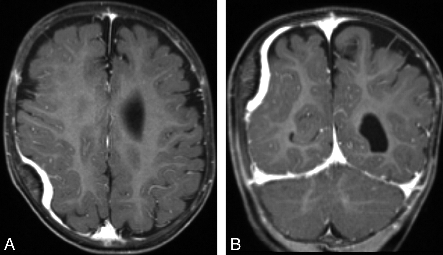
Axial (A) and coronal (B) T1 post-gadolinium-enhanced sequences demonstrating the avidly enhancing abnormal dural thickening underlying the hyperostosis.

A CT scan confirmed the focal expansion of the diploic bone and endosteal thickening (Fig 3). A benign bone lesion with an abnormal dural reaction, specifically a hemangioma, was favored, but the lack of diploic enhancement was thought atypical for such a lesion. The appearance of the dura raised suspicions of a more sinister process necessitating histopathologic analysis; hence, excisional biopsy, along with resection of abnormal bone, was performed. On histopathologic examination, the bone segment demonstrated symmetric expansion of cancellous bone covered by an unremarkable cortex with normocellular intervening marrow consistent with reactive bone. The dural biopsy demonstrated numerous irregular small blood vessels permeating the mature fibrous tissue (Fig 4A), the endothelium of which stained strongly for Glut-1 by immunohistochemistry (Fig 4B).

Fig 3.
  • Download figure
  • Open in new tab
  • Download powerpoint
Fig 3.

CT scan demonstrating benign-appearing focal diploic expansion and hyperostosis.

Fig 4.
  • Download figure
  • Open in new tab
  • Download powerpoint
Fig 4.

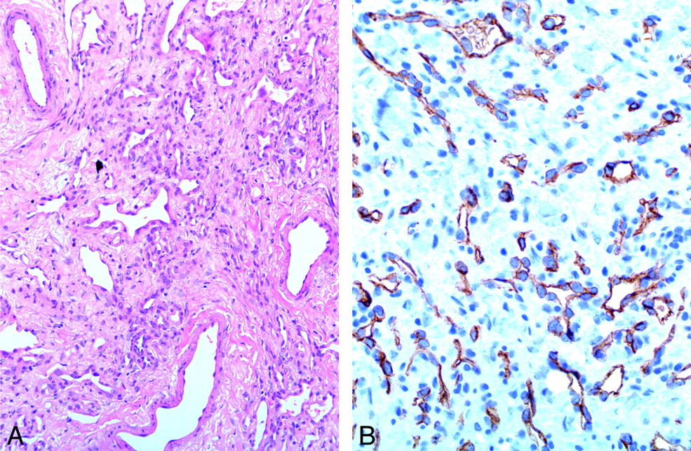
A, Mature fibrous tissue permeated by blood vascular channels of various sizes (hematoxylin-eosin, original magnification ×20 objective). B, Strong, uniform staining of endothelial cells for Glut-1 (immunohistochemical stain, original magnification ×40 objective).

MR imaging at 6-month follow-up revealed reduction in dural thickening consistent with spontaneous involution and appropriate bony healing. Clinically, without further treatment, the patient is achieving milestones with plateauing of her head growth. Dedicated review revealed no cutaneous lesions.

Discussion

IHs (previously termed “capillary hemangiomas”) are the most common vascular tumors of infancy. They are benign neoplasms of endothelial cells that rapidly proliferate initially, usually in the first 5 months of life, followed by a plateau phase and slow involution.1 On histologic examination, these lesions comprise lobules of tightly packed endothelial cells forming capillary-like structures, separated by septa of fibrous connective tissue.1,2 Identification of the Glut-1 protein on immunohistochemical staining is used to distinguish these lesions from other vascular lesions.1 More commonly, they involve the skin and soft tissues. Involvement of the neural axis is rare.

There have been 28 cases of intracranial IH published in the literature.3⇓⇓⇓⇓⇓⇓⇓⇓⇓⇓⇓⇓–16 Classically, these cases involve the extra-axial basal cisterns, subarachnoid and ventricular spaces, and cavernous sinus.7⇓⇓–10,12⇓–14 Two articles reported dural attachment,10,16 and only 4 cases were intra-axial.3,4 Most intracranial IHs (71%)4,7,9,12,14 were associated with extracranial soft-tissue extension or cutaneous hemangiomas.

While spinal dural capillary hemangiomas have been described, this is the first case of cranial dural IH. Cranial IH typically presents in childhood (86%),3⇓⇓⇓⇓⇓–9,12,14⇓–16 and in a few cases, in adolescence,3,10,11,13 while spinal IH usually presents in an older population, (range, 40–62 years).17

The imaging characteristics of IH within the neural axis are consistent with those found elsewhere, with iso- to low-T1 signal intensity, high T2 signal intensity, and avid contrast enhancement typical on MR imaging.14,17 Intralesional flow voids are a common feature,10,14 and foci of high T1 signal intensity are also described.4,10,17 This may represent thrombosis in some of the small vascular spaces or hemorrhage.

Our initial interpretation of the imaging findings was that this was primarily a bony lesion, an intradiploic IH, with hyperostosis and secondary dural reaction. However, the features were atypical and of concern. There was hyperostosis but no abnormal diploic space enhancement or large abnormal diploic space vessels. The dural reaction was too extensive and florid to be a secondary dural reaction to an intradiploic IH or an essentially benign-appearing bone lesion. Meningioma was considered a possibility, given the florid hyperostosis and dural thickening; however, the appearance was also atypical for this condition. The extensive dural thickening on imaging, therefore, raised concern of a primary dural lesion of sinister etiology.

Hydrocephalus was been described in 10% of intracranial IHs. It is uncertain whether this represents an association with or a consequence of the hemangioma. It could be postulated that thrombosis of small venous structures associated with the hemangioma or the direct mass effect of the lesion alters local CSF flow dynamics, resulting in hydrocephalus. The mild ventricular dilation in our case may reflect such a prior CSF flow alteration because we postulated that this lesion was already involuting at diagnosis.

Given that IHs are self-limited, management is dependent on size, location, and any associated complications, which include visual and neurologic deficits for the intracranial lesions. Contemporary management of IH is with propranolol; however, systemic steroid and interferon therapy has been described.12,13 In the absence of complicating features, observation in expectation of involution is favored.

Conclusions

We report a new entity of intracranial dural hemangioma, histologically confirmed by Glut-1 staining. The description of the imaging findings and pitfalls in diagnosing this lesion should be considered in assessing durally based lesions in childhood and should help to guide the management of this benign self-limiting lesion.

Footnotes

  • Disclosures: Alison Wray—Unrelated: Travel/Accommodations/Meeting Expenses: Medtronic,* Details: educational grant to attend intrathecal baclofen users' group meeting. * Money paid to institution.

References

  1. 1.↵
    1. Holland KE,
    2. Drolet BA
    . Infantile hemangioma. Pediatr Clin North Am 2010; 57: 1069– 83
    CrossRefPubMed
  2. 2.↵
    1. Puttgen KB,
    2. Pearl M,
    3. Tekes A,
    4. et al
    . Update on pediatric extracranial vascular anomalies of the head and neck. Childs Nerv Syst 2010; 26: 1417– 33
    CrossRefPubMed
  3. 3.↵
    1. Abe M,
    2. Tabuchi K,
    3. Tanaka S,
    4. et al
    . Capillary hemangioma of the central nervous system. J Neurosurg 2004; 101: 73– 81
    CrossRefPubMed
  4. 4.↵
    1. Balaci E,
    2. Sumner TE,
    3. Auringer ST,
    4. et al
    . Diffuse neonatal hemangiomatosis with extensive involvement of the brain and cervical spinal cord. Pediatr Radiol 1999; 29: 441– 43
    CrossRefPubMed
  5. 5.↵
    1. Bihannic A,
    2. Michot C,
    3. Heckly A,
    4. et al
    . Capillary haemangioma arising from the anterior choroidal artery. Child's Nervous System 2005; 21: 265– 71. Epub 2005 Jan 20
    CrossRefPubMed
  6. 6.↵
    1. Daenekindt T,
    2. Weyns F,
    3. Kho KH,
    4. et al
    . Giant intracranial capillary hemangioma associated with enlarged head circumference in a newborn. J Neurosurg Pediatr 2008; 1: 488– 92
    CrossRefPubMed
  7. 7.↵
    1. Ersoy S,
    2. Mancini AJ
    . Hemifacial infantile hemangioma with intracranial extension: a rare entity. Pediatr Dermatol 2005; 22: 309– 13
    CrossRefPubMed
  8. 8.↵
    1. Karikari IO,
    2. Selznick LA,
    3. Cummings TJ,
    4. et al
    . Capillary hemangioma of the fourth ventricle in an infant: case report and review of the literature. J Neurosurg 2006; 104 (3 suppl): 188– 91
    CrossRefPubMed
  9. 9.↵
    1. Poindexter G,
    2. Metry DW,
    3. Barkovich AJ,
    4. et al
    . PHACE syndrome with intracerebral hemangiomas, heterotopia, and endocrine dysfunction. Pediatr Neurol 2007; 36: 402– 06
    CrossRefPubMed
  10. 10.↵
    1. Simon SL,
    2. Moonis G,
    3. Judkins AR,
    4. et al
    . Intracranial capillary hemangioma: case report and review of the literature. Surg Neurol 2005; 64: 154– 59
    CrossRefPubMed
  11. 11.↵
    1. Smith I,
    2. Skelton V
    . An unusual intracranial tumour presenting in pregnancy. Int J Obstet Anesth 2007; 16: 82– 85
    CrossRefPubMed
  12. 12.↵
    1. Tortori-Donati P,
    2. Fondelli MP,
    3. Rossi A,
    4. et al
    . Intracranial contrast-enhancing masses in infants with capillary haemangioma of the head and neck: intracranial capillary haemangioma? Neuroradiology 1999; 41: 369– 75
    CrossRefPubMed
  13. 13.↵
    1. Tsao MN,
    2. Schwartz ML,
    3. Bernstein M,
    4. et al
    . Capillary hemangioma of the cavernous sinus: report of two cases. J Neurosurg 2003; 98: 169– 74
    CrossRefPubMed
  14. 14.↵
    1. Viswanathan V,
    2. Smith ER,
    3. Mulliken JB,
    4. et al
    . Infantile hemangiomas involving the neuraxis: clinical and imaging findings. AJNR Am J Neuroradiol 2009; 30: 1005– 13
    Abstract/FREE Full Text
  15. 15.↵
    1. Watanabe M,
    2. Kubo N,
    3. Hakozaki S,
    4. et al
    . Intracranial capillary hemangioma: a case report [in Japanese]. No Shinkei Geka 2001; 29: 1049– 54
    PubMed
  16. 16.↵
    1. Willing SJ,
    2. Faye-Petersen O,
    3. Aronin P,
    4. et al
    . Radiologic-pathologic correlation: capillary hemangioma of the meninges. AJNR Am J Neuroradiol 1993; 14: 529– 36
    Abstract/FREE Full Text
  17. 17.↵
    1. Nowak DA,
    2. Widenka DC
    . Spinal intradural capillary haemangioma: a review. Eur Spine J 2001; 10: 464– 72
    CrossRefPubMed
  • Received April 23, 2011.
  • Accepted after revision May 17, 2011.
  • © 2012 by American Journal of Neuroradiology
View Abstract
PreviousNext
Back to top

In this issue

American Journal of Neuroradiology: 33 (6)
American Journal of Neuroradiology
Vol. 33, Issue 6
1 Jun 2012
  • Table of Contents
  • Index by author
Advertisement
Print
Download PDF
Email Article

Thank you for your interest in spreading the word on American Journal of Neuroradiology.

NOTE: We only request your email address so that the person you are recommending the page to knows that you wanted them to see it, and that it is not junk mail. We do not capture any email address.

Enter multiple addresses on separate lines or separate them with commas.
Dural Infantile Hemangioma Masquerading as a Skull Vault Lesion
(Your Name) has sent you a message from American Journal of Neuroradiology
(Your Name) thought you would like to see the American Journal of Neuroradiology web site.
CAPTCHA
This question is for testing whether or not you are a human visitor and to prevent automated spam submissions.
Cite this article
C. Philpott, A. Wray, D. MacGregor, L. Coleman
Dural Infantile Hemangioma Masquerading as a Skull Vault Lesion
American Journal of Neuroradiology Jun 2012, 33 (6) E85-E87; DOI: 10.3174/ajnr.A2752

Citation Manager Formats

  • BibTeX
  • Bookends
  • EasyBib
  • EndNote (tagged)
  • EndNote 8 (xml)
  • Medlars
  • Mendeley
  • Papers
  • RefWorks Tagged
  • Ref Manager
  • RIS
  • Zotero
0 Responses
Respond to this article
Share
Bookmark this article
Dural Infantile Hemangioma Masquerading as a Skull Vault Lesion
C. Philpott, A. Wray, D. MacGregor, L. Coleman
American Journal of Neuroradiology Jun 2012, 33 (6) E85-E87; DOI: 10.3174/ajnr.A2752
del.icio.us logo Twitter logo Facebook logo Mendeley logo
  • Tweet Widget
  • Facebook Like
  • Google Plus One
Purchase

Jump to section

  • Article
    • Abstract
    • ABBREVIATIONS:
    • Case Report
    • Discussion
    • Conclusions
    • Footnotes
    • References
  • Figures & Data
  • Info & Metrics
  • Responses
  • References
  • PDF

Related Articles

  • No related articles found.
  • PubMed
  • Google Scholar

Cited By...

  • No citing articles found.
  • Crossref (10)
  • Google Scholar

This article has been cited by the following articles in journals that are participating in Crossref Cited-by Linking.

  • Primary Intraosseous Cavernous Hemangioma in the Skull
    Yi Yang, Jian Guan, Wenbin Ma, Yongning Li, Bing Xing, Zuyuan Ren, Changbao Su, Renzhi Wang
    Medicine 2016 95 11
  • Beta Blockade as Treatment for Intracranial Infantile Hemangioma: Case Report and Literature Review
    Elise Kang, Neil Friedman, Ihsan Mamoun, Joan Tamburro, Alex Golden
    Pediatric Neurology 2016 59
  • Safe and Effective Treatment of Intracranial Infantile Hemangiomas with Beta-Blockers
    Aoife Naughton, Ariel Yuhan Ong, Goran Darius Hildebrand
    Pediatric Reports 2021 13 3
  • Intracranial capillary hemangiomas: literature review in pediatric and adult population
    Giorgio Santoro, Manolo Piccirilli, Vito Chiarella, Nicoletta Greco, Luigi Valentino Berra, Antonio Santoro
    Neurosurgical Review 2021 44 4
  • Large Orbital Pediatric Intraosseous Hemangioma
    Colin Bacorn, Lily Koo Lin
    Case Reports in Ophthalmological Medicine 2020 2020
  • A potential VEGF-driven hypothesis of calvarial centripetal proliferation in cerebral proliferative angiopathy
    Shigeta Miyake, Timo Krings, Tze Phei Kee, Thierry A. G. M. Huisman
    Scientific Reports 2025 15 1
  • An infant with posterior fossa hemangioma with aortic stenosis: Case report and review of the literature
    Yigit Can Senol, Ergun Daglioglu, Ozge Aydogdu Basaran, Ahmet Deniz Belen
    Journal of Pediatric Neurosciences 2022 17 4
  • Intracranial Infantile Hemangioma: Highlighting a Rare Presentation With a Case Report and Literature Review
    Sultan F Albalawi, Badr E Hafiz, Alanoud Turki, Murad Alturkustani, Salwa I Bakhsh, Abdulrahman J Sabbagh
    Cureus 2024
  • Nursing Care of the Pediatric Neurosurgery Patient
    Theresa M. Gabay, Davonna Ledet
    2017
  • Pediatric Vascular Neurosurgery
    Evan Winograd, Renée M. Reynolds, Veetai Li, L. Nelson Hopkins
    2021

More in this TOC Section

  • Atypical Diffusion-Restricted Lesion in 5-Fluorouracil Encephalopathy
  • Multimodal Imaging of Spike Propagation: A Technical Case Report
Show more Case Reports

Similar Articles

Advertisement

Indexed Content

  • Current Issue
  • Accepted Manuscripts
  • Article Preview
  • Past Issues
  • Editorials
  • Editor's Choice
  • Fellows' Journal Club
  • Letters to the Editor
  • Video Articles

Cases

  • Case Collection
  • Archive - Case of the Week
  • Archive - Case of the Month
  • Archive - Classic Case

Special Collections

  • AJNR Awards
  • ASNR Foundation Special Collection
  • Most Impactful AJNR Articles
  • Photon-Counting CT
  • Spinal CSF Leak Articles (Jan 2020-June 2024)

More from AJNR

  • Trainee Corner
  • Imaging Protocols
  • MRI Safety Corner

Multimedia

  • AJNR Podcasts
  • AJNR Scantastics

Resources

  • Turnaround Time
  • Submit a Manuscript
  • Submit a Video Article
  • Submit an eLetter to the Editor/Response
  • Manuscript Submission Guidelines
  • Statistical Tips
  • Fast Publishing of Accepted Manuscripts
  • Graphical Abstract Preparation
  • Imaging Protocol Submission
  • Evidence-Based Medicine Level Guide
  • Publishing Checklists
  • Author Policies
  • Become a Reviewer/Academy of Reviewers
  • News and Updates

About Us

  • About AJNR
  • Editorial Board
  • Editorial Board Alumni
  • Alerts
  • Permissions
  • Not an AJNR Subscriber? Join Now
  • Advertise with Us
  • Librarian Resources
  • Feedback
  • Terms and Conditions
  • AJNR Editorial Board Alumni

American Society of Neuroradiology

  • Not an ASNR Member? Join Now

© 2025 by the American Society of Neuroradiology All rights, including for text and data mining, AI training, and similar technologies, are reserved.
Print ISSN: 0195-6108 Online ISSN: 1936-959X

Powered by HighWire