
Abstract
BACKGROUND AND PURPOSE: The flow-diverter stent has been proved a feasible, safe, and efficient technique, particularly for the treatment of large and broad-neck carotid siphon aneurysms. Wide-neck bifurcation aneurysms remain, in some cases, a challenge for neurointerventionalists. We report the outcomes of the treatment of saccular middle cerebral artery bifurcation aneurysms with flow diversion in our institution.
MATERIALS AND METHODS: From the institution data base, all saccular, nondissecting MCA bifurcation aneurysms, treated with flow-diverter stents, were retrospectively reviewed. Technical issues, immediate posttreatment and follow-up angiographic findings, and clinical outcomes were assessed.
RESULTS: Fourteen patients with 15 aneurysms were included in the study. Ischemic complications, as confirmed by MR imaging, occurred in 6 patients (43%). Procedure-related morbidity and mortality at last follow-up were 21% and 0%, respectively. Angiographic follow-up was available for 13 aneurysms, with a mean follow-up of 16 months. Complete occlusion was obtained for 8 aneurysms (62%).
CONCLUSIONS: Compared with other available therapeutic options, the flow-diverter stent does not appear to be a suitable solution for the treatment of saccular MCA bifurcation aneurysms.
ABBREVIATIONS:
- EVT
- endovascular treatment
- FDS
- flow-diverter stent
Endovascular treatment (EVT) of middle cerebral artery aneurysms is considered safe and effective.1⇓–3 However the choice between surgical and endovascular approaches for the treatment of aneurysms in this location is still a matter of debate.4 Furthermore, wide-neck MCA bifurcation lesions can sometimes prove challenging for traditional EVT approaches, and numerous techniques have been developed to address these cases. Simple coil embolization and remodeling techniques, including single-balloon- or multiple-balloon-assisted coiling5,6; the dual-catheter technique7; stent-assisted coiling8⇓–10; the “Y-stent placement” technique with and without coiling11,12; the “waffle cone” technique13,14; neck-bridge devices15,16; and intra-aneurysmal flow disrupters17,18 have all been trialed.
Flow-diverter stents (FDSs) have proved feasible, safe, and efficient for the treatment of large and broad-neck carotid siphon aneurysms.19 They have been designed to induce aneurysm sac thrombosis through flow disruption at the level of the neck, while preserving normal flow into parent vessels and adjacent branches.20 Few studies have been published concerning FDS use for bifurcation lesion treatment, to our knowledge. In this report, our aim was to evaluate the feasibility, safety, and efficacy of the use of the FDSs for MCA saccular bifurcation aneurysms, with special consideration regarding the effects on the covered branches.
Materials and Methods
Population
Between May 2013 and June 2014, 77 FDS procedures for intracranial aneurysm treatment were performed in the Beaujon Hospital (APHP, Paris). From our prospectively maintained data base, we extracted all MCA bifurcation saccular aneurysms treated with an FDS and collected the following data for each patient: age, sex, aneurysm location and biometry (maximum diameter, neck size, aspect ratio), rupture status, previous treatment, the modified Rankin Scale scores at the time of admission and discharge, modalities of treatment, intraoperative complications (aneurysm rupture, thromboembolic events, deployment failure), and postoperative complications (delayed bleeding, thromboembolic events). Immediate postoperative DSA, 3D rotational angiography, and VasoCT images (Philips Healthcare, Best, the Netherlands)21,22 were collected along with any available follow-up imaging data. The last mRS evaluation during follow-up was also recorded.
In this study, only saccular aneurysms were included, and nonsaccular aneurysms (fusiform, blood blister–like, or dissecting) were excluded. Those lesions were considered very difficult to treat for both interventionists and neurosurgeons. Conventional EVT approaches were judged not suitable. Some relatively small lesions were treated because of a patient history of multiple aneurysms and previous rupture. In each case, the decision to treat was made by a multidisciplinary team, which included interventionists and surgeons. Informed consent was obtained in each case.
Endovascular Treatment
All patients were treated preoperatively with a dual antiplatelet therapy of 160 mg of aspirin and 75 mg of clopidogrel per day for 7 days before treatment. The effectiveness of the platelet inhibition therapy was tested by hematologic analysis, and in cases of insufficiency, loading doses were administrated.
Endovascular treatment was performed with the patient under general anesthesia and systemic heparinization. The protocol for heparinization was administration of a 50-U/kg bolus at the beginning of the procedure, followed by a continuous intravenous injection of 35–50 U/kg to maintain an activated clotting time of 2–2.5 times the baseline.
We used 3 types of commercial FDSs: the Silk flow diverter (Balt, Montmorency, France), the Pipeline Embolization Device (Covidien, Irvine, California), and the FRED stent (MicroVention, Tustin, California). The selection of the specific FDS in each case depended on operator preference, and sizing was performed on the basis of artery measurement, acquired from 3D rotational angiography data.
For the postprocedural medication regimen, aspirin and clopidogrel were both maintained for 3 months and aspirin was continued for a further 9 months.
Procedural Assessment and Follow-Up
Follow-up angiographies were performed at 3–6 months then at 1.5 and 3.5 years after treatment, depending on the previous results. Standard projections, 3D rotational angiography, and VasoCT analyses were used to evaluate residual flow within the aneurysm and to assess the patency of both the FDS and MCA bifurcation branches. Aneurysm occlusion was graded as either complete, residual neck, residual aneurysm, or unchanged. Permanent morbidity and mortality rates subsequent to treatment were evaluated at discharge and at follow-up when possible. MR imaging studies were performed only in cases of acute neurologic deficit. Morbidity was defined as an mRS of >1. When the preoperative mRS was >1, morbidity was defined by any increase in the mRS score.
Results
Population
Fourteen patients (9 women and 5 men; age range, 40–67 years; median age, 57 years), with 15 MCA bifurcation aneurysms were included in this study. Five aneurysms had a history of rupture and recanalization after coil treatment, but none were treated with an FDS at the acute phase of bleeding. In 2 cases, the reason to treat was incomplete occlusion or recanalization after WEB aneurysm embolization system (Sequent Medical, Aliso Viejo, California) treatment. Twelve aneurysms (80%) were located on the right side. Initial aneurysm size varied from 2.6 to 14 mm (mean, 6.1 ± 3.6 mm), neck size varied from 2.3 to 7 mm (mean, 4.1 ± 1.5 mm), and the aspect ratio varied from 0.6 to 1.2 mm (mean, 0.8 ± 0.2 mm).
Feasibility
FDS delivery was possible in all cases. All patients except 1 were treated with only 1 FDS. The remaining patient had 2 aneurysms on the same bifurcation; stent shortening was perceived during follow-up and a new FDS was secondarily implanted, in a telescopic fashion, because one of the aneurysm necks was no longer sufficiently covered.
Additional sac coiling was performed during 1 procedure in 1 patient.
Complication Rate
One patient had intraoperative occlusion of the covered branch; however, collateral flow was excellent and the patient was asymptomatic on waking after the operation. The day 2 angiogram demonstrated reopening of the artery. This patient was also the only one who presented with a weak sensitivity to antiplatelet medications, thus requiring a loading dose of 300 mg of clopidogrel (Plavix) before treatment.
In another case, slight flow modification was depicted at the end of the treatment in the covered branch, and hemiparesis was evident when the patient awoke. A new angiogram was immediately obtained, and covered branch occlusion leading to neurologic deficit was confirmed, despite efficient abciximab infusion and artery reopening.
For 1 patient, a second FDS, positioned in a telescopic fashion, was deemed necessary due to original FDS shortening. Within 20 hours of the second intervention, the patient presented with left hemiplegia and DSA analysis showed dual-FDS occlusions, extending into both bifurcation branches. Intra-arterial thrombolysis allowed efficient reopening of both FDS devices and bifurcation branches. The patient recovered from all motor deficits within days and was able to return to work, having only minor cognitive impairment. In this case, treatment with aspirin and a double dose of Plavix was continued for 6 months, though biologic tests did not demonstrate any antiplatelet resistance. After 6 months, Plavix therapy was stopped. Two years after EVT, on termination of aspirin treatment, the patient reported a transient ischemic attack in the MCA territory, so aspirin was re-instated for life.

A 58-year-old woman was previously treated in our institution for a right posterior communicating artery ruptured aneurysm. Three months later, she was scheduled for the treatment of a second aneurysm. A, The right internal carotid angiogram shows a saccular MCA bifurcation aneurysm. B, The unsubtracted picture after FDS placement in the inferior bifurcation branch. C, The final angiogram shows slow flow in the covered branch (arrow). Five hours after the treatment, the patient presented with transient right hemiparesis when the mean blood pressure dropped to <80 mm Hg. D, MR imaging shows limited DWI-positive findings in the corresponding territory. The patient then remained asymptomatic. E, Six-month follow-up shows a reduction in the size of the aneurysm. F–H, DSA, 3D rotational angiography, and VasoCT at 2.5-year follow-up demonstrate complete occlusion of the aneurysm and the unchanged aspect of the covered branch.
Slow flow was observed in the covered branch on the final angiograms in 4 patients. This led to postoperative presentation of no deficits in 1 case, transient deficits in 1 case, and permanent deficits in 2 cases.
One of those 4 patients exhibited covered branch occlusion during follow-up. This patient presented with slight left hemiparesis due to slow flow in the covered branch after treatment and was admitted to our institution 1 month later with complete left-sided motor deficits. The patient had discontinued clopidogrel medication a few days before admission, and DSA showed complete occlusion of the stent, though the collateral flow was relatively good. The patient partially overcame the deficit within days.
Overall in this study, no hemorrhagic complications occurred. Immediate flow modifications in the covered branch were not identified in only 7 patients (50%).
Clinical ischemic complications, defined as new neurologic deficits and confirmed by MR imaging, occurred in 43% of patients, though most were small DWI lesions caused by flow restriction in the covered branch. Procedure-related morbidity and mortality at last follow-up were 21% and 0%, respectively.
Angiographic Follow-Up
Angiographic follow-up data were available for 12 patients and 13 aneurysms (87%). The remaining 2 patients have yet to undergo their first follow-up DSA. The average length of angiographic follow-up for this cohort was 16 months (range, 3–41; median, 8 months).
On the basis of the most recent angiogram in each case, we observed, in total, 2 covered branch occlusions and 6 other cases (50%) presented with a significantly reduced caliber. Overall, covered branch modifications were present in 66% of cases.
We noted complete aneurysm occlusion in 62% of patients, aneurysm remnants with partial occlusion in 31% of patients, and a neck remnant in 8% of patients.
Discussion
Wide-neck bifurcation aneurysms can, in some cases, be difficult and challenging for traditional EVT approaches. Most can be successfully treated with the balloon-remodeling technique,5 especially in ruptured cases. However, attenuated and complete coil occlusion is not always possible, and recurrence rates are not negligible.23,24
In these cases, stent-assisted coiling has been increasingly favored,10,11 but although efficiency rates are good, complication rates appear high for “X” and “Y” configurations, with a 10.0% occurrence of procedure-related permanent neurologic deficits and a 1.0% death rate reported in the study of Bartolini et al.25
The use of FDSs in the treatment of intracranial aneurysms is gradually increasing. The FDS concept is to induce intra-aneurysmal thrombosis by disrupting the flow near the aneurysmal neck while preserving the patency of the parent vessels and the covered branches.19 It has been shown that it is safe to cover the supraclinoid internal carotid branch while treating internal carotid aneurysms with FDSs and that in these cases, the occlusion rate of the covered branches is low.26,27
A small number of articles describe the safety and efficiency of FDSs in the treatment of bifurcation aneurysms. Very recently, some articles investigating their use in the treatment of MCA aneurysms were published, though study populations were sometimes heterogeneous and included saccular and fusiform aneurysms and both bifurcation and nonbifurcation types.28⇓–30

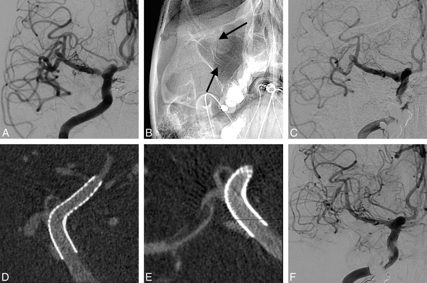
A 44-year-old woman who presented with an incidentally found aneurysm was treated in our institution. Although the aneurysm was small, the patient requested treatment. A, The right internal carotid angiogram shows a saccular MCA bifurcation aneurysm. B, The unsubtracted view after FDS placement in the superior bifurcation branch. C, The final internal carotid angiogram confirms the partial thrombosis but shows very good collateral flow. After stent delivery, VasoCT curved reconstructions of the superior (D) and inferior branches (E) show good patency of the FDS but immediate partial thrombosis in the covered branch and aneurysm. The patient was kept under a double dose of antiplatelet therapy, heparin, and noradrenaline for 48 hours. She did not develop any symptoms, and early follow-up DSA showed the normal aspect of the covered branch. However, the angiogram obtained at 2-year follow-up (F), demonstrates asymptomatic complete occlusion of both the covered branch and aneurysm.
From 25 MCA aneurysms (including 21 bifurcation lesions) treated with FDSs, Yavuz et al28 reported good results, with 84% complete occlusion at follow-up. Although the last follow-up showed that 43% of covered branches had reduced caliber or were occluded, no permanent neurologic deficits occurred. Subsequent articles did not show similar results.
In the Saleme et al29 study of FDS-treated bifurcation aneurysms, 19 were located at the MCA bifurcation (89% saccular type). The complete occlusion rate was good (92%), but the overall procedure-related complication rate was 8%, with new permanent neurologic deficits occurring in 9.4% of patients during hospitalization and new DWI-positive findings reported in 64.8% of cases. Middle cerebral artery bifurcation covered branch modifications remained unchanged in only 37% of cases, and symptomatic narrowing or occlusion occurred in 16% of cases.
In a small MCA aneurysm study, with 15 subjects, Briganti et al30 claimed that EVT with FDS was a relatively safe treatment for those lesions, though they described high rates of permanent postprocedural neurologic deficits (27% of cases).
In our study, the ischemic complication rate was also very high. In 43% of cases, neurologic deficits with DWI-positive findings were reported. We did not encounter perforator branch occlusion. The infarct zones were usually relatively small and were due to flow impairment inside the covered branch. Infarctions were limited because potential collateral flow was always tested with balloon MCA occlusion before FDS treatment, though existing, collateral flow was sometimes insufficient. Balloon test occlusion in these situations obviously cannot be used to guarantee sufficient collateral flow because of the absence of a clinical evaluation during the test. However, balloon test occlusion was used to determine cases in which balloon test occlusion would clearly be insufficient. Thus, if no collateral flow was perceived, the FDS technique was rejected in favor of other approaches.
Also, because 80% of the lesions were located in the nondominant hemisphere, clinical consequences were not as devastating as one might anticipate. Nonetheless, 21% of patients had an mRS score of >1 at last follow-up.
Antiplatelet therapy certainly plays a crucial role in ensuring the safety of FDS treatment, particularly in cases of bifurcation aneurysms. Individual antiplatelet sensitivity was not related to thromboembolic complications in our study because only 1 patient was shown to be weakly sensitive, in which case a loading dose had been administrated. It is conceivable that more intensive antiplatelet therapy with prasugrel or ticagrelor could lower the complication rates. Apart from 1 incident of TIA after complete interruption of antiplatelet therapy, no posthospitalization complications were reported for our cohort. Therefore, it seems that hemodynamic changes in the covered branch are most likely during the very acute phase, and medical management of coagulation and blood pressure should be optimized at that time to avoid ischemic strokes. Because most complications occurred at the acute phase, there is no evidence that an extension of the double-antiplatelet regimen would modify the result. On the other hand, a life-long aspirin therapy could be discussed in this population at risk of ischemia. Furthermore, our patients did not experience any hemorrhagic complications.
Angiographic follow-up demonstrated complete occlusion in only 62% of the patients in our study. The aneurysms treated with this technique were very challenging, and though anatomic results were superior in other FDS studies (80%–92%),28⇓–30 better results could be achieved with other techniques.
Recently, numerous devices for the EVT of wide-neck bifurcation aneurysms have been developed. Most have not yet been clinically evaluated. The WEB device is intended for the treatment of the same population as seen in our study. Some midterm follow-up studies have been published17,31,32 and show good safety with morbidity rates of between 1.3% and 3.1%, with no mortality reported. Midterm anatomic results17,31,33 have shown complete occlusion in 26.7%–56.9% of aneurysms.
However, it is imperative that new EVT techniques compare favorably with neurosurgical results. In a recent, large study of unruptured MCA aneurysms, the mortality and morbidity rates of surgical repair were 0.4% and 4.9%, respectively.34 In another large series,4 5.4% of patients were worse after surgery for nonruptured MCA aneurysms, with a 1.9% mortality rate. However, anatomic results were excellent, with complete occlusion in 97.4% of patients.
Our study has several limitations. First, it was limited by a relatively small number of patients, though numbers were sufficient to show high complication rates. Our angiographic follow-up was short (median, 8 months), so longer term follow-up is needed to evaluate the effect of FDSs for the treatment of MCA bifurcation aneurysms, not in the least because the effect of this treatment is based on progressive flow reduction inside the sac and side branch and occlusion rates could improve with time. We did not include dissecting MCA aneurysms in this study. For this very specific population, results would possibly favor FDS treatment because other therapeutic options, including surgery, present high complication risks.
Conclusions
From our experience and compared with other therapeutic options, flow-diverter stents do not appear to be suitable for the treatment of saccular MCA bifurcation aneurysms.
Footnotes
Disclosures: Jacques Moret—UNRELATED: Consultancy: Covidien, MicroVention; OTHER RELATIONSHIPS: My son is an employee of MicroVention. Laurent Spelle—UNRELATED: Consultancy: Sequent Medical, Stryker, Medtronic.
References
- Received April 3, 2015.
- Accepted after revision June 12, 2015.
- © 2016 by American Journal of Neuroradiology


Jump to section
Related Articles
Cited By...
- Introducing the Caliber-Flow Status Scale (CFSS): a novel tool for assessing covered cortical branch status after flow diverter treatment of middle cerebral artery aneurysms
- Two year follow-up of distal unruptured intracranial aneurysms treated with a surface modified flow diverter under prasugrel monotherapy
- Flow diversion for the treatment of intracranial bifurcation aneurysms: a systematic review and meta-analysis
- Magnetic resonance perfusion imaging findings following flow diversion in patients with complex middle cerebral artery bifurcation aneurysms: a single-center analysis regarding the jailed cortical branches
- Endovascular treatment of wide-necked intracranial aneurysms using the Nautilus Intrasaccular System: initial case series of 41 patients at a single center
- Endoluminal flow diverting stents for middle cerebral artery bifurcation aneurysms: multicenter cohort
- Treatment of distal unruptured intracranial aneurysms using a surface-modified flow diverter under prasugrel monotherapy: a pilot safety trial
- Wide neck bifurcation aneurysms: what is the optimal endovascular treatment?
- Aspirin monotherapy in the treatment of distal intracranial aneurysms with a surface modified flow diverter: a pilot study
- Outcome of intracranial flow diversion according to the antiplatelet regimen used: a systematic review and meta-analysis
- Flow diversion treatment of aneurysms of the complex region of the anterior communicating artery: which stent placement strategy should 'I use? A single center experience
- Periprocedural safety and technical outcomes of the new Silk Vista Baby flow diverter for the treatment of intracranial aneurysms: results from a multicenter experience
- Treatment of Unruptured Distal Anterior Circulation Aneurysms with Flow-Diverter Stents: A Meta-Analysis
- Toward Better Understanding of Flow Diversion in Bifurcation Aneurysms
- Phosphorylcholine surface modified flow diverter associated with reduced intimal hyperplasia
- PED Flex with Shield Technology: a feasible alternative for fusiform MCA aneurysms
- The Role of Hemodynamics in Intracranial Bifurcation Arteries after Aneurysm Treatment with Flow-Diverter Stents
- Treatment of Middle Cerebral Artery Aneurysms with Flow-Diverter Stents: A Systematic Review and Meta-Analysis
- Flow changes in the posterior communicating artery related to flow-diverter stents in carotid siphon aneurysms
- Use of flow diverters in the treatment of unruptured saccular aneurysms of the anterior cerebral artery
- Flow diversion of bifurcation aneurysms is more effective when the jailed branch is occluded: an experimental study in a novel canine model
- Treatment of complex anterior cerebral artery aneurysms with Pipeline flow diversion: mid-term results
- Middle Cerebral Artery Bifurcation Aneurysms Treated by Extrasaccular Flow Diverters: Midterm Angiographic Evolution and Clinical Outcome
- Are Anatomic Results Influenced by WEB Shape Modification? Analysis in a Prospective, Single-Center Series of 39 Patients with Aneurysms Treated with the WEB
- Endovascular Coiling of Wide-Neck and Wide-Neck Bifurcation Aneurysms: A Systematic Review and Meta-Analysis
- Reply:
- Flow-Diverter Stents for the Treatment of Saccular Middle Cerebral Artery Bifurcation Aneurysms: Is "Unsuitable" the Right Conclusion?