Advertisement

Main menu

  • Home
  • Content
    • Current Issue
    • Accepted Manuscripts
    • Article Preview
    • Past Issue Archive
    • Video Articles
    • AJNR Case Collection
    • Case of the Week Archive
    • Case of the Month Archive
    • Classic Case Archive
  • Special Collections
    • AJNR Awards
    • ASNR Foundation Special Collection
    • Most Impactful AJNR Articles
    • Photon-Counting CT
    • Spinal CSF Leak Articles (Jan 2020-June 2024)
  • Multimedia
    • AJNR Podcasts
    • AJNR SCANtastic
    • Trainee Corner
    • MRI Safety Corner
    • Imaging Protocols
  • For Authors
    • Submit a Manuscript
    • Submit a Video Article
    • Submit an eLetter to the Editor/Response
    • Manuscript Submission Guidelines
    • Statistical Tips
    • Fast Publishing of Accepted Manuscripts
    • Graphical Abstract Preparation
    • Imaging Protocol Submission
    • Author Policies
  • About Us
    • About AJNR
    • Editorial Board
    • Editorial Board Alumni
  • More
    • Become a Reviewer/Academy of Reviewers
    • Subscribers
    • Permissions
    • Alerts
    • Feedback
    • Advertisers
    • ASNR Home

User menu

  • Alerts
  • Log in

Search

  • Advanced search
American Journal of Neuroradiology

American Journal of Neuroradiology

ASHNR American Society of Functional Neuroradiology ASHNR American Society of Pediatric Neuroradiology ASSR
  • Alerts
  • Log in

Advanced Search

  • Home
  • Content
    • Current Issue
    • Accepted Manuscripts
    • Article Preview
    • Past Issue Archive
    • Video Articles
    • AJNR Case Collection
    • Case of the Week Archive
    • Case of the Month Archive
    • Classic Case Archive
  • Special Collections
    • AJNR Awards
    • ASNR Foundation Special Collection
    • Most Impactful AJNR Articles
    • Photon-Counting CT
    • Spinal CSF Leak Articles (Jan 2020-June 2024)
  • Multimedia
    • AJNR Podcasts
    • AJNR SCANtastic
    • Trainee Corner
    • MRI Safety Corner
    • Imaging Protocols
  • For Authors
    • Submit a Manuscript
    • Submit a Video Article
    • Submit an eLetter to the Editor/Response
    • Manuscript Submission Guidelines
    • Statistical Tips
    • Fast Publishing of Accepted Manuscripts
    • Graphical Abstract Preparation
    • Imaging Protocol Submission
    • Author Policies
  • About Us
    • About AJNR
    • Editorial Board
    • Editorial Board Alumni
  • More
    • Become a Reviewer/Academy of Reviewers
    • Subscribers
    • Permissions
    • Alerts
    • Feedback
    • Advertisers
    • ASNR Home
  • Follow AJNR on Twitter
  • Visit AJNR on Facebook
  • Follow AJNR on Instagram
  • Join AJNR on LinkedIn
  • RSS Feeds

AJNR is seeking candidates for the AJNR Podcast Editor. Read the position description.

Research ArticleINTERVENTIONAL

Collar Sign in Incompletely Occluded Aneurysms after Pipeline Embolization: Evaluation with Angiography and Optical Coherence Tomography

C.J. Griessenauer, R. Gupta, S. Shi, A. Alturki, R. Motiei-Langroudi, N. Adeeb, C.S. Ogilvy and A.J. Thomas
American Journal of Neuroradiology February 2017, 38 (2) 323-326; DOI: https://doi.org/10.3174/ajnr.A5010
C.J. Griessenauer
aFrom the Neurosurgical Service, Beth Israel Deaconess Medical Center, Harvard Medical School, Boston, Massachusetts.
  • Find this author on Google Scholar
  • Find this author on PubMed
  • Search for this author on this site
  • ORCID record for C.J. Griessenauer
R. Gupta
aFrom the Neurosurgical Service, Beth Israel Deaconess Medical Center, Harvard Medical School, Boston, Massachusetts.
  • Find this author on Google Scholar
  • Find this author on PubMed
  • Search for this author on this site
  • ORCID record for R. Gupta
S. Shi
aFrom the Neurosurgical Service, Beth Israel Deaconess Medical Center, Harvard Medical School, Boston, Massachusetts.
  • Find this author on Google Scholar
  • Find this author on PubMed
  • Search for this author on this site
  • ORCID record for S. Shi
A. Alturki
aFrom the Neurosurgical Service, Beth Israel Deaconess Medical Center, Harvard Medical School, Boston, Massachusetts.
  • Find this author on Google Scholar
  • Find this author on PubMed
  • Search for this author on this site
  • ORCID record for A. Alturki
R. Motiei-Langroudi
aFrom the Neurosurgical Service, Beth Israel Deaconess Medical Center, Harvard Medical School, Boston, Massachusetts.
  • Find this author on Google Scholar
  • Find this author on PubMed
  • Search for this author on this site
  • ORCID record for R. Motiei-Langroudi
N. Adeeb
aFrom the Neurosurgical Service, Beth Israel Deaconess Medical Center, Harvard Medical School, Boston, Massachusetts.
  • Find this author on Google Scholar
  • Find this author on PubMed
  • Search for this author on this site
  • ORCID record for N. Adeeb
C.S. Ogilvy
aFrom the Neurosurgical Service, Beth Israel Deaconess Medical Center, Harvard Medical School, Boston, Massachusetts.
  • Find this author on Google Scholar
  • Find this author on PubMed
  • Search for this author on this site
  • ORCID record for C.S. Ogilvy
A.J. Thomas
aFrom the Neurosurgical Service, Beth Israel Deaconess Medical Center, Harvard Medical School, Boston, Massachusetts.
  • Find this author on Google Scholar
  • Find this author on PubMed
  • Search for this author on this site
  • ORCID record for A.J. Thomas
  • Article
  • Figures & Data
  • Supplemental
  • Info & Metrics
  • Responses
  • References
  • PDF
Loading

Abstract

BACKGROUND AND PURPOSE: Flow diversion with the Pipeline Embolization Device has emerged as an attractive treatment for cerebral aneurysms. Processes involved in aneurysm occlusion include changes in intra-aneurysmal hemodynamics and endothelialization of the device. Here, we call attention to a radiographic sign not previously reported that is detected in incompletely occluded aneurysms after treatment with the Pipeline Embolization Device at angiographic follow-up and referred to as the “collar sign.”

MATERIALS AND METHODS: A retrospective review of all patients who underwent placement of a Pipeline Embolization Device for cerebral aneurysms between January 2014 and May 2016 was performed. All aneurysms found to show the collar sign at follow-up were included. Optical coherence tomography was performed in 1 case.

RESULTS: One hundred thirty-five aneurysms were treated in 115 patients. At angiographic follow-up, 17 (10.7%) aneurysms were found to be incompletely occluded. Ten (58.8%) of these aneurysms (average diameter, 7.9 ± 5.0 mm) were found to have the collar sign at angiographic follow-up (average, 5.5 ± 1.0 months). Four (40.0%) of the aneurysms underwent a second angiographic follow-up (average, 11.0 ± 0.9 months) after treatment, and again were incompletely occluded and showing the collar sign. Two patients underwent retreatment with a second Pipeline Embolization Device. Optical coherence tomography showed great variability of endothelialization at the proximal end of the Pipeline Embolization Device.

CONCLUSIONS: The collar sign appears to be indicative of endothelialization, but continued blood flow into the aneurysm. This is unusual given the processes involved in aneurysm occlusion after placement of the Pipeline Embolization Device and has not been previously reported.

ABBREVIATIONS:

OCT
optical coherence tomography
PED
Pipeline Embolization Device

Flow diversion with the Pipeline Embolization Device (PED; Covidien, Irvine, California) has emerged as an attractive treatment option for cerebral aneurysms, resulting in 6-month occlusion rates higher than 90%.1⇓–3 Serving as a scaffold for endothelialization, the PED is believed to divert blood flow away from the aneurysm, inducing thrombosis and collagenization within its sac.4 The relative contribution of the 2 processes, namely endothelialization and blood stasis followed by clot formation, in leading to aneurysm occlusion and shrinkage remains poorly understood. However, visualization of complete obliteration of the lesion via DSA is believed to coincide with endothelialization across the neck of the lesion. Here, we report on a series of incompletely occluded aneurysms after PED placement where a gap in contrast opacification between parent vessel and aneurysm dome, or “collar sign,” was observed upon angiographic follow-up. Optical coherence tomography (OCT) was performed in 1 case.

Materials and Methods

After obtaining institutional review board approval, a retrospective review of all patients who underwent flow-diversion treatment for intracranial aneurysms with the PED at our institution between January 2014 and May 2016 was performed. All aneurysms were followed up with DSA, generally at 6 months after PED placement, by using standard anteroposterior and lateral projections and high-magnification views. Injections were performed from the ICA in all cases. All aneurysms found to show the collar sign at angiographic follow-up were included. Follow-up images were reviewed by at least 2 physicians with subspecialty training in neuroendovascular surgery.

Optical Coherence Tomography Procedure

OCT was performed in 1 case (case 10). A 6F guide catheter was placed in the right ICA. Baseline intracranial angiograms were obtained and 5000 U of IV heparin were administered. A Navien intracranial support catheter (Covidien) was positioned in the cavernous ICA. A 0.014-inch Synchro Standard microwire (Stryker Neurovascular, Kalamazoo, Michigan) was advanced through the Navien, past the aneurysm, and positioned in the MCA. Next, a 2.7F Dragonfly OCT imaging catheter (St. Jude Medical, St. Paul, Minnesota) was advanced over the microwire. Attempts to negotiate the carotid siphon past the aneurysm with the Dragonfly OCT catheter failed. These attempts included alternative distal access catheters and stiffer microwires, such as the 0.014-inch Transend (Stryker Neurovascular). After several attempts, there was profound spasm in the ICA, and the decision was made to only perform OCT of the proximal end of the PED in the horizontal segment of the cavernous ICA. The guide catheter was connected to a power injector. An OCT acquisition over 2.7 seconds was done during power injection of contrast to trigger the internalized automated “pull-back” method that renders catheter manipulation unnecessary during image acquisition and to transiently clear red blood cells from the imaging area.5,6 Image processing and data analysis were done using a commercially available OCT system (Ilumien System; St. Jude Medical).

Results

A total of 135 aneurysms were treated at our institution with PEDs in 115 patients between January 2014 and May 2016. Ninety of these aneurysms (66.7%) underwent angiographic follow-up, at which point 73 (81.1%) were found to be completely occluded and 5 (5.6%) were >90% occluded. Of 17 aneurysms that were incompletely occluded, 10 (58.8%) aneurysms (average diameter, 7.9 ± 5.0 mm) in 9 patients (average age, 54.2 ± 14.5 years) were found to have a collar sign with variable thickness at first angiographic follow-up (average, 5.5 ± 1.0 months). Two aneurysms underwent retreatment with a second PED device. Seven (70.0%) aneurysms were paraophthalmic ICA aneurysms, but the collar sign was also observed in MCA, ICA bifurcation, and posterior communicating artery aneurysms (1 aneurysm each). No procedural complications occurred. Satisfactory wall apposition as demonstrated on fluoroscopy was achieved in all cases. Four (40.0%) of the aneurysms underwent a second angiographic follow-up (average, 11.0 ± 0.9 months) after treatment, and again were incompletely occluded showing the collar sign (Fig 1). Opacification of parent vessel and aneurysm occurred simultaneously with injection of the ICA, reducing the possibility for retrograde aneurysm filling via external carotid artery or other collaterals. The collar sign was observed were in both clopidogrel responders and nonresponders treated with ticagrelor (On-line Table).

Fig 1.
  • Download figure
  • Open in new tab
  • Download powerpoint
Fig 1.

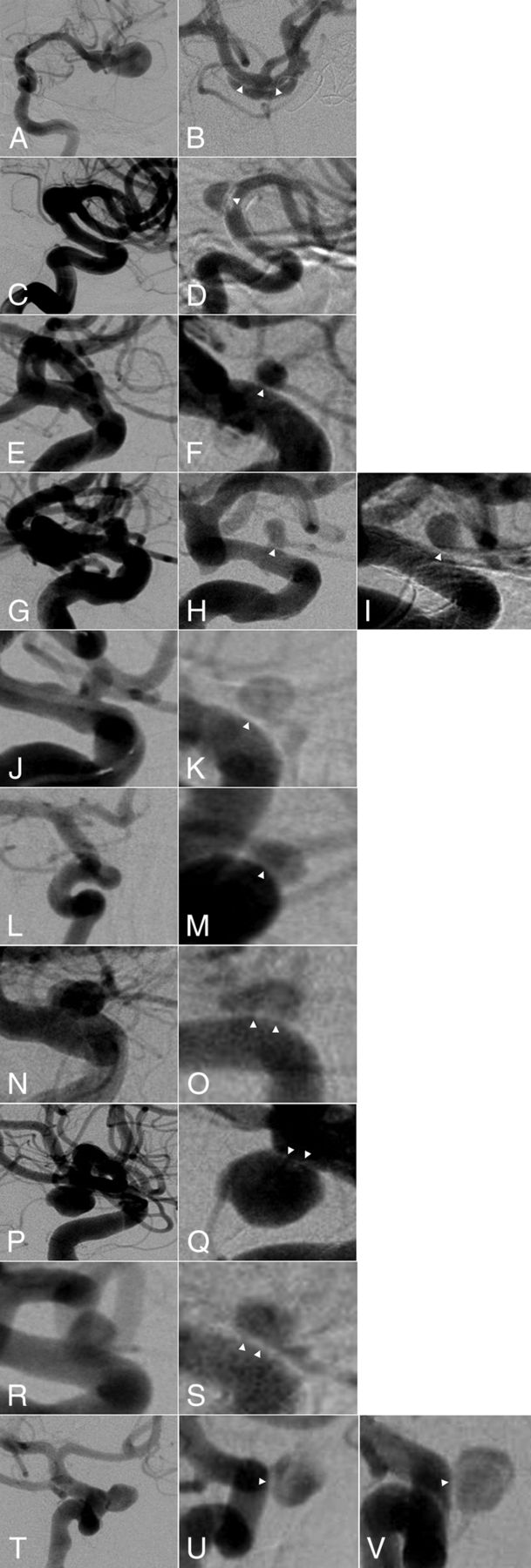
DSA showing collar sign in incompletely occluded aneurysms after Pipeline embolization. Each line represents 1 aneurysm corresponding to the On-line Table. The left column shows the aneurysm before Pipeline embolization. The right column shows the residual aneurysm with the collar sign (white arrowheads) after Pipeline placement at last follow-up. For aneurysms 4 and 10, both first and second angiographic follow-up are shown.

Optical Coherence Tomography Findings

Because of inability to negotiate the carotid siphon with the OCT catheter past the aneurysm, only the proximal end of the PED in the horizontal segment of the cavernous ICA could be imaged with OCT (Fig 2). Interestingly, there was great variability of endothelialization of the PED. There were portions where the PED struts laid bare or were covered by thin endothelium or robust neointima. The degree of endothelialization was even variable on the same cross-section.

Fig 2.
  • Download figure
  • Open in new tab
  • Download powerpoint
Fig 2.

Case 10. DSA showing collar sign in incompletely occluded paraophthalmic ICA aneurysm after Pipeline embolization (A). Lateral DSA of the ICA showing the aneurysm (white dashed line) and position of the tip (arrowhead) and the beginning of the scanning portion (arrow) of the OCT catheter (B). OCT images showing variable degree of endothelialization (C–E). There are portions where the PED struts lay bare (arrowheads) or are covered with thin endothelium (arrow) or robust neointima (double arrow and white line showing the distance from lumen to PED struts). After treatment with a second PED, there is stasis of contrast within the aneurysm (F).

Discussion

Here, we report on a series of patients with incompletely occluded cerebral aneurysms after PED placement where the parent vessel appeared to be separate from the aneurysm on angiographic follow-up. We decided to use the term “collar sign” to describe this observation. The main purpose of this article was to report this interesting observation and spark discussion among neurointerventionalists about its significance.

Potential Mechanisms Underlying the Collar Sign

Given the contiguous nature of this sign with the endothelial lining of the parent vessel, it appears to represent endothelialization on the surface of the stent adjacent to the neck of the aneurysm. Why there is continued blood flow into the aneurysm, however, is unclear. Small channels, not readily apparent on DSA, must remain, connecting the parent vessel with the aneurysm. It has been postulated that endothelialization, rather than intra-aneurysmal hemodynamic changes, are primarily responsible for aneurysm obliteration after PED placement. After performing immunohistochemical analysis in rabbit models, Kadirvel et al7 found that endothelial cells derived from the parent vessel rather than from progenitor cells from the bone marrow are responsible for neointimal growth after flow-diverter placement, supporting previous results in mice models. They further found that smooth muscle cells derived from the parent vessel and the aneurysm itself also served as the scaffold for neointimal formation. The presence of inflammatory cells, including macrophages and monocytes, within the portion of the device covering the aneurysm neck was observed in cases of incomplete obliteration and was associated with disorganized thrombus formation at 8 weeks after flow-diverter placement. The macrophages and monocytes formed islands at the neck of the aneurysms without associated endothelial, smooth muscle, and progenitor cells. Similar processes may, in part, be responsible for the collar sign observed on follow-up DSA in humans, and it may represent an angiographic correlate for inflammatory-driven aberrant endothelialization. Along the same lines, Dai et al8 reported a case of a patient who underwent PED placement for a basilar tip aneurysm. At 4 months, the aneurysm was occluded, but at 1 year, it had recanalized, and the patient died from brain stem compression. Microscopic analysis of the aneurysm revealed channels extending through thick, newly formed islands of intima from the parent vessel to the sac of the aneurysm containing fresh blood clot. The aneurysm was filled with disorganized thrombus. The collar sign on the DSAs obtained in the present series of patients were mostly homogeneous in appearance with variable thickness, even though they contained rare regions speckled with contrast, indicative of blood flow through this layer. OCT performed in 1 case showed variable degrees of endothelialization at the proximal end of the PED, ranging from no coverage of the PED struts to thick neointima. Unfortunately, the area of the collar sign could not be imaged because of inability to negotiate the carotid siphon with the OCT catheter. In animal models, OCT has been used to assess PED wall apposition9 and side branches covered by the PED.10,11 We are not aware of any prior use of OCT after PED placement in humans.

Clinical Implications of the Collar Sign

The implications of the collar sign are unclear. Initially, we assumed that incompletely occluded aneurysms with this associated sign at 6 months would likely be occluded at subsequent follow-up. However, all patients who had additional angiographic follow-up past the 6-month mark continued to have aneurysm filling, with no notable change in the collar sign appearance. Given that flow diversion is associated with high rates of complete occlusion at 6-month follow-up,2,12⇓–14 these aneurysms only represent a small fraction of all aneurysms treated with the PED. Nevertheless, it is important to consider. If the collar sign at 6 months is not predictive of occlusion in the future, early treatment with a second PED may be warranted. In the current series, this approach was pursued in 2 aneurysms, with close follow-up on the others.

Conclusions

Pipeline embolization is associated with high rates of occlusion of cerebral aneurysms. Two mechanisms underlying occlusion have been proposed for the device: 1) diversion of blood flow away from the fundus of the lesion, resulting in subsequent thrombus formation and aneurysm shrinkage; and 2) endothelialization of the neck of the aneurysm over time. Here, we report on a series of patients who underwent PED placement and were found to have incomplete occlusion of their aneurysms at angiographic follow-up, with a characteristic collar sign visible on angiography. They appear to represent endothelialization along the neck of the aneurysm with continued filling of the lesions. Subsequent follow-ups in a subset of these patients demonstrated residual blood flow into these aneurysms and continued incomplete endothelialization across the neck, warranting retreatment.

Footnotes

  • Disclosures: Ajith Thomas—UNRELATED: Consultancy: Stryker, Comments: data safety monitoring board of SCENT trial*; Expert Testimony: testimony related to aneurysm treatment. *Money paid to the institution.

References

  1. 1.↵
    1. Alderazi YJ,
    2. Shastri D,
    3. Kass-Hout T, et al
    . Flow diverters for intracranial aneurysms. Stroke Res Treat 2014;2014:415653 doi:10.1155/2014/415653 pmid:24967131
    CrossRefPubMed
  2. 2.↵
    1. Szikora I,
    2. Berentei Z,
    3. Kulcsar Z, et al
    . Treatment of intracranial aneurysms by functional reconstruction of the parent artery: the Budapest experience with the Pipeline embolization device. AJNR Am J Neuroradiol 2010;31:1139–47 doi:10.3174/ajnr.A2023 pmid:20150304
    Abstract/FREE Full Text
  3. 3.↵
    1. Griessenauer CJ,
    2. Ogilvy CS,
    3. Foreman PM, et al
    . Pipeline Embolization Device for small paraophthalmic artery aneurysms with an emphasis on the anatomical relationship of ophthalmic artery origin and aneurysm. J Neurosurg 2016 Mar 4:1–8. [Epub ahead of print] doi:10.3171/2015.12.JNS152499 pmid:22948199
    CrossRefPubMed
  4. 4.↵
    1. D'Urso PI,
    2. Lanzino G,
    3. Cloft HJ, et al
    . Flow diversion for intracranial aneurysms: a review. Stroke 2011;42:2363–68 doi:10.1161/STROKEAHA.111.620328 pmid:21737793
    Abstract/FREE Full Text
  5. 5.↵
    1. Attizzani GF,
    2. Jones MR,
    3. Given CA 2nd., et al
    . Frequency-domain optical coherence tomography assessment of very late vascular response after carotid stent implantation. J Vasc Surg 2013;58:201–04 doi:10.1016/j.jvs.2012.11.038 pmid:23357518
    CrossRefPubMed
  6. 6.↵
    1. Griessenauer CJ,
    2. Foreman PM,
    3. Deveikis JP, et al
    . Optical coherence tomography of traumatic aneurysms of the internal carotid artery: report of 2 cases. J Neurosurg 2016;124:305–09 doi:10.3171/2015.1.JNS142840 pmid:26252460
    CrossRefPubMed
  7. 7.↵
    1. Kadirvel R,
    2. Ding YH,
    3. Dai D, et al
    . Cellular mechanisms of aneurysm occlusion after treatment with a flow diverter. Radiology 2014;270:394–99 doi:10.1148/radiol.13130796 pmid:24086073
    CrossRefPubMed
  8. 8.↵
    1. Dai D,
    2. Ding YH,
    3. Kelly M, et al
    . Histopathological findings following Pipeline embolization in a human cerebral aneurysm at the basilar tip. Interv Neuroradiol 2016;22:153–57 doi:10.1177/1591019915622165 pmid:26842611
    Abstract/FREE Full Text
  9. 9.↵
    1. van der Marel K,
    2. Gounis MJ,
    3. Weaver JP, et al
    . Grading of Regional Apposition after Flow-Diverter Treatment (GRAFT): a comparative evaluation of VasoCT and intravascular OCT. J Neurointerv Surg 2016;8:847–52 doi:10.1136/neurintsurg-2015-011843 pmid:26220411
    Abstract/FREE Full Text
  10. 10.↵
    1. Iosif C,
    2. Saleme S,
    3. Ponsonnard S, et al
    . Intravascular optical coherence tomography for the evaluation of arterial bifurcations covered by flow diverters. J Neurointerv Surg 2016 Jan 27. [Epub ahead of print] doi:10.1136/neurintsurg-2015-012084 pmid:26819448
    Abstract/FREE Full Text
  11. 11.↵
    1. Lopes DK,
    2. Johnson AK
    . Evaluation of cerebral artery perforators and the Pipeline embolization device using optical coherence tomography. J Neurointerv Surg 2012;4:291–94 doi:10.1136/neurintsurg-2011-010102 pmid:21990536
    Abstract/FREE Full Text
  12. 12.↵
    1. Kan P,
    2. Siddiqui AH,
    3. Veznedaroglu E, et al
    . Early postmarket results after treatment of intracranial aneurysms with the Pipeline embolization device: a U.S. multicenter experience. Neurosurgery 2012;71:1080–87, discussion 1087–88 doi:10.1227/NEU.0b013e31827060d9 pmid:22948199
    CrossRefPubMed
  13. 13.↵
    1. O'Kelly CJ,
    2. Spears J,
    3. Chow M, et al
    . Canadian experience with the Pipeline embolization device for repair of unruptured intracranial aneurysms. AJNR Am J Neuroradiol 2013;34:381–87 doi:10.3174/ajnr.A3224 pmid:22859284
    Abstract/FREE Full Text
  14. 14.↵
    1. Yu SC,
    2. Kwok CK,
    3. Cheng PW, et al
    . Intracranial aneurysms: midterm outcome of Pipeline embolization device–a prospective study in 143 patients with 178 aneurysms. Radiology 2012;265:893–901 doi:10.1148/radiol.12120422 pmid:22996749
    CrossRefPubMed
  • Received August 12, 2016.
  • Accepted after revision September 15, 2016.
  • © 2017 by American Journal of Neuroradiology
View Abstract
PreviousNext
Back to top

In this issue

American Journal of Neuroradiology: 38 (2)
American Journal of Neuroradiology
Vol. 38, Issue 2
1 Feb 2017
  • Table of Contents
  • Index by author
  • Complete Issue (PDF)
Advertisement
Print
Download PDF
Email Article

Thank you for your interest in spreading the word on American Journal of Neuroradiology.

NOTE: We only request your email address so that the person you are recommending the page to knows that you wanted them to see it, and that it is not junk mail. We do not capture any email address.

Enter multiple addresses on separate lines or separate them with commas.
Collar Sign in Incompletely Occluded Aneurysms after Pipeline Embolization: Evaluation with Angiography and Optical Coherence Tomography
(Your Name) has sent you a message from American Journal of Neuroradiology
(Your Name) thought you would like to see the American Journal of Neuroradiology web site.
CAPTCHA
This question is for testing whether or not you are a human visitor and to prevent automated spam submissions.
Cite this article
C.J. Griessenauer, R. Gupta, S. Shi, A. Alturki, R. Motiei-Langroudi, N. Adeeb, C.S. Ogilvy, A.J. Thomas
Collar Sign in Incompletely Occluded Aneurysms after Pipeline Embolization: Evaluation with Angiography and Optical Coherence Tomography
American Journal of Neuroradiology Feb 2017, 38 (2) 323-326; DOI: 10.3174/ajnr.A5010

Citation Manager Formats

  • BibTeX
  • Bookends
  • EasyBib
  • EndNote (tagged)
  • EndNote 8 (xml)
  • Medlars
  • Mendeley
  • Papers
  • RefWorks Tagged
  • Ref Manager
  • RIS
  • Zotero
0 Responses
Respond to this article
Share
Bookmark this article
Collar Sign in Incompletely Occluded Aneurysms after Pipeline Embolization: Evaluation with Angiography and Optical Coherence Tomography
C.J. Griessenauer, R. Gupta, S. Shi, A. Alturki, R. Motiei-Langroudi, N. Adeeb, C.S. Ogilvy, A.J. Thomas
American Journal of Neuroradiology Feb 2017, 38 (2) 323-326; DOI: 10.3174/ajnr.A5010
del.icio.us logo Twitter logo Facebook logo Mendeley logo
  • Tweet Widget
  • Facebook Like
  • Google Plus One
Purchase

Jump to section

  • Article
    • Abstract
    • ABBREVIATIONS:
    • Materials and Methods
    • Results
    • Discussion
    • Conclusions
    • Footnotes
    • References
  • Figures & Data
  • Supplemental
  • Info & Metrics
  • Responses
  • References
  • PDF

Related Articles

  • PubMed
  • Google Scholar

Cited By...

  • Prevalence of intracerebral thrombus detected by optical coherence tomography in patients with posterior circulation stroke or transient ischemic attack
  • Endovascular Cerebral Venous Sinus Imaging with Optical Coherence Tomography
  • Optical coherence tomography evaluation of vertebrobasilar artery stenosis: case series and literature review
  • The Collar Sign in Pipeline Embolization Device-Treated Aneurysms
  • Implications of the Collar Sign in Incompletely Occluded Aneurysms after Pipeline Embolization Device Implantation: A Follow-Up Study
  • A new set of eyes: development of a novel microangioscope for neurointerventional surgery
  • Endothelialization following Flow Diversion for Intracranial Aneurysms: A Systematic Review
  • European Multicenter Study for the Evaluation of a Dual-Layer Flow-Diverting Stent for Treatment of Wide-Neck Intracranial Aneurysms: The European Flow-Redirection Intraluminal Device Study
  • Predictors of Incomplete Occlusion following Pipeline Embolization of Intracranial Aneurysms: Is It Less Effective in Older Patients?
  • Crossref (38)
  • Google Scholar

This article has been cited by the following articles in journals that are participating in Crossref Cited-by Linking.

  • European Multicenter Study for the Evaluation of a Dual-Layer Flow-Diverting Stent for Treatment of Wide-Neck Intracranial Aneurysms: The European Flow-Redirection Intraluminal Device Study
    M. Killer-Oberpfalzer, N. Kocer, C.J. Griessenauer, H. Janssen, T. Engelhorn, M. Holtmannspötter, J.H. Buhk, T. Finkenzeller, G. Fesl, J. Trenkler, W. Reith, A. Berlis, K. Hausegger, M. Augustin, C. Islak, B. Minnich, M. Möhlenbruch
    American Journal of Neuroradiology 2018 39 5
  • Predictors of Incomplete Occlusion following Pipeline Embolization of Intracranial Aneurysms: Is It Less Effective in Older Patients?
    N. Adeeb, J.M. Moore, M. Wirtz, C.J. Griessenauer, P.M. Foreman, H. Shallwani, R. Gupta, A.A. Dmytriw, R. Motiei-Langroudi, A. Alturki, M.R. Harrigan, A.H. Siddiqui, E.I. Levy, A.J. Thomas, C.S. Ogilvy
    American Journal of Neuroradiology 2017 38 12
  • Endothelialization following Flow Diversion for Intracranial Aneurysms: A Systematic Review
    K. Ravindran, M.M. Salem, A.Y. Alturki, A.J. Thomas, C.S. Ogilvy, J.M. Moore
    American Journal of Neuroradiology 2019 40 2
  • Intravascular Optical Coherence Tomography for Neurointerventional Surgery
    Matthew J. Gounis, Giovanni J. Ughi, Miklos Marosfoi, Demetrius K. Lopes, David Fiorella, Hiram G. Bezerra, Conrad W. Liang, Ajit S. Puri
    Stroke 2019 50 1
  • Comparison of Pipeline Embolization Device and Flow Re-Direction Endoluminal Device Flow Diverters for Internal Carotid Artery Aneurysms: A Propensity Score-Matched Cohort Study
    Christoph J Griessenauer, Ajith J Thomas, Alejandro Enriquez-Marulanda, Aviraj Deshmukh, Abhi Jain, Christopher S Ogilvy, Naci Kocer, Tobias Engelhorn, Markus Möhlenbruch, Markus Holtmannspötter, Hendrik Janssen, Thomas Finkenzeller, Wolfgang Reith, Michael Sonnberger, Jan-Hendrik Buhk, Clemens M Schirmer, Monika Killer-Oberpfalzer
    Neurosurgery 2019 85 2
  • Experience With the Pipeline Embolization Device for Posterior Circulations Aneurysms: A Multicenter Cohort Study
    Christoph J Griessenauer, Alejandro Enriquez-Marulanda, Philipp Taussky, Arundhati Biswas, Ramesh Grandhi, Sissi Xiang, Tao Hong, Lorenzo Rinaldo, Giuseppe Lanzino, Waleed Brinjikji, Jan-Karl Burkhardt, Peter Kan, Mandeep Ghuman, Victor X D Yang, Karen Chen, Mohammad A Aziz-Sultan, Mohammad Ghorbani, Clemens M Schirmer, Oded Goren, Shamsher S Dalal, Monika Killer-Oberpfalzer, Erasmia Müller-Thies-Broussalis, Matthew J Koch, Christopher J Stapleton, Aman B Patel, Paul M Foreman, Marshall C Cress, Robert A Hirschl, Timo Krings, Hongqi Zhang, Adam A Dmytriw
    Neurosurgery 2020 87 6
  • Flow diversion for treatment of intracranial aneurysms: Mechanism and implications
    Melissa M. J. Chua, Luke Silveira, Justin Moore, Vitor M. Pereira, Ajith J. Thomas, Adam A. Dmytriw
    Annals of Neurology 2019 85 6
  • Optical Coherence Tomography for Neurovascular Disorders
    Vania Anagnostakou, Giovanni J. Ughi, Ajit S. Puri, Matthew J. Gounis
    Neuroscience 2021 474
  • Optical coherence tomography evaluation of vertebrobasilar artery stenosis: case series and literature review
    Xiaohui Xu, Min Li, Rui Liu, Qin Yin, Xuan Shi, Fang Wang, Jie Gao, Gelin Xu, Ruidong Ye, Xinfeng Liu
    Journal of NeuroInterventional Surgery 2020 12 8
  • Clinical impact of Sim & Size® simulation software in the treatment of patients with cerebral aneurysms with flow-diverter Pipeline stents
    Daniel Mantilla, Carlos A Ferreira-Prada, Melquisedec Galvis, Oliverio Vargas, Carolina Valenzuela-Santos, Piergiorgio Canci, Miguel Ochoa, Franck Nicoud, Vincent Costalat
    Interventional Neuroradiology 2023 29 1

More in this TOC Section

  • SAVE vs. Solumbra Techniques for Thrombectomy
  • Contrast-Induced Encephalopathy after NeuroIR
  • CT Perfusion&Reperfusion in Acute Ischemic Stroke
Show more Interventional

Similar Articles

Advertisement

Indexed Content

  • Current Issue
  • Accepted Manuscripts
  • Article Preview
  • Past Issues
  • Editorials
  • Editor's Choice
  • Fellows' Journal Club
  • Letters to the Editor
  • Video Articles

Cases

  • Case Collection
  • Archive - Case of the Week
  • Archive - Case of the Month
  • Archive - Classic Case

Special Collections

  • AJNR Awards
  • ASNR Foundation Special Collection
  • Most Impactful AJNR Articles
  • Photon-Counting CT
  • Spinal CSF Leak Articles (Jan 2020-June 2024)

More from AJNR

  • Trainee Corner
  • Imaging Protocols
  • MRI Safety Corner

Multimedia

  • AJNR Podcasts
  • AJNR Scantastics

Resources

  • Turnaround Time
  • Submit a Manuscript
  • Submit a Video Article
  • Submit an eLetter to the Editor/Response
  • Manuscript Submission Guidelines
  • Statistical Tips
  • Fast Publishing of Accepted Manuscripts
  • Graphical Abstract Preparation
  • Imaging Protocol Submission
  • Evidence-Based Medicine Level Guide
  • Publishing Checklists
  • Author Policies
  • Become a Reviewer/Academy of Reviewers
  • News and Updates

About Us

  • About AJNR
  • Editorial Board
  • Editorial Board Alumni
  • Alerts
  • Permissions
  • Not an AJNR Subscriber? Join Now
  • Advertise with Us
  • Librarian Resources
  • Feedback
  • Terms and Conditions
  • AJNR Editorial Board Alumni

American Society of Neuroradiology

  • Not an ASNR Member? Join Now

© 2025 by the American Society of Neuroradiology All rights, including for text and data mining, AI training, and similar technologies, are reserved.
Print ISSN: 0195-6108 Online ISSN: 1936-959X

Powered by HighWire