
Abstract
BACKGROUND AND PURPOSE: The causal effect of early febrile convulsions (FC) on later-onset temporal lobe epilepsy (TLE) remains unclear. In this study, we sought to examine the hippocampal alterations in epileptic children with or without FC history by using MR spectroscopy and volumetry.
METHODS: Fifty-five children ranging in age from 18 months to 15 years were enrolled in this study. Subjects were divided into three groups: the control group without either TLE or history of FC (n = 16), the TLE group with early history of FC (TLE + FC; n = 22), and the TLE group without FC history (n = 17). Measurement of hippocampal volume (HV) was performed on thin section T1-weighted images acquired with a 3D gradient echo MR image and normalized by the intracranial volume. Each individual subject had two measures of lateralization; one gives the smaller side of HV and the other the contralateral larger side of HV, assuming that the side with smaller HV is the possible primary site of seizure focus and the contralateral larger HV the secondary or normal site. Single-voxel proton MR spectroscopy of the hippocampi was performed, with metabolic ratio n-acetylaspartate (NAA)/choline (Cho) + creatine plus phosphocreatine (Cr) calculated and grouped separately as were with volumetry.
RESULTS: The overall mean HV for the control group was 2.61 ± 0.21 cm3 at an average intracranial volume of 965 ± 241 cm3, and the asymmetry index for hippocampal volume was (2.32 ± 1.58)%. The overall mean HV was 2.30 ± 0.33 cm3 for TLE + FC group and 2.34 ± 0.33 cm3 for TLE group. Mean HV differed significantly for the three groups (P < .01). When the small and large sides were analyzed separately, significant differences were found between control and TLE as well as between control and TLE + FC for the smaller side (P < .05), whereas for the larger side significant differences were found only between control and TLE + FC. In MR spectroscopic measurements, the mean NAA/(Cr + Cho) of bilateral hippocampi was 0.77 ± 0.06 for control group, 0.62 ± 0.12 for TLE + FC group, and 0.66 ± 0.11 for TLE group. In terms of statistically significant difference between groups, spectroscopic results were similar to volumetric measurements, except that there was no significant interaction effect between groups and measures of asymmetrical indices (P = .272).
CONCLUSION: Children with TLE and early history of FC tend to have lower hippocampal volumes and NAA/(Cr + Cho) ratios than do TLE children without FC history. The TLE + FC group seems to have increased vulnerability of the contralateral hippocampus as compared with TLE group. MR volumetry and spectroscopy are equally capable of showing the trends of hippocampal alternations in children with TLE with or without FC history.
Febrile convulsion (FC) is the most common convulsive disorder in young children, affecting 2–4% before the age of 5 years (1, 2). One-third of those with FC have recurrent FC by age 6 years, and the incidence of subsequent epilepsy is 2–8% (1, 2). The causal relationship between FC and subsequent epilepsy has been a focus of intense investigation. Population studies have not revealed the associations between early-life FC and temporal lobe epilepsy (1). Nonetheless, a history of prolonged FC was commonly elicited in individuals with mesial temporal lobe epilepsy (1–3).
Hippocampal sclerosis (HS) is the most common pathologic finding in adults with intractable temporal lobe epilepsy (TLE), reported in approximately 65% of adult temporal lobectomy series (4). According to Falconer’s investigations, TLE with surgical verified mesial temporal sclerosis often occurs in adult patients whose onset traces back to childhood (5). Several investigators have reported a link between the presence of HS in adults and a history of FC in early childhood (6–8), although the causal nature of this relation remains unclear. A frequently proposed hypothesis is that a subtle, pre-existing malformation due to embryogenesis, perinatal trauma, or other intracranial lesions of the medial temporal lobe facilitates FC and acts as a crystallization point for the development of subsequent TLE and HS (9–11). On the other hand, the findings of VanLandingham et al (12 ) and Lynch et al (13 ) suggest that focal prolonged or repetitive FC can produce an acute insult to the hippocampus that may evolve to HS. Whether HS is a cause or consequence of FC is still a controversial issue in epilepsy study (14, 15).
Although the relationship of HS with respect to TLE in children has been reported in previous MR imaging studies (16–18), to the best of our knowledge the added complexity of FC to TLE in school-age children and their causal nature has not been investigated. The purpose of our study was to examine the differences of hippocampal alterations of TLE in school-age children with and those without FC in early childhood by using MR spectroscopy and volumetry. Normative data on hippocampal volume and n-acetylaspartate (NAA)/(Cr + Cho) ratio were also measured for children <15 years of age.
Methods
Subjects
Children with TLE who visited outpatient clinic for epilepsy at the National Cheng-Kung University Hospital from January 1999 to July 2001 were enrolled. The hospital ethics committee approved the study, and informed consent was obtained from all participants before their inclusion. A thorough neurologic assessment was conducted by one of the investigators (C.-C.H.). In the neurologic evaluation, data on the patients’ previous medical histories and seizure presentations were collected from the medical records and by interviewing the patients and their parents. Seizure type and the site of seizure onset were determined by a comprehensive evaluation, including detailed clinical seizure semiology, neurologic examination, neuropsychological evaluation, and interictal scalp EEG localization. The diagnosis of TLE was based on seizure semiology; that is, definitive interictal epileptiform discharges (spikes or sharp waves) on one or both temporal regions by repeated interictal electroencephalographic (EEG) recordings. The epilepsies and epileptic syndromes were grouped according to the classification proposed by the International League against Epilepsy in 1989. Patients were defined according to the type of epilepsies as partial and generalized and also according to their syndromic characteristics. Twenty four-hour in-hospital ictal EEG was not available in our hospital and thus was not performed in our patients. None of the patients with temporal lobe seizure had received surgical treatment for seizure control. All patients were evaluated for the following parameters: age at onset of FC, type of FC (simple versus complex, partial versus generalized), age at onset of TLE, interval between the first FC and TLE, recurrence rate of FC and TLE, and family history of epilepsy and FC.
Among the patients with TLE, those children with a history of early childhood FC were age matched to those without FC for MR imaging studies.
Hippocampal Volume Measurement
High-spatial-resolution MR images for hippocampal volumetry were acquired by using 3D T1-weighted gradient-echo technique on a 1.5-T unit (Siemens Vision+, Erlangen, Germany) with all sections obtained perpendicular to the long-axis of the hippocampus (TR/TE/NEX = 9.7/4.0/2, flip angle = 9°, resolution = 0.59 × 0.78 × 2.0 mm3). The boundary of the hippocampus was manually traced by one rater (W.-C.W.) who received thorough training from an experienced radiologist (C.-Y.C.) before undertaking this study. The anterior boundary was the first section where the amygdala was not seen. The in-plane boundary, adapted from the definition proposed by Jack (19), was traced laterally along the temporal horn, superiorly by CSF in the choroidal fissure, medially by the CSF in the uncal and ambient cistern, and inferiorly by the gray-white matter in the parahippocampal gyrus. The posterior boundary, an intrinsic landmark proposed by Watson et al (20) in 1992, was the section before the crus of the fornix.
Segmentation was histogram-based and was performed by using National Institutes of Health Image 1.60 software on a Macintosh computer. The threshold, defined by the rater, was kept the same during the measurement of image series in one subject but redefined among different subjects. The selected area was then segmented into CSF, gray matter, and white matter. Only gray matter pixels were counted (Fig 1). To compare the hippocampal volume (HV) measured from different subjects, the measured volume was normalized according to the following equation:  where IV̄ is the mean intracranial volume of all subjects included in this study, IVi is the intracranial volume of the ith children, HVi is the measured HV of the ith children and HVi,n is the normalized HV of the ith children. For intracranial volume, conventional spin-echo T1-wighted MR images were obtained in each subject (TR/TE/NEX, 700/14/1; resolution, 0.59 × 0.78 × 5 mm3; transverse with 2-mm gap). Intracranial volume measurements provide an estimate of maximum premorbid brain size and, unlike cerebral volume, should be unaffected by atrophy due to neurodegeneration or aging (21). Some studies have either used simple estimates of size that correlate with intracranial volume, like cross-sectional area (22), or modeled the head as an oblate spheroid (23). These measures, however, cannot satisfactorily account for the individual difference in head shape. In this study, one rater (W.-C.W.) performed all intracranial volume measurements. In addition, volumetric asymmetry index (AIv), defined as the absolute difference between the two hippocampal volumes divided by their mean, was also calculated. To test the reproducibility and precision of the volume calculation, repeated measurements of the hippocampal volume by using our approach were done by the same rater (W.-C.W.).
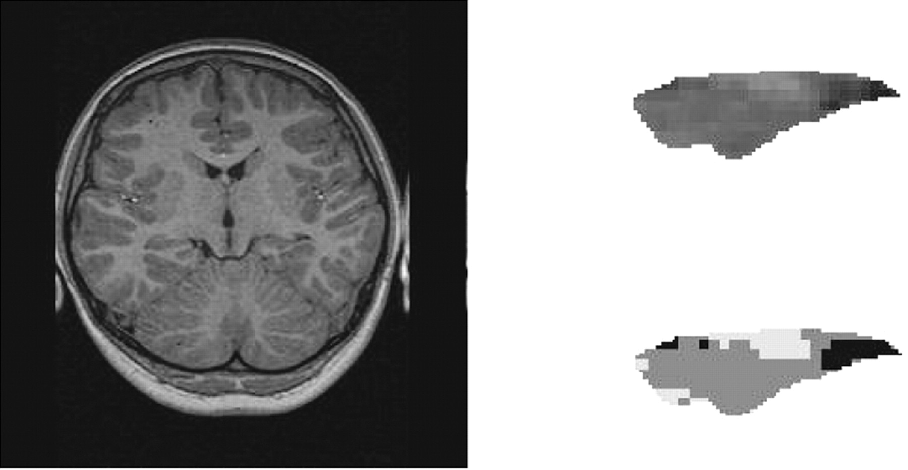
Segmentation procedure. After manually tracing the in-plane boundaries, histogram-based segmentation was used to categorize the region of interest into CSF (black), gray matter (gray), and white matter (white). Only pixels of gray matter are counted to exclude the region of gliosis and atrophy.
Each individual subject had two measures of hippocampi; one gives the smaller side of HV and the other the contralateral, larger side of HV, assuming that the side with smaller HV is the possible primary site of seizure focus and the contralateral larger HV the secondary or normal site.
MR Spectroscopy
Single-voxel proton MR spectra were obtained following localization with scout MR images obtained in three orthogonal planes by using fast low-angle shot gradient echo sequence. Shimming on the water proton was performed to obtain spectra with full-width-at-half-maximum <25 Hz and 10 Hz for global and localized shimming, respectively. Single-voxel proton MR spectroscopy was performed by using a point-resolved spectroscopic sequence technique (TR/TE/NEX, 1500/135/128; 1 × 1 × 2 cm3) with region of interest specifically conforming to the volume of each hippocampus. Brain spectra contain three peaks: one primarily from the n-trimethyl protons of choline-containing metabolites at 3.2 ppm (Cho), one from the n-methyl protons of creatine and phosphocreatine at 3.0 ppm (Cr), and one primarily from the methyl protons of NAA at 2.0 ppm. Relative quantification of NAA, Cho, and Cr signals were performed after Gaussian curve fitting by using standard spectroscopic analysis software (Siemens Numaris, Erlangen, Germany). NAA/(Cho + Cr) ratios were calculated from both sides of hippocampi, following which the spectral asymmetry index (AIs) was derived using similar definition as the volumetric asymmetry index (i.e., absolute difference divided by their mean value). Similar to volumetric measurements, the precision of NAA/(Cho+Cr) value was assessed by comparing repeated estimations with one operator (W.-C.W.).
Statistical Analysis
According to previous studies, there exists asymmetry between the two measures of human hippocampal volume. Without being complicated by the asymmetry, the analysis of variance (ANOVA) with repeated measures was used to test statistical significance for interaction effect between groups and volume measures and for the main effect of group differences. The ANOVA design had subjects nested within groups, each with two volume measures distinguished as larger and small sides. The post hoc analysis was conducted by using the Tukey wholly significant difference test, which had better control over the family wise risk for type I error at the level of significance. By analogy, the same analyses were applied to the spectroscopic study in which the NAA/(Cho + Cr) ratios were compared between groups. For each subject, the ratios measured on both sides of hippocampi are also distinguished as larger and small sides.
Results
Patient Characteristics
A cohort of 55 subjects ranging in age from 18 months to 15 years was included in this study (28 boys and 27 girls). Children were divided into three groups (Table 1): the control group, without either TLE or history of FC (n = 16); the TLE group, with early history of FC (TLE + FC; n = 22); and the TLE group, without FC history (n = 17).
55 children were included for MR volumetry and MR spectroscopy study
By visual inspection, MR imaging characteristics of mesial temporal sclerosis were noted in 27.3% (6/22) of the TLE + FC group, which showed no significant difference from that of 17.7% (3/17) in the TLE group. No clinically significant correlations were found between hippocampal volumes and age at onset of first afebrile seizure, duration of temporal lobe epilepsy, or complex partial and secondarily generalized seizure frequency, in patients with and those without FC.
Volumetry
To test reproducibility, repeated measurements of the hippocampal volume by using our approach were done. The variation was within 6.0%. In the control group, the mean normalized HV of both sides was 2.61 ± 0.21 cm3 at an average intracranial volume of 965 ± 241 cm3. In addition, AIv was (2.32 ± 1.58)%. The mean normalized HV of both sides was 2.30 ± 0.33 cm3 in TLE + FC and 2.34 ± 0.33 cm3 in TLE. The plots in Figure 2 suggest that there was an interaction effect between groups and volume measures (P < .01)—that is, the trends of group means were not parallel for the two volume measures. The overall means for the three groups also differed significantly (P < .01). Because of the interaction effect, the post-hoc comparisons were analyzed at each level of volume measures. The results indicate that, for the mean hippocampal volume for the smaller side, there were significant differences between the control and TLE groups (P < .01) and between the control and TLE + FC groups (P < .01). For the larger side, the control and TLE + FC groups also differed significantly in mean hippocampal volume (P < .05). The mean values in AIv were significantly larger in the TLE + FC and TLE groups than in the control group (Student t test, P < .01), whereas no statistical AIv difference was found between the TLE + FC and TLE groups (Table 2).

For each subject, two sides of HV are divided into large and small sides. In the control group, the asymmetry index (AIv) is 2.2% on average. The plot shows different relationships among three groups in large and small sides. The interaction between groups and sides is statistically significant (P < .01).
Values of asymmetry indices in three groups
MR Spectroscopy
Repeated estimations of NAA/(Cr + Cho) yielded a variation <3.8%, showing highly reproducible measurements. In the control group, the mean NAA/(Cr + Cho) of the bilateral hippocampi was 0.77 ± 0.06. The asymmetry index AIs varied in a rather divergent range, with mean and standard deviation as (8.46 ± 5.05)%. The overall mean NAA/(Cr + Cho) of the bilateral hippocampi was 0.62 ± 0.12 in TLE + FC and 0.66 ± 0.11 in TLE. For the spectroscopy results plotted in Figure 3, the interaction effect between groups and ratios was not significant (P = .272), but the overall test for mean differences between groups was significant (P < .01). The post hoc analysis for the overall differences in the mean values of NAA/(Cr + Cho) between groups indicated that the difference between the control and TLE+FC groups was significant (P < .01) regardless of their sides, but the difference between the control and TLE groups was significant only on the smaller side. Compared with the control group, AIs was significantly larger in the TLE group (P < .05); however, AIs was larger in a nonsignificant trend in the TLE + FC group (P = .074). On the other hand, no statistical AIs difference was found between TLE + FC and TLE groups (Table 2).

For each subject, two sides of NAA/(Cho + Cr) ratios are divided into large and small sides. The asymmetry index (AIs) scatters in a wide range. In the control group, the AIs is 0.77 on the average. The plot shows that the relationships among three groups in large and small sides are quite parallel. There is a trend of lowering NAA/(Cho + Cr) ratios in TLE + FC, but no statistical interaction between groups and sides was found (P = .272).
Discussion
In this study we sought to examine the possible influence of FC on TLE in children. Retrospective studies of adult patients who underwent surgery for refractory TLE found a relation between history of early FC, TLE, and the development of HS. There are at least three hypotheses that explain the causal relation (24–26): First, mesial temporal sclerosis precedes infantile FC (27). This hypothesis implies a prior insult producing the HS, such as prenatal or perinatal injury, meningitis, encephalitis, or head trauma. Second, prolonged or focal FC produces acute hippocampal injury that evolves into HS (28). Third, dual disease in TLE includes cerebral dysgenesis and mesial temporal sclerosis (29). In other words, complex FC produces additional injury to a preexisting hippocampal malformation. Kalviainen and Salmenpera proposed that recurrent seizures might cause progressive damage to the hippocampus throughout the lifetime of the TLE patients (30). In contrast, a recent MR volumetric study on hippocampal formation and amygdala in adult epilepsy patients with childhood FC found that no occurrence of HS in this group after a mean follow-up time of 12.3 years by comparing age-matched control (31). Our study showed that, although there was no difference in the MR evidence of mesial temporal sclerosis between the TLE + FC and TLE groups, the HV in the control group is significantly larger than in the TLE + FC or TLE group on the impaired hippocampi. Moreover, significant difference on the contralateral side is also found between the control and TLE + FC groups. In addition, our results demonstrated statistical interaction between groups and sides of hippocampus. This indicates the different impacts of TLE and FC on the volume of the impaired and contralateral hippocampi. Children with TLE and FC history seem to have increased vulnerability of the contralateral hippocampi. In other words, unilateral focal onset of TLE following early childhood FC appears to have more likelihood of causing damage to the contralateral hippocampus, compared with children with TLE but without FC history.
More recently, MR spectroscopy was proposed to provide useful information for detection or lateralization of TLE (32). Our results showed that the NAA/(Cho + Cr) ratio is significantly higher in the control and TLE + FC groups on both sides of hippocampi, whereas difference between the control and TLE was significant only on the smaller side. It should be noted, however, that the sensitivity of MR spectroscopy might have been underestimated because of the partial volume effect in our spectroscopic data that might include the whole hippocampus where most areas are intact.
In our study, both MR volumetry and spectroscopy fail to show the difference between TLE + FC and TLE groups. It may be that the differences between TLE + FC and TLE may not lie in HV or NAA/(Cho + Cr) themselves at all but in the interaction between sides and groups instead. Nevertheless, MR spectroscopy and volumetry still likely provide the trends of hippocampal alterations in the evaluation of childhood TLE and FC. It is also noted that, although both FC and TLE cause changes in metabolite concentration and HV reduction, the alterations seem to be independent of each other.
Traditionally, visual discrimination of a normal from an abnormal hippocampus with MR images is straightforward when one is clearly normal and the other is grossly abnormal. This visual binary paradigm, however, may break down in the presence of mild unilateral disease, bilateral disease, or both. Therefore, absolute quantitative measurements are necessary to accurately determine the presence and severity of hippocampal atrophy in both hippocampi. This concept is also reflected by several previous reports that the presence and severity of HS in both hippocampi may provide useful prognostic information about postoperative seizure control and memory outcome (33–35). In our study, quantitative information was obtained from the noninvasive assessment of hippocampi in children with or without TLE. In addition to the cut-off value of NAA/(Cr + Cho) ratio and HV, AIv and AIs were also used as a measure of bilateral or unilateral atrophy in light of the interaction between groups and measures of lateralization as demonstrated in our results. As compared with the control group, AIv revealed marked asymmetry in the TLE + FC and TLE groups, which coincided with our analysis indicating the interaction of statistical groups with sides in the volumetric method. On the other hand, AIs, varying in a wide range, was also found to be significantly larger in TLE than the control groups, although the groups’ interaction was less significant with sides by metabolic ratio. Indeed, on the basis of structural symmetry, the asymmetry indices, AIv and AIs, should be able to detect the “unilateral” lesion. Unfortunately, FC followed by TLE sometimes brings about damage to both sides of hippocampi, as shown in the results, which makes it difficult to come up with a concise conclusion for asymmetry indexes. Low AIv (< 5%) or AIs (< 19%) may exist in bilateral alteration as well as in normal subjects. Nevertheless, in case of mild damage of hippocampus where HV and NAA/(Cho + Cr) are both in normal ranges, asymmetry indexes may potentially help lateralize the lesion.
The finding of HS on MR images in young children with a brief history of seizure challenges the view that HS is primarily an older child or adult lesion that results from a long period of recurrent seizures during early childhood and adolescence. Moreover, studies on younger children with HS apparently require reliable normative measurements from control. In our study, we measured both normative hippocampal volume (2.61 ± 0.21 cm2, normalized to intracranial volume of 965 ± 241 cm2) and NAA/(Cr + Cho) ratio (0.77 ± 0.06) for children younger than 15 years. Because quantitative MR imaging studies on controls and children with TLE were rare (36), this normative volume and NAA/(Cr + Cho) ratio, although small in subject size, might be a helpful reference for epileptic researches in children. One weakness of this study, however, is the relatively small patient population, which may not meet the criteria of normal distribution even though statistical difference has been obtained. Further study, including a larger number of patients, is necessary.
Conclusion
Although there is no statistical significance on HV or metabolic ratio means between TLE + FC and TLE groups, our study shows that children with TLE and history of FC in early childhood tend to have lower hippocampal volumes and lower NAA/(Cr + Cho) ratios than do TLE children without FC history. The TLE + FC group seems to have increased vulnerability of the contralateral hippocampus compared with TLE group. MR volumetry and spectroscopy are equally capable of showing the different trends of hippocampal alterations in children with TLE with or without FC history. Further studies with more sophisticated tools are needed to unravel the link between early childhood FC and later-onset TLE.
References
- Received June 8, 2004.
- Accepted after revision September 24, 2004.
- Copyright © American Society of Neuroradiology
In this issue


Jump to section
Related Articles
Cited By...
- No citing articles found.