Advertisement

Main menu

  • Home
  • Content
    • Current Issue
    • Accepted Manuscripts
    • Article Preview
    • Past Issue Archive
    • Video Articles
    • AJNR Case Collection
    • Case of the Week Archive
    • Case of the Month Archive
    • Classic Case Archive
  • Special Collections
    • AJNR Awards
    • ASNR Foundation Special Collection
    • Most Impactful AJNR Articles
    • Photon-Counting CT
    • Spinal CSF Leak Articles (Jan 2020-June 2024)
  • Multimedia
    • AJNR Podcasts
    • AJNR SCANtastic
    • Trainee Corner
    • MRI Safety Corner
    • Imaging Protocols
  • For Authors
    • Submit a Manuscript
    • Submit a Video Article
    • Submit an eLetter to the Editor/Response
    • Manuscript Submission Guidelines
    • Statistical Tips
    • Fast Publishing of Accepted Manuscripts
    • Graphical Abstract Preparation
    • Imaging Protocol Submission
    • Author Policies
  • About Us
    • About AJNR
    • Editorial Board
    • Editorial Board Alumni
  • More
    • Become a Reviewer/Academy of Reviewers
    • Subscribers
    • Permissions
    • Alerts
    • Feedback
    • Advertisers
    • ASNR Home

User menu

  • Alerts
  • Log in

Search

  • Advanced search
American Journal of Neuroradiology

American Journal of Neuroradiology

ASHNR American Society of Functional Neuroradiology ASHNR American Society of Pediatric Neuroradiology ASSR
  • Alerts
  • Log in

Advanced Search

  • Home
  • Content
    • Current Issue
    • Accepted Manuscripts
    • Article Preview
    • Past Issue Archive
    • Video Articles
    • AJNR Case Collection
    • Case of the Week Archive
    • Case of the Month Archive
    • Classic Case Archive
  • Special Collections
    • AJNR Awards
    • ASNR Foundation Special Collection
    • Most Impactful AJNR Articles
    • Photon-Counting CT
    • Spinal CSF Leak Articles (Jan 2020-June 2024)
  • Multimedia
    • AJNR Podcasts
    • AJNR SCANtastic
    • Trainee Corner
    • MRI Safety Corner
    • Imaging Protocols
  • For Authors
    • Submit a Manuscript
    • Submit a Video Article
    • Submit an eLetter to the Editor/Response
    • Manuscript Submission Guidelines
    • Statistical Tips
    • Fast Publishing of Accepted Manuscripts
    • Graphical Abstract Preparation
    • Imaging Protocol Submission
    • Author Policies
  • About Us
    • About AJNR
    • Editorial Board
    • Editorial Board Alumni
  • More
    • Become a Reviewer/Academy of Reviewers
    • Subscribers
    • Permissions
    • Alerts
    • Feedback
    • Advertisers
    • ASNR Home
  • Follow AJNR on Twitter
  • Visit AJNR on Facebook
  • Follow AJNR on Instagram
  • Join AJNR on LinkedIn
  • RSS Feeds

AJNR is seeking candidates for the AJNR Podcast Editor. Read the position description.

OtherSPINE

Epidural Blood Patch at C2: Diagnosis and Treatment of Spontaneous Intracranial Hypotension

Ansaar Rai, Charles Rosen, Jeffrey Carpenter and Vincent Miele
American Journal of Neuroradiology November 2005, 26 (10) 2663-2666;
Ansaar Rai
  • Find this author on Google Scholar
  • Find this author on PubMed
  • Search for this author on this site
Charles Rosen
  • Find this author on Google Scholar
  • Find this author on PubMed
  • Search for this author on this site
Jeffrey Carpenter
  • Find this author on Google Scholar
  • Find this author on PubMed
  • Search for this author on this site
Vincent Miele
  • Find this author on Google Scholar
  • Find this author on PubMed
  • Search for this author on this site
  • Article
  • Figures & Data
  • Info & Metrics
  • Responses
  • References
  • PDF
Loading

Abstract

Summary: Spontaneous intracranial hypotension in a 37-year-old man with intractable headaches was diagnosed on MR imaging. A generous CSF leak was identified at C2 on CT myelography. Successful treatment was performed with CT-guided blood patch at the leakage site after the patient had failed 2 lumbar blood patches. Imaging-guided precise placement of the blood patch is safe and recommended when a lumbar blood patch away from the leakage site could be ineffective.

Spontaneous CSF leak is a recognized cause of intracranial hypotension, and the syndrome of spontaneous intracranial hypotension has been described as a distinct entity (1–3). The symptoms may include postural headaches, neck stiffness, nausea, vomiting, visual symptoms, and cranial nerve pareses (typically VI) (3). Different diagnostic techniques have been used, and the imaging findings have been described on CT, MR imaging, and radionucleotide imaging (1–4). Angiographic features associated with the syndrome have also been documented (5). The treatment options range from conservative supportive measures to epidural blood patch (3). Epidural blood patch has traditionally been performed in the lumbar area, with few cases reported in the thoracic and lower cervical spine (1, 3, 4). We report the clinical and imaging presentation of a case of intracranial hypotension caused by spontaneous CSF leak originating at the C2 level, treated by an epidural blood patch performed at the site of the leak.

Case Report

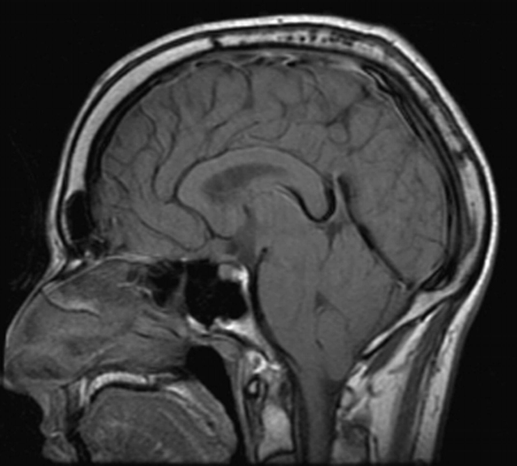
The patient was a 37-year-old man with a 1-month history of progressive intractable headaches and neck stiffness. The headaches were relatively relieved in the supine position. MR imaging of the brain performed at that time showed tonsillar ectopia of at least 8 mm, extending almost to the arch of C1 with a peglike appearance (Fig 1) and diffuse dural enhancement (Fig 2). A thin subdural hygroma was seen over the high right frontal and parietal convexity. A diagnosis of intracranial hypotension was considered. The patient underwent CT myelography of the entire spinal column, which localized a generous leak at the atlantoaxial level with contrast extravasation in the lateral epidural space on the left side (Fig 3). The patient did not have a history of trauma, and there were no historical or physical findings of an underlying connective tissue disorder. Initial treatment included bedrest and supportive measures. He also underwent 2 lumbar epidural blood patches, separated by 1 month, with no relief in his symptoms. Because of the large size of the leak and persistent progressive symptoms despite 2 lumbar blood patches, a direct cervical blood patch was planned with the final option being surgical repair in case of failure.

Fig 1.
  • Download figure
  • Open in new tab
  • Download powerpoint
Fig 1.

Midsection sagittal T1-weighted image obtained without contrast material shows tonsillar descent almost to the arch of C1.

Fig 2.
  • Download figure
  • Open in new tab
  • Download powerpoint
Fig 2.

Axial postcontrast T1-weighted (A) and coronal postcontrast fast spoiled gradient-recalled echo (B) sequences show diffuse meningeal enhancement.

Fig 3.
  • Download figure
  • Open in new tab
  • Download powerpoint
Fig 3.

Axial thin-section CT images obtained through the C1-C2 (A) and the C2 (B) levels show epidural contrast material accumulation on the left side, with the possible site of leakage at the C1-C2 level.

The procedure was performed under CT guidance. After placing the patient prone on the CT gantry, we guided a 22-gauge coaxial needle to the left lateral epidural compartment at the mid-C2 level. The needle was positioned adjacent to the thecal sac, matching the site of the leak seen on the CT myelogram (Fig 4). Needle position was also confirmed with injection of 0.2 mL of iohexol (Omnipaque 240, Nycomed Amersham, Oslo, Norway). The blood patch was then performed with injection of the patient’s autologous blood obtained from the right brachial vein. The injection was stopped at 5 mL, at which point the patient reported an increased pressure sensation in the neck. Findings of a neurologic examination performed after the procedure were normal. The patient was kept overnight in the hospital to monitor for signs of spinal compression and was discharged the next day in a stable condition. At the time of discharge, he reported total pain relief.

Fig 4.
  • Download figure
  • Open in new tab
  • Download powerpoint
Fig 4.

Two axial CT images obtained with the patient in the prone position show needle placement in the left lateral epidural compartment at the upper C2 level (A) followed by administration of the blood patch; contrast material injected to confirm the epidural location is identified with mild flattening of the lateral thecal sac margin (B).

MR imaging of the brain performed 3 weeks after the first procedure showed near-complete resolution of the dural enhancement; however, the tonsillar ectopia was unchanged. About 1 month and a half after the initial cervical blood patch, his headache returned, though it was of a much lower intensity than the initial intractable headaches. A second cervical blood patch was then performed at the C2 level, and a total of 8 mL of autologous blood was slowly injected. The patient reported complete symptomatic relief without any recurrence up to 6 months after the procedure. Additionally, MR imaging of the brain and cervical spine performed 3 months after the procedure showed resolution of the inferior tonsillar displacement (Fig 5) as well as near-complete resolution of the dural enhancement (Fig 6).

Fig 5.
  • Download figure
  • Open in new tab
  • Download powerpoint
Fig 5.

Midsection sagittal T1-weighted image obtained without contrast material shows resolution of the tonsillar descent. This measured less than 3 mm compared with the initial measurement of 8 mm. Compare with Fig 1.

Fig 6.
  • Download figure
  • Open in new tab
  • Download powerpoint
Fig 6.

Axial postcontrast T1-weighted (A) and coronal postcontrast fast spoiled gradient-recalled echo (B) sequences show resolution of the diffuse dural enhancement. Compare with Fig 2.

Discussion

Spontaneous intracranial hypotension, as the name implies, is caused by low CSF pressure, usually secondary to an occult leak (6, 7). The most common presenting symptom is postural headaches. Association with Marfan syndrome and other connective tissue disorders has been described (8). MR imaging is the study of choice to depict the intracranial manifestations; the neuroimaging features include diffuse meningeal enhancement, acquired Chiari malformation, and subdural fluid collections (7, 9, 10).

In cases of spontaneous intracranial hypotension, the cause of the CSF leak is predominantly in the cervical or thoracic region, and the diagnosis is typically established by CT myelography or radionuclide imaging (11–13). In our patient, CT myelography was instrumental in identifying the leak site. Although supportive measures and medical therapy such as hydration, bedrest, caffeine, and parenteral fluid may provide temporary relief, a more durable treatment is to seal the site of the leak. A blood patch performed in the lumbar region, though technically easier, offers less likelihood of controlling a large leak in the cervical region. Furthermore, the volume of blood injected is much larger than if the blood patch is performed at the precise site of the leakage with imaging guidance.

Apart from raising the epidural pressure by injecting blood or saline, we believe imaging-guided blood patch at the site of the leakage enhances the sealant effect of the coagulated blood in controlling the leak. Although a lumbar blood patch performed away from the leak may temporarily control the leak by raising the epidural pressure, the leak may reopen once the pressure is dissipated. Large leaks may be difficult to control, and surgical treatment may be ultimately required. Our patient underwent 2 lumbar blood patches without any relief, and it took 2 attempts with CT guidance to achieve symptomatic and imaging resolution. In cases such as these, it is very important to precisely localize the site of leak. With current technology, imaging-guided procedures in the spine can be performed with relative safety and minimal discomfort to the patient, and in cases of cervical leaks, when time can be lost by pursuing other less effective therapies, it is reasonable to offer a cervical blood patch as the initial treatment.

References

  1. ↵
    Cousins MJ, Brazier D, Cook R. Intracranial hypotension caused by cervical cerebrospinal fluid leak: treatment with epidural blood patch. Anesth Analg 2004;98:1794–1797
    CrossRefPubMed
  2. Nowak DA, Rodiek SO, Zinner J, Guhlmann A, Topka H. Broadening the clinical spectrum: unusual presentation of spontaneous cerebrospinal fluid hypovolemia—case report. J Neurosurg 2003;98:903–907
    PubMed
  3. ↵
    Schievink WI, Meyer FB, Atkinson JL, Mokri B. Spontaneous spinal cerebrospinal fluid leaks and intracranial hypotension. J Neurosurg 1996;84:598–605
    CrossRefPubMed
  4. ↵
    Dillo W, Hollenhorst J, Brassel F, von Hof-Strobach K, Heidenreich F, Johannes S. Successful treatment of a spontaneous cervical cerebrospinal fluid leak with a CT guided epidural blood patch. J Neurol 2002;249:224–225
    PubMed
  5. ↵
    Koss SA, Ulmer JL, Hacein-Bey L. Angiographic features of spontaneous intracranial hypotension. AJNR Am J Neuroradiol 2003;24:704–706
    Abstract/FREE Full Text
  6. ↵
    Khurana RK. Intracranial hypotension. Semin Neurol 1996;16:5–10
    CrossRefPubMed
  7. ↵
    Grimaldi D, Mea E, Chiapparini L, et al. Spontaneous low cerebrospinal pressure: a mini review. Neurol Sci 2004;25(suppl 3):S135–137
  8. ↵
    Mokri B, Maher CO, Sencakova D. Spontaneous CSF leaks: underlying disorder of connective tissue. Neurology 2002;58:814–816
    Abstract/FREE Full Text
  9. ↵
    Spelle L, Boulin A, Tainturier C, Visot A, Graveleau P, Pierot L. Neuroimaging features of spontaneous intracranial hypotension. Neuroradiology 2001;43:622–627
    CrossRefPubMed
  10. ↵
    Atkinson JL, Weinshenker BG, Miller GM, Piepgras DG, Mokri B. Acquired Chiari I malformation secondary to spontaneous spinal cerebrospinal fluid leakage and chronic intracranial hypotension syndrome in seven cases. J Neurosurg 1998;88:237–242
    PubMed
  11. ↵
    Burtis MT, Ulmer JL, Miller GA, Barboli AC, Koss SA, Brown WD. Intradural spinal vein enlargement in craniospinal hypotension. AJNR Am J Neuroradiol 2005;26:34–38
    Abstract/FREE Full Text
  12. Fujimaki H, Saito N, Tosaka M, Tanaka Y, Horiguchi K, Sasaki T. Cerebrospinal fluid leak demonstrated by three-dimensional computed tomographic myelography in patients with spontaneous intracranial hypotension. Surg Neurol 2002;58:280–284; discussion 284–285
    PubMed
  13. ↵
    Bai J, Yokoyama K, Kinuya S, Konishi S, Michigishi T, Tonami N. Radionuclide cisternography in intracranial hypotension syndrome. Ann Nucl Med 2002;16:75–78
    CrossRefPubMed
  • Received March 27, 2005.
  • Accepted after revision April 25, 2005.
  • Copyright © American Society of Neuroradiology
View Abstract
PreviousNext
Back to top

In this issue

American Journal of Neuroradiology: 26 (10)
American Journal of Neuroradiology
Vol. 26, Issue 10
1 Nov 2005
  • Table of Contents
  • Index by author
Advertisement
Print
Download PDF
Email Article

Thank you for your interest in spreading the word on American Journal of Neuroradiology.

NOTE: We only request your email address so that the person you are recommending the page to knows that you wanted them to see it, and that it is not junk mail. We do not capture any email address.

Enter multiple addresses on separate lines or separate them with commas.
Epidural Blood Patch at C2: Diagnosis and Treatment of Spontaneous Intracranial Hypotension
(Your Name) has sent you a message from American Journal of Neuroradiology
(Your Name) thought you would like to see the American Journal of Neuroradiology web site.
CAPTCHA
This question is for testing whether or not you are a human visitor and to prevent automated spam submissions.
Cite this article
Ansaar Rai, Charles Rosen, Jeffrey Carpenter, Vincent Miele
Epidural Blood Patch at C2: Diagnosis and Treatment of Spontaneous Intracranial Hypotension
American Journal of Neuroradiology Nov 2005, 26 (10) 2663-2666;

Citation Manager Formats

  • BibTeX
  • Bookends
  • EasyBib
  • EndNote (tagged)
  • EndNote 8 (xml)
  • Medlars
  • Mendeley
  • Papers
  • RefWorks Tagged
  • Ref Manager
  • RIS
  • Zotero
0 Responses
Respond to this article
Share
Bookmark this article
Epidural Blood Patch at C2: Diagnosis and Treatment of Spontaneous Intracranial Hypotension
Ansaar Rai, Charles Rosen, Jeffrey Carpenter, Vincent Miele
American Journal of Neuroradiology Nov 2005, 26 (10) 2663-2666;
del.icio.us logo Twitter logo Facebook logo Mendeley logo
  • Tweet Widget
  • Facebook Like
  • Google Plus One

Jump to section

  • Article
    • Abstract
    • Case Report
    • Discussion
    • References
  • Figures & Data
  • Info & Metrics
  • Responses
  • References
  • PDF

Related Articles

  • No related articles found.
  • PubMed
  • Google Scholar

Cited By...

  • CT Myelography for the Planning and Guidance of Targeted Epidural Blood Patches in Patients with Persistent Spinal CSF Leakage
  • CT-Guided Epidural Blood Patching of Directly Observed or Potential Leak Sites for the Targeted Treatment of Spontaneous Intracranial Hypotension
  • Crossref
  • Google Scholar

This article has not yet been cited by articles in journals that are participating in Crossref Cited-by Linking.

More in this TOC Section

  • Bern Score Validity for SIH
  • MP2RAGE 7T in MS Lesions of the Cervical Spine
  • Deep Learning for STIR Spine MRI Quality
Show more Spine

Similar Articles

Advertisement

Indexed Content

  • Current Issue
  • Accepted Manuscripts
  • Article Preview
  • Past Issues
  • Editorials
  • Editor's Choice
  • Fellows' Journal Club
  • Letters to the Editor
  • Video Articles

Cases

  • Case Collection
  • Archive - Case of the Week
  • Archive - Case of the Month
  • Archive - Classic Case

Special Collections

  • AJNR Awards
  • ASNR Foundation Special Collection
  • Most Impactful AJNR Articles
  • Photon-Counting CT
  • Spinal CSF Leak Articles (Jan 2020-June 2024)

More from AJNR

  • Trainee Corner
  • Imaging Protocols
  • MRI Safety Corner

Multimedia

  • AJNR Podcasts
  • AJNR Scantastics

Resources

  • Turnaround Time
  • Submit a Manuscript
  • Submit a Video Article
  • Submit an eLetter to the Editor/Response
  • Manuscript Submission Guidelines
  • Statistical Tips
  • Fast Publishing of Accepted Manuscripts
  • Graphical Abstract Preparation
  • Imaging Protocol Submission
  • Evidence-Based Medicine Level Guide
  • Publishing Checklists
  • Author Policies
  • Become a Reviewer/Academy of Reviewers
  • News and Updates

About Us

  • About AJNR
  • Editorial Board
  • Editorial Board Alumni
  • Alerts
  • Permissions
  • Not an AJNR Subscriber? Join Now
  • Advertise with Us
  • Librarian Resources
  • Feedback
  • Terms and Conditions
  • AJNR Editorial Board Alumni

American Society of Neuroradiology

  • Not an ASNR Member? Join Now

© 2025 by the American Society of Neuroradiology All rights, including for text and data mining, AI training, and similar technologies, are reserved.
Print ISSN: 0195-6108 Online ISSN: 1936-959X

Powered by HighWire