Advertisement

Main menu

  • Home
  • Content
    • Current Issue
    • Accepted Manuscripts
    • Article Preview
    • Past Issue Archive
    • Video Articles
    • AJNR Case Collection
    • Case of the Week Archive
    • Case of the Month Archive
    • Classic Case Archive
  • Special Collections
    • AJNR Awards
    • ASNR Foundation Special Collection
    • Most Impactful AJNR Articles
    • Photon-Counting CT
    • Spinal CSF Leak Articles (Jan 2020-June 2024)
  • Multimedia
    • AJNR Podcasts
    • AJNR SCANtastic
    • Trainee Corner
    • MRI Safety Corner
    • Imaging Protocols
  • For Authors
    • Submit a Manuscript
    • Submit a Video Article
    • Submit an eLetter to the Editor/Response
    • Manuscript Submission Guidelines
    • Statistical Tips
    • Fast Publishing of Accepted Manuscripts
    • Graphical Abstract Preparation
    • Imaging Protocol Submission
    • Author Policies
  • About Us
    • About AJNR
    • Editorial Board
    • Editorial Board Alumni
  • More
    • Become a Reviewer/Academy of Reviewers
    • Subscribers
    • Permissions
    • Alerts
    • Feedback
    • Advertisers
    • ASNR Home

User menu

  • Alerts
  • Log in

Search

  • Advanced search
American Journal of Neuroradiology

American Journal of Neuroradiology

ASHNR American Society of Functional Neuroradiology ASHNR American Society of Pediatric Neuroradiology ASSR
  • Alerts
  • Log in

Advanced Search

  • Home
  • Content
    • Current Issue
    • Accepted Manuscripts
    • Article Preview
    • Past Issue Archive
    • Video Articles
    • AJNR Case Collection
    • Case of the Week Archive
    • Case of the Month Archive
    • Classic Case Archive
  • Special Collections
    • AJNR Awards
    • ASNR Foundation Special Collection
    • Most Impactful AJNR Articles
    • Photon-Counting CT
    • Spinal CSF Leak Articles (Jan 2020-June 2024)
  • Multimedia
    • AJNR Podcasts
    • AJNR SCANtastic
    • Trainee Corner
    • MRI Safety Corner
    • Imaging Protocols
  • For Authors
    • Submit a Manuscript
    • Submit a Video Article
    • Submit an eLetter to the Editor/Response
    • Manuscript Submission Guidelines
    • Statistical Tips
    • Fast Publishing of Accepted Manuscripts
    • Graphical Abstract Preparation
    • Imaging Protocol Submission
    • Author Policies
  • About Us
    • About AJNR
    • Editorial Board
    • Editorial Board Alumni
  • More
    • Become a Reviewer/Academy of Reviewers
    • Subscribers
    • Permissions
    • Alerts
    • Feedback
    • Advertisers
    • ASNR Home
  • Follow AJNR on Twitter
  • Visit AJNR on Facebook
  • Follow AJNR on Instagram
  • Join AJNR on LinkedIn
  • RSS Feeds

AJNR is seeking candidates for the AJNR Podcast Editor. Read the position description.

Research ArticleHead & Neck

MR Thermography–Guided Head and Neck Lesion Laser Ablation

D.T. Ginat, S. Sammet and G. Christoforidis
American Journal of Neuroradiology September 2018, 39 (9) 1593-1596; DOI: https://doi.org/10.3174/ajnr.A5726
D.T. Ginat
aFrom the Department of Radiology, University of Chicago, Pritzker School of Medicine, Chicago, Illinois.
  • Find this author on Google Scholar
  • Find this author on PubMed
  • Search for this author on this site
  • ORCID record for D.T. Ginat
S. Sammet
aFrom the Department of Radiology, University of Chicago, Pritzker School of Medicine, Chicago, Illinois.
  • Find this author on Google Scholar
  • Find this author on PubMed
  • Search for this author on this site
  • ORCID record for S. Sammet
G. Christoforidis
aFrom the Department of Radiology, University of Chicago, Pritzker School of Medicine, Chicago, Illinois.
  • Find this author on Google Scholar
  • Find this author on PubMed
  • Search for this author on this site
  • ORCID record for G. Christoforidis
  • Article
  • Figures & Data
  • Info & Metrics
  • Responses
  • References
  • PDF
Loading

Abstract

SUMMARY: Interstitial laser ablation has been successfully used as a minimally invasive treatment option for tumors in many parts of the body, including the head and neck. In this article, we describe the use of MR imaging guidance and mapping sequences for accurate localization of the target lesion, percutaneous interstitial laser ablation methods, and the use of MR thermography for temperature monitoring during laser ablation, with a focus on applications in the head and neck region.

Laser ablation, also known as interstitial laser ablation/thermal therapy or stereotactically guided interstitial laser thermal ablation, is a minimally invasive treatment technique that can be performed as an outpatient percutaneous procedure. The technique uses the phenomenon that heat-induced protein denaturation and coagulative necrosis occur at temperatures above 43°C, such that the time to cell death varies exponentially with temperature.1 This is accomplished with light at wavelengths of 800–1064 nm from diode or neodymium-doped yttrium aluminum garnet lasers, which is absorbed by chromophores in tissues, leading to the release of thermal energy.2

Commercial fiber-optic laser tips can be cooled using fluids, which allow higher initial power use without damaging the fiber-optic tip. The cooling process also removes heat from the probe-tissue interface, thereby preventing carbonization or vaporization and enabling the creation of larger ablation volumes. Furthermore, cooling prevents the probe tip from adhering to the ablated tissue.1 Laser ablation of soft tissues results in 5 distinct concentric zones: 1) a core that corresponds to the probe tract; 2) a central zone of coagulation necrosis that contains damaged cell membranes and stains positive for markers of apoptosis; 3) a peripheral zone that contains thrombosed vessels and distended cell bodies, followed by liquefaction necrosis; 4) a peripheral rim of leaky vasculature; and 5) a marginal zone of reversible edema.3,4 The volume of the necrosis induced by laser ablation depends on the probe size, duration of the procedure, and temperature setting. Larger lesions may require multiple probes or serial laser ablation with repositioning of the probe. Ultimately, laser ablation produces a sharp transition zone between dead and viable tissue.

Interstitial laser ablation has several advantages over other thermal therapy methods. Unlike high-intensity focused sonography, laser ablation is rapid and is less affected by target motion and heats more slowly. Unlike radiofrequency ablation, the laser fiber does not produce large MR imaging artifacts caused by electromagnetic interferences.5,6 Thus, laser ablation can be used simultaneously with real-time MR temperature monitoring. Furthermore, patients who undergo percutaneous interstitial laser ablation tend to be discharged after a shorter stay compared with open surgical procedures.2,7⇓⇓⇓⇓–12 Consequently, interstitial laser ablation has been found to be a cost-effective option for treating brain tumors. For example, this technique is less costly than craniotomy for patients with brain metastases.13 Furthermore, interstitial laser ablation improves survival of patients with high-grade gliomas at a cost that appears to be of good value to society.14 Although there is a paucity of data regarding the cost-effectiveness of interstitial laser ablation for head and neck lesions, we believe that in many cases, the procedure can provide good value as well.

Head and Neck Laser Ablation Indications and Complications

Laser ablation has been successfully performed for treating various lesions in the head and neck, brain, breast, liver, prostate, and colon among other sites.2,7⇓⇓⇓⇓–12,15⇓⇓⇓⇓⇓⇓–22 Regarding the head and neck in particular, percutaneous laser ablation has been implemented for treating benign solid thyroid nodules, lymphatic malformations, and various malignant neoplasms.8,9,16⇓⇓⇓⇓⇓–22 Laser ablation performed to shrink symptomatic benign solid thyroid nodules has been reported to yield a 47%–82% reduction in volume, a minor complication rate of 38%, and a major complication rate of 3%.22 Major complications related to laser ablation of benign solid thyroid nodules include dysphonia, nodule rupture, hypothyroidism, and brachial plexus injury, while minor complications include hematoma, vomiting, skin burn, pain, edema, fever, and coughing,22 though many of the procedures were not performed under MR thermography guidance.

In a small study of lymphangiomas in the tongue and neck, MR imaging–guided laser ablation performed via a percutaneous approach using a multiapplicator technique resulted in a considerable decrease in the size of all lesions by 3 months, with improvement in functions such as speech and swallowing in most patients.20 In particular, the cystic components showed the most change as the treated lymphatic channels become surrounded by fibrous tissue. Recurrence of the lymphatic malformations is nevertheless a possibility, but laser ablation of these lesions can be performed to facilitate subsequent debulking surgery.

Recurrent head and neck cancers after major surgery and/or radiation therapy are difficult to manage via conventional treatment methods. Laser ablation is a relatively safe treatment option for recurrent head and neck cancers for which surgery is limited by the proximity of vital neurovascular structures and the aggressive nature of these tumors.19 A 78% partial or complete response and a median survival of 19 months was achieved for recurrent head and neck cancers treated with laser ablation, which surpasses the results obtained via a standard regimen of cisplatin/fluorouracil (5-FU).8 In particular, oral cavity squamous cell carcinomas showed the greatest response to the procedure.8 Furthermore, laser ablation reduces clinical symptoms in most patients. Infusion of chemotherapy into the ablated tumor can potentially augment the effects of laser thermal therapy while mitigating systemic toxicity and is the subject of investigation.8

MR Imaging Guidance for Head and Neck Laser Ablation

Various navigation techniques and devices have been developed for MR imaging–guided interventions.23⇓–25 The advantages and disadvantages mainly depend on the type of the MR imaging scanner and the interventional environment.23 For example, open MR imaging scanners require minimal patient transfer during the procedure, which facilitates the interventional workflow. On the other hand, while most high-field-strength closed-bore MR imaging scanners provide better image quality, they do not enable instruments to be inserted within the bore of the magnet. Consequently, interventions in closed-bore scanners require the patient being moved out of the bore, though multimodal overlays, augmented reality displays, and robotic assistance devices can facilitate the process.23

Insertion of the laser probe can be accomplished under MR imaging using freehand or stereotactic approaches. In either case, the device can be advanced into the target lesion under MR imaging “fluoroscopic” guidance, in which the choice of tissue contrast weighting can be tailored to maximize lesion conspicuity on the basis of preprocedural imaging. Furthermore, MR imaging provides the ability to continuously visualize vessels throughout a procedure without using intravenous contrast.

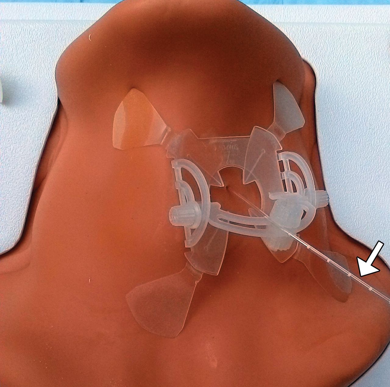
Lesion targeting has traditionally been performed using “in-and-out” techniques in closed-bore MR imaging scanners with control imaging performed inside the bore, and needle adjustments, outside the bore. Alternatively, the process of targeting the lesion can be expedited using guidance and navigation systems, such as virtual 3D-MR imaging with real-time overlay of an optically tracked biopsy needle using reference markers or the interventional MR imaging suite (iSuite) image guidance and mapping system (Philips Research Labs, Hamburg, Germany), which facilitates triangulation of the laser probe insertion trajectory (Fig 1).26 The biopsy needles and laser probe used for the procedure can be secured in position using commercially available skin-adhesive holder devices, such as the Simplify needle holder (NeoRad, Oslo, Norway) or Cradles Needle Localization Wire Protectors (Beekley Medical, Norcross, Georgia) (Fig 2).

Fig 1.
  • Download figure
  • Open in new tab
  • Download powerpoint
Fig 1.

MR imaging guidance for laser applicator insertion. 3D T1-weighted fast-field echo image shows saturation bands (arrowheads) that are used to triangulate the target “lesion” (arrow) in the neck phantom.

Fig 2.
  • Download figure
  • Open in new tab
  • Download powerpoint
Fig 2.

Photograph shows the laser applicator (Medtronic, Minneapolis, Minnesota) (arrow) inserted into the neck phantom (CIRS, Norfolk, Virginia) and supported by a skin-adhesive device (NeoRad).

MR Thermography

In general, the success of laser interstitial thermal ablation depends on accurate insertion of the laser fiber into the target lesion, real-time monitoring of the effects of the treatment, and subsequent evaluation of the extent of thermal tissue damage.19 MR thermography is a validated noninvasive imaging technique that can provide accurate monitoring during laser ablation.19,27 Several MR thermography methods have been developed, including sequences based on the proton resonance frequency, the diffusion coefficient, T1 and T2 relaxation times, magnetization transfer, proton density, and temperature-sensitive contrast agents.4,24,27 In particular, proton resonance frequency techniques use temperature-induced effects of chemical shift, in which the temperature difference is directly proportional to the phase difference. This method provides accurate temperature measurements in the temperature range of interest for thermal ablation.24

A thermal dose can be calculated from a map of temperature as a function of time and is used for the prediction of tissue destruction (Fig 3).7 Ultimately, continuous MR thermometry during thermal ablation procedures can help optimize safe thermal therapy delivery. Although the adequacy of ablation is estimated on the basis of the laser ablation system–generated damage model, it is important to obtain an immediate postablation MR imaging scan to ensure adequate ablation of the lesion.

Fig 3.
  • Download figure
  • Open in new tab
  • Download powerpoint
Fig 3.

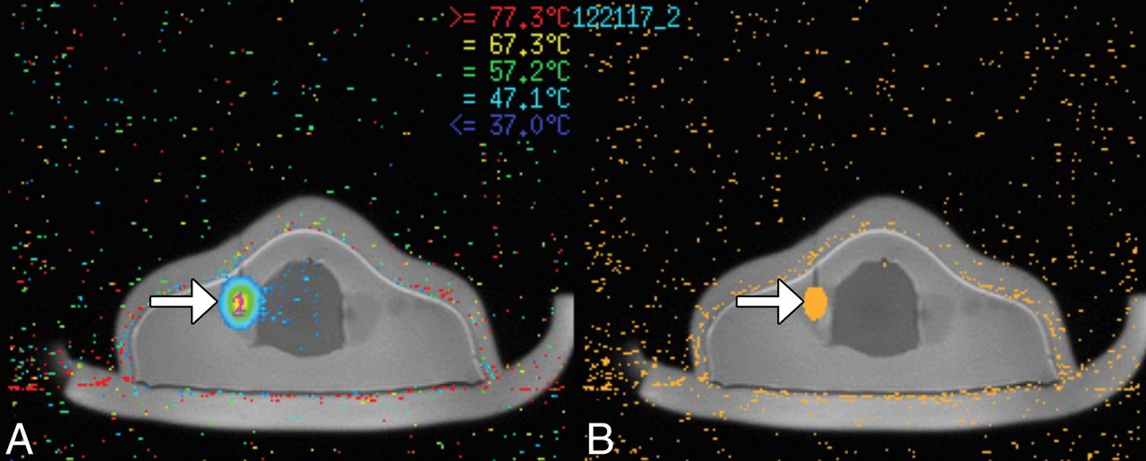
MR thermography during laser ablation. Temperature map acquired using the proton resonance frequency shift (A) and heat-damage model in a neck phantom (B) shows the ablation zone surrounding the tip of the probe (arrows).

Important technical considerations for performing MR imaging–guided laser ablation include coil selection and assessing the quality of MR imaging sequences in the head and neck region. For example, the use of whole-body coils contained within the closed bore of a high-field-strength MR imaging scanner is an ergonomic option with fewer obstacles between the interventionalist and the patient. Although these types of coils yield lower signal-to-noise compared with head and neck neurovascular coils, for example, the image quality may be generally adequate for clinical use.28

Furthermore, the quality of MR thermography can be limited by the presence of susceptibility artifacts from hemorrhage, calcifications, or underlying surgical hardware; the excessive presence of fat surrounding the lesion; and misregistration artifacts related to motion.3 Ultimately, interstitial laser ablation should be performed only with MR thermography if a lesion can be adequately discerned on MR thermography images obtained as part of the pretreatment MR imaging.

Conclusions

Interstitial laser ablation is a minimally invasive and potentially cost-effect technique that can be used to treat a variety of head and neck lesions, including benign thyroid nodules, vascular malformations, and cancers with low complication rates. In particular, the use of MR imaging guidance for inserting the laser applicator and the use of MR thermography for monitoring temperature changes during the procedure can help optimize safety and efficacy.

Acknowledgments

We thank Amy Schneider of Medtronic for assistance in performing the laser ablation procedure.

Footnotes

  • This work was supported by Toshiba America Medical Systems/RSNA Research Seed Grant.

References

  1. 1.↵
    1. Mensel B,
    2. Weigel C,
    3. Hosten N
    . Laser-induced thermotherapy. Recent Results Cancer Res 2006;167:69–75 doi:10.1007/3-540-28137-1_5 pmid:17044297
    CrossRefPubMed
  2. 2.↵
    1. Stafford RJ,
    2. Fuentes D,
    3. Elliott AA, et al
    . Laser-induced thermal therapy for tumor ablation. Crit Rev Biomed Eng 2010;38:79–100 doi:10.1615/CritRevBiomedEng.v38.i1.70 pmid:21175405
    CrossRefPubMed
  3. 3.↵
    1. Medvid R,
    2. Ruiz A,
    3. Komotar RJ, et al
    . Current applications of MRI-guided laser interstitial thermal therapy in the treatment of brain neoplasms and epilepsy: a radiologic and neurosurgical overview. AJNR Am J Neuroradiol 2015;36:1998–2006 doi:10.3174/ajnr.A4362 pmid:26113069
    Abstract/FREE Full Text
  4. 4.↵
    1. Schober R,
    2. Bettag M,
    3. Sabel M, et al
    . Fine structure of zonal changes in experimental Nd:YAG laser-induced interstitial hyperthermia. Lasers Surg Med 1993;13:234–41 doi:10.1002/lsm.1900130212 pmid:8464310
    CrossRefPubMed
  5. 5.↵
    1. Kickhefel A,
    2. Rosenberg C,
    3. Weiss CR, et al
    . Clinical evaluation of MR temperature monitoring of laser-induced thermotherapy in human liver using the proton-resonance-frequency method and predictive models of cell death. J Magn Reson Imaging 2011;33:704–12 doi:10.1002/jmri.22499 pmid:21563256
    CrossRefPubMed
  6. 6.↵
    1. Boss A,
    2. Graf H,
    3. Müller-Bierl B, et al
    . Magnetic susceptibility effects on the accuracy of MR temperature monitoring by the proton resonance frequency method. J Magn Reson Imaging 2005;22:813–20 doi:10.1002/jmri.20438 pmid:16270289
    CrossRefPubMed
  7. 7.↵
    1. Oto A,
    2. Sethi I,
    3. Karczmar G, et al
    . MR imaging-guided focal laser ablation for prostate cancer: phase I trial. Radiology 2013;267:932–40 doi:10.1148/radiol.13121652 pmid:23440319
    CrossRefPubMed
  8. 8.↵
    1. Sercarz JA,
    2. Bublik M,
    3. Joo J, et al
    . Outcomes of laser thermal therapy for recurrent head and neck cancer. Otolaryngol Head Neck Surg 2010;142:344–50 doi:10.1016/j.otohns.2009.11.019 pmid:20172378
    CrossRefPubMed
  9. 9.↵
    1. Joo J,
    2. Sercarz JA,
    3. Paolini AA, et al
    . Laser-induced thermal therapy and cisplatin for recurrent head and neck cancer: a case characterized by an unusually long disease-free survival. Ear Nose Throat J 2009;88:E13–E16 pmid:19924650
    PubMed
  10. 10.↵
    1. Banerjee C,
    2. Snelling B,
    3. Berger MH, et al
    . The role of magnetic resonance-guided laser ablation in neurooncology. Br J Neurosurg 2015;29:192–96 doi:10.3109/02688697.2014.996527 pmid:25665599
    CrossRefPubMed
  11. 11.↵
    1. Carpentier A,
    2. McNichols RJ,
    3. Stafford RJ, et al
    . Laser thermal therapy: real-time MRI-guided and computer-controlled procedures for metastatic brain tumors. Lasers Surg Med 2011;43:943–50 doi:10.1002/lsm.21138 pmid:22109661
    CrossRefPubMed
  12. 12.↵
    1. Carpentier A,
    2. McNichols RJ,
    3. Stafford RJ, et al
    . Real-time magnetic resonance-guided laser thermal therapy for focal metastatic brain tumors. Neurosurgery 2008;63:ONS21–28; discussion ONS28–29 pmid:18728600
    PubMed
  13. 13.↵
    1. Leuthardt EC,
    2. Voigt J,
    3. Kim AH, et al
    . A single-center cost analysis of treating primary and metastatic brain cancers with either brain Laser Interstitial Thermal Therapy (LITT) or craniotomy. Pharmacoecon Open 2017;1:53–63 doi:10.1007/s41669-016-0003-2 pmid:29442297
    CrossRefPubMed
  14. 14.↵
    1. Voigt JD,
    2. Barnett G
    . The value of using a brain laser interstitial thermal therapy (LITT) system in patients presenting with high grade gliomas where maximal safe resection may not be feasible. Cost Eff Resour Alloc 2016;14:6 doi:10.1186/s12962-016-0055-2 pmid:27006643
    CrossRefPubMed
  15. 15.↵
    1. Eckardt A,
    2. Barth EL,
    3. Kokemueller H, et al
    . Recurrent carcinoma of the head and neck: treatment strategies and survival analysis in a 20-year period. Oral Oncol 2004;40:427–32 doi:10.1016/j.oraloncology.2003.09.019 pmid:14969822
    CrossRefPubMed
  16. 16.↵
    1. Pacella CM,
    2. Bizzarri G,
    3. Spiezia S, et al
    . Thyroid tissue: US-guided percutaneous laser thermal ablation. Radiology 2004;232:272–80 doi:10.1148/radiol.2321021368 pmid:15155898
    CrossRefPubMed
  17. 17.↵
    1. Shahrzad MK
    . Laser thermal ablation of thyroid benign nodules. J Lasers Med Sci 2015;6:151–56 doi:10.15171/jlms.2015.10 pmid:26705459
    CrossRefPubMed
  18. 18.↵
    1. Achille G,
    2. Zizzi S,
    3. Di Stasio E, et al
    . Ultrasound-guided percutaneous laser ablation in treating symptomatic solid benign thyroid nodules: our experience in 45 patients. Head Neck 2016;38:677–82 doi:10.1002/hed.23957 pmid:25522303
    CrossRefPubMed
  19. 19.↵
    1. Mack MG,
    2. Vogl TJ
    . MR-guided ablation of head and neck tumors. Neuroimaging Clin N Am 2004;14:853–59 doi:10.1016/j.nic.2004.07.013 pmid:15489155
    CrossRefPubMed
  20. 20.↵
    1. Eyrich GK,
    2. Bruder E,
    3. Hilfiker P, et al
    . Temperature mapping of magnetic resonance-guided laser interstitial thermal therapy (LITT) in lymphangiomas of the head and neck. Lasers Surg Med 2000;26:467–76 doi:10.1002/1096-9101(2000)26:5<467::AID-LSM6>3.0.CO%3B2-8 pmid:10861702
    CrossRefPubMed
  21. 21.↵
    1. Feyh J,
    2. Gutmann R,
    3. Leunig A, et al
    . MRI-guided laser interstitial thermal therapy (LITT) of head and neck tumors: progress with a new method. J Clin Laser Med Surg 1996;14:361–66 pmid:9467326
    PubMed
  22. 22.↵
    1. Mainini AP,
    2. Monaco C,
    3. Pescatori LC, et al
    . Image-guided thermal ablation of benign thyroid nodules. J Ultrasound 2017;20:11–22 doi:10.1007/s40477-016-0221-6 pmid:28298940
    CrossRefPubMed
  23. 23.↵
    1. Moche M,
    2. Trampel R,
    3. Kahn T, et al
    . Navigation concepts for MR image-guided interventions. J Magn Reson Imaging 2008;27:276–91 doi:10.1002/jmri.21262 pmid:18219682
    CrossRefPubMed
  24. 24.↵
    1. Weiss CR,
    2. Nour SG,
    3. Lewin JS
    . MR-guided biopsy: a review of current techniques and applications. J Magn Reson Imaging 2008;27:311–25 doi:10.1002/jmri.21270 pmid:18219685
    CrossRefPubMed
  25. 25.↵
    1. Cleary K,
    2. Peters TM
    . Image-guided interventions: technology review and clinical applications. Annu Rev Biomed Eng 2010;12:119–42 doi:10.1146/annurev-bioeng-070909-105249 pmid:20415592
    CrossRefPubMed
  26. 26.↵
    1. Busse H,
    2. Riedel T,
    3. Garnov N, et al
    . Targeting accuracy, procedure times and user experience of 240 experimental MRI biopsies guided by a clinical add-on navigation system. PLoS One 2015;10:e0134370 doi:10.1371/journal.pone.0134370 pmid:26222443
    CrossRefPubMed
  27. 27.↵
    1. Quesson B,
    2. de Zwart JA,
    3. Moonen CT
    . Magnetic resonance temperature imaging for guidance of thermotherapy. J Magn Reson Imaging 2000;12:525–33 doi:10.1002/1522-2586(200010)12:4<525::AID-JMRI3>3.0.CO%3B2-V pmid:11042633
    CrossRefPubMed
  28. 28.↵
    1. Ginat DT,
    2. Anthony GJ,
    3. Christoforidis G, et al
    . Comparison between whole-body and head and neck neurovascular coils for 3-T magnetic resonance proton resonance frequency shift thermography guidance in the head and neck region. Lasers Med Sci 2018;33:369–73 doi:10.1007/s10103-017-2397-1 pmid:29224048
    CrossRefPubMed
  • Received January 1, 2018.
  • Accepted after revision April 19, 2018.
  • © 2018 by American Journal of Neuroradiology
View Abstract
PreviousNext
Back to top

In this issue

American Journal of Neuroradiology: 39 (9)
American Journal of Neuroradiology
Vol. 39, Issue 9
1 Sep 2018
  • Table of Contents
  • Index by author
  • Complete Issue (PDF)
Advertisement
Print
Download PDF
Email Article

Thank you for your interest in spreading the word on American Journal of Neuroradiology.

NOTE: We only request your email address so that the person you are recommending the page to knows that you wanted them to see it, and that it is not junk mail. We do not capture any email address.

Enter multiple addresses on separate lines or separate them with commas.
MR Thermography–Guided Head and Neck Lesion Laser Ablation
(Your Name) has sent you a message from American Journal of Neuroradiology
(Your Name) thought you would like to see the American Journal of Neuroradiology web site.
CAPTCHA
This question is for testing whether or not you are a human visitor and to prevent automated spam submissions.
Cite this article
D.T. Ginat, S. Sammet, G. Christoforidis
MR Thermography–Guided Head and Neck Lesion Laser Ablation
American Journal of Neuroradiology Sep 2018, 39 (9) 1593-1596; DOI: 10.3174/ajnr.A5726

Citation Manager Formats

  • BibTeX
  • Bookends
  • EasyBib
  • EndNote (tagged)
  • EndNote 8 (xml)
  • Medlars
  • Mendeley
  • Papers
  • RefWorks Tagged
  • Ref Manager
  • RIS
  • Zotero
0 Responses
Respond to this article
Share
Bookmark this article
MR Thermography–Guided Head and Neck Lesion Laser Ablation
D.T. Ginat, S. Sammet, G. Christoforidis
American Journal of Neuroradiology Sep 2018, 39 (9) 1593-1596; DOI: 10.3174/ajnr.A5726
del.icio.us logo Twitter logo Facebook logo Mendeley logo
  • Tweet Widget
  • Facebook Like
  • Google Plus One
Purchase

Jump to section

  • Article
    • Abstract
    • Conclusions
    • Acknowledgments
    • Footnotes
    • References
  • Figures & Data
  • Info & Metrics
  • Responses
  • References
  • PDF

Related Articles

  • No related articles found.
  • PubMed
  • Google Scholar

Cited By...

  • No citing articles found.
  • Crossref (5)
  • Google Scholar

This article has been cited by the following articles in journals that are participating in Crossref Cited-by Linking.

  • Proton Resonance Frequency Shift Thermometry: A Review of Modern Clinical Practices
    James Blackwell, Marcin J. Kraśny, Aoife O'Brien, Keyoumars Ashkan, Josette Galligan, Michel Destrade, Niall Colgan
    Journal of Magnetic Resonance Imaging 2022 55 2
  • Computation of thermophysical properties for magnetite-based hyperthermia treatment simulations using infrared thermography
    I. Garrido, S. Lagüela, J.V. Román, E.M. Martín-del Valle, D. González-Aguilera
    International Journal of Heat and Mass Transfer 2020 154
  • Assessment of Proton Resonance Frequency Shift Magnetic Resonance Thermography Imaging Quality for Head and Neck Tumors
    Daniel T. Ginat, Steffen Sammet
    Ear, Nose & Throat Journal 2024 103 3
  • Effectiveness and Safety of Ultrasound-Guided Microwave Ablation for the Treatment of Primary Hyperparathyroidism with Parathyroid Adenoma
    若琳 吕
    Advances in Clinical Medicine 2023 13 04
  • MR-guided percutaneous microwave coagulation of small breast tumors
    Ying Ji, Yaoyao Zhuo, Ting Li, Jingge Lian, Zilin Wang, Xinyu Guo, Dexing Kong, Kangan Li
    Insights into Imaging 2024 15 1

More in this TOC Section

  • WHO Classification Update: Nasal&Skull Base Tumors
  • Peritumoral Signal in Vestibular Schwannomas
  • NI-RADS for HEAD&NECK Cancer Recurrence
Show more HEAD & NECK

Similar Articles

Advertisement

Indexed Content

  • Current Issue
  • Accepted Manuscripts
  • Article Preview
  • Past Issues
  • Editorials
  • Editor's Choice
  • Fellows' Journal Club
  • Letters to the Editor
  • Video Articles

Cases

  • Case Collection
  • Archive - Case of the Week
  • Archive - Case of the Month
  • Archive - Classic Case

Special Collections

  • AJNR Awards
  • ASNR Foundation Special Collection
  • Most Impactful AJNR Articles
  • Photon-Counting CT
  • Spinal CSF Leak Articles (Jan 2020-June 2024)

More from AJNR

  • Trainee Corner
  • Imaging Protocols
  • MRI Safety Corner

Multimedia

  • AJNR Podcasts
  • AJNR Scantastics

Resources

  • Turnaround Time
  • Submit a Manuscript
  • Submit a Video Article
  • Submit an eLetter to the Editor/Response
  • Manuscript Submission Guidelines
  • Statistical Tips
  • Fast Publishing of Accepted Manuscripts
  • Graphical Abstract Preparation
  • Imaging Protocol Submission
  • Evidence-Based Medicine Level Guide
  • Publishing Checklists
  • Author Policies
  • Become a Reviewer/Academy of Reviewers
  • News and Updates

About Us

  • About AJNR
  • Editorial Board
  • Editorial Board Alumni
  • Alerts
  • Permissions
  • Not an AJNR Subscriber? Join Now
  • Advertise with Us
  • Librarian Resources
  • Feedback
  • Terms and Conditions
  • AJNR Editorial Board Alumni

American Society of Neuroradiology

  • Not an ASNR Member? Join Now

© 2025 by the American Society of Neuroradiology All rights, including for text and data mining, AI training, and similar technologies, are reserved.
Print ISSN: 0195-6108 Online ISSN: 1936-959X

Powered by HighWire