Advertisement

Main menu

  • Home
  • Content
    • Current Issue
    • Accepted Manuscripts
    • Article Preview
    • Past Issue Archive
    • Video Articles
    • AJNR Case Collection
    • Case of the Week Archive
    • Case of the Month Archive
    • Classic Case Archive
  • Special Collections
    • AJNR Awards
    • ASNR Foundation Special Collection
    • Most Impactful AJNR Articles
    • Photon-Counting CT
    • Spinal CSF Leak Articles (Jan 2020-June 2024)
  • Multimedia
    • AJNR Podcasts
    • AJNR SCANtastic
    • Trainee Corner
    • MRI Safety Corner
    • Imaging Protocols
  • For Authors
    • Submit a Manuscript
    • Submit a Video Article
    • Submit an eLetter to the Editor/Response
    • Manuscript Submission Guidelines
    • Statistical Tips
    • Fast Publishing of Accepted Manuscripts
    • Graphical Abstract Preparation
    • Imaging Protocol Submission
    • Author Policies
  • About Us
    • About AJNR
    • Editorial Board
    • Editorial Board Alumni
  • More
    • Become a Reviewer/Academy of Reviewers
    • Subscribers
    • Permissions
    • Alerts
    • Feedback
    • Advertisers
    • ASNR Home

User menu

  • Alerts
  • Log in

Search

  • Advanced search
American Journal of Neuroradiology

American Journal of Neuroradiology

ASHNR American Society of Functional Neuroradiology ASHNR American Society of Pediatric Neuroradiology ASSR
  • Alerts
  • Log in

Advanced Search

  • Home
  • Content
    • Current Issue
    • Accepted Manuscripts
    • Article Preview
    • Past Issue Archive
    • Video Articles
    • AJNR Case Collection
    • Case of the Week Archive
    • Case of the Month Archive
    • Classic Case Archive
  • Special Collections
    • AJNR Awards
    • ASNR Foundation Special Collection
    • Most Impactful AJNR Articles
    • Photon-Counting CT
    • Spinal CSF Leak Articles (Jan 2020-June 2024)
  • Multimedia
    • AJNR Podcasts
    • AJNR SCANtastic
    • Trainee Corner
    • MRI Safety Corner
    • Imaging Protocols
  • For Authors
    • Submit a Manuscript
    • Submit a Video Article
    • Submit an eLetter to the Editor/Response
    • Manuscript Submission Guidelines
    • Statistical Tips
    • Fast Publishing of Accepted Manuscripts
    • Graphical Abstract Preparation
    • Imaging Protocol Submission
    • Author Policies
  • About Us
    • About AJNR
    • Editorial Board
    • Editorial Board Alumni
  • More
    • Become a Reviewer/Academy of Reviewers
    • Subscribers
    • Permissions
    • Alerts
    • Feedback
    • Advertisers
    • ASNR Home
  • Follow AJNR on Twitter
  • Visit AJNR on Facebook
  • Follow AJNR on Instagram
  • Join AJNR on LinkedIn
  • RSS Feeds

AJNR is seeking candidates for the AJNR Podcast Editor. Read the position description.

OtherBRAIN

Richter Transformation of Chronic Lymphocytic Leukemia Presenting as a Dural-Based Non-Hodgkin Lymphoma Mass

M. Ghofrani, B. Tantiwongkosi, A.S. Smith and D.A. Wasdahl
American Journal of Neuroradiology February 2007, 28 (2) 318-320;
M. Ghofrani
  • Find this author on Google Scholar
  • Find this author on PubMed
  • Search for this author on this site
B. Tantiwongkosi
  • Find this author on Google Scholar
  • Find this author on PubMed
  • Search for this author on this site
A.S. Smith
  • Find this author on Google Scholar
  • Find this author on PubMed
  • Search for this author on this site
D.A. Wasdahl
  • Find this author on Google Scholar
  • Find this author on PubMed
  • Search for this author on this site
  • Article
  • Figures & Data
  • Info & Metrics
  • Responses
  • References
  • PDF
Loading

Abstract

SUMMARY: Richter transformation is defined as a diffuse large cell lymphoma, occurring by transformation of chronic lymphocytic leukemia (CLL). We present a 64-year-old man with a history of CLL and a left parieto-occipital transtentorial extra-axial mass. The patient underwent CT and MR imaging, demonstrating a large dural-based mass with extracranial extension, which occluded the left transverse sinus. Biopsy of the mass proved a pathologic diagnosis of diffuse large cell non-Hodgkin lymphoma, consistent with Richter transformation of CLL.

Central nervous system lymphomatous involvement in the setting of chronic lymphocytic leukemia (CLL), known as Richter transformation, is rare. Thirteen cases have been reported,1–7 only 5 of which had isolated leptomeningeal involvement,4,5,8 and the other 8 demonstrated parenchymal involvement. We report a case of central nervous system Richter transformation, presenting as a large left occipital dural-based mass. It was investigated by means of conventional radiography, CT, MR imaging, and MR angiography and was surgically biopsied.

Case Report

A 64-year-old man presented to the emergency department with headaches, 2–3 days of vomiting, and a left occipital mass present for 3–4 weeks. He had a medical history of rheumatoid arthritis, immune thrombocytopenic purpura, and a 5-year history of chronic lymphocytic leukemia (CD5+, CD20+, and CD23+), treated through multiple chemotherapy courses (including 3 cycles of fludarabine 4 years prior). The patient described the headaches as “nagging” and running across his forehead, and he also stated that he had experienced some occasional blurry vision and “floaters” during the past weeks, but no visual loss. He had episodes of dizziness and balance difficulty, but no history of falls or loss of consciousness. Physical examination revealed an alert, well-nourished man, with a nontender mass in the left occipital region. A retinal hemorrhage was noted in the right eye on fundoscopic examination, and the left pupil appeared larger. An egg-sized lymph node was palpable in the right axilla. Findings of a neurologic examination were grossly nonfocal.

Skull radiographs did not show any osseous abnormality or soft-tissue calcification, but mild prominence of the left occipital soft tissues was noted. CT and MR imaging demonstrated a large 8.0 × 5.5 × 8.5 cm oval dural-based transtentorial mass with a dural tail in the left occipital-to-parietal region, with both extracranial and intracranial components. The dural tail extended along the left tentorium to the incisura. The intracranial component was 6.5 × 3.5 × 8.0 cm. The mass was hyperattenuated compared with normal brain parenchyma on unenhanced CT and enhanced homogeneously (Fig 1). There was localized effacement of sulci and ventricles, without parenchymal edema. The mass was abutting the confluence of dural sinuses and eroding the overlying left occipital and posterior parietal bones. MR imaging showed obliteration of the left transverse sinus, but the left sigmoid sinus was unaffected (Fig 2). CT studies of the chest, abdomen, and pelvis were unremarkable.

Fig 1.
  • Download figure
  • Open in new tab
  • Download powerpoint
Fig 1.

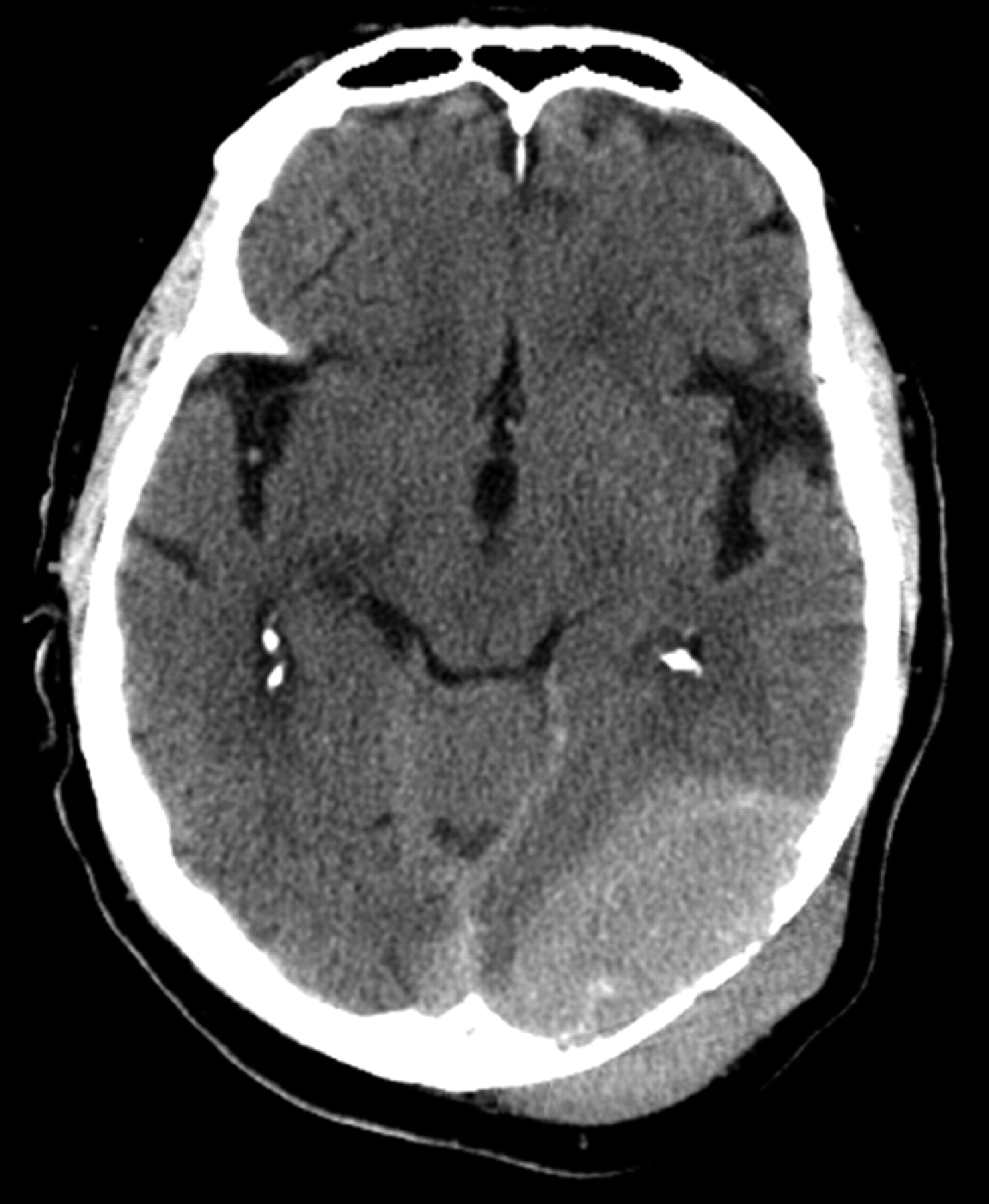
Noncontrast CT scan of the brain shows a large heterogeneously attenuated dural-based mass extending across the tentorium and the calvaria.

Fig 2.
  • Download figure
  • Open in new tab
  • Download powerpoint
Fig 2.

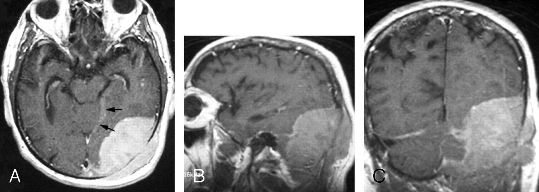
Gadolinium-enhanced axial (A), sagittal (B), and coronal (C) fast spin-echo T1-weighted images (400/18/1 [TR/TE/excitations]) through the left occipital region show a large enhanced dural-based mass measuring 8.0 × 5.5 × 8.5 cm. A part of the dural tail is seen along the incisura (arrows). Laterally, the mass erodes the occipital bone and demonstrates a mass effect on the adjacent occipital lobe and cerebellum without invasion.

The images presented a differential consideration of metastatic disease, meningeal hemangiopericytoma, malignant meningioma, and plasmacytoma.

The mass was surgically biopsied. An incision of the left posterior scalp revealed a grayish tumor under the fascia, from which multiple biopsy specimens were obtained and sent for frozen and permanent histologic section and flow cytometry. The pathology report indicated diffuse large cell non-Hodgkin lymphoma, consistent with Richter transformation involving the dura, skull, and brain, as well as the extracranial soft tissue (Fig 3).

Fig 3.
  • Download figure
  • Open in new tab
  • Download powerpoint
Fig 3.

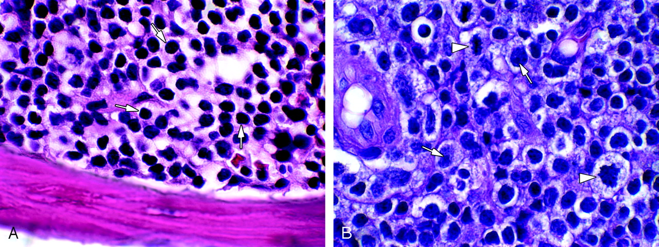
A, Bone marrow hematoxylin-eosin (H&E) 1000× (oil), bone marrow in CLL, shows replacement of normal marrow elements by sheets of small mature lymphocytes (arrows). B, Brain H&E 1000× (oil), large cell non-Hodgkin lymphoma (Richter) transformation of CLL. Note nucleoli (arrows) and mitotic figures (arrowheads).

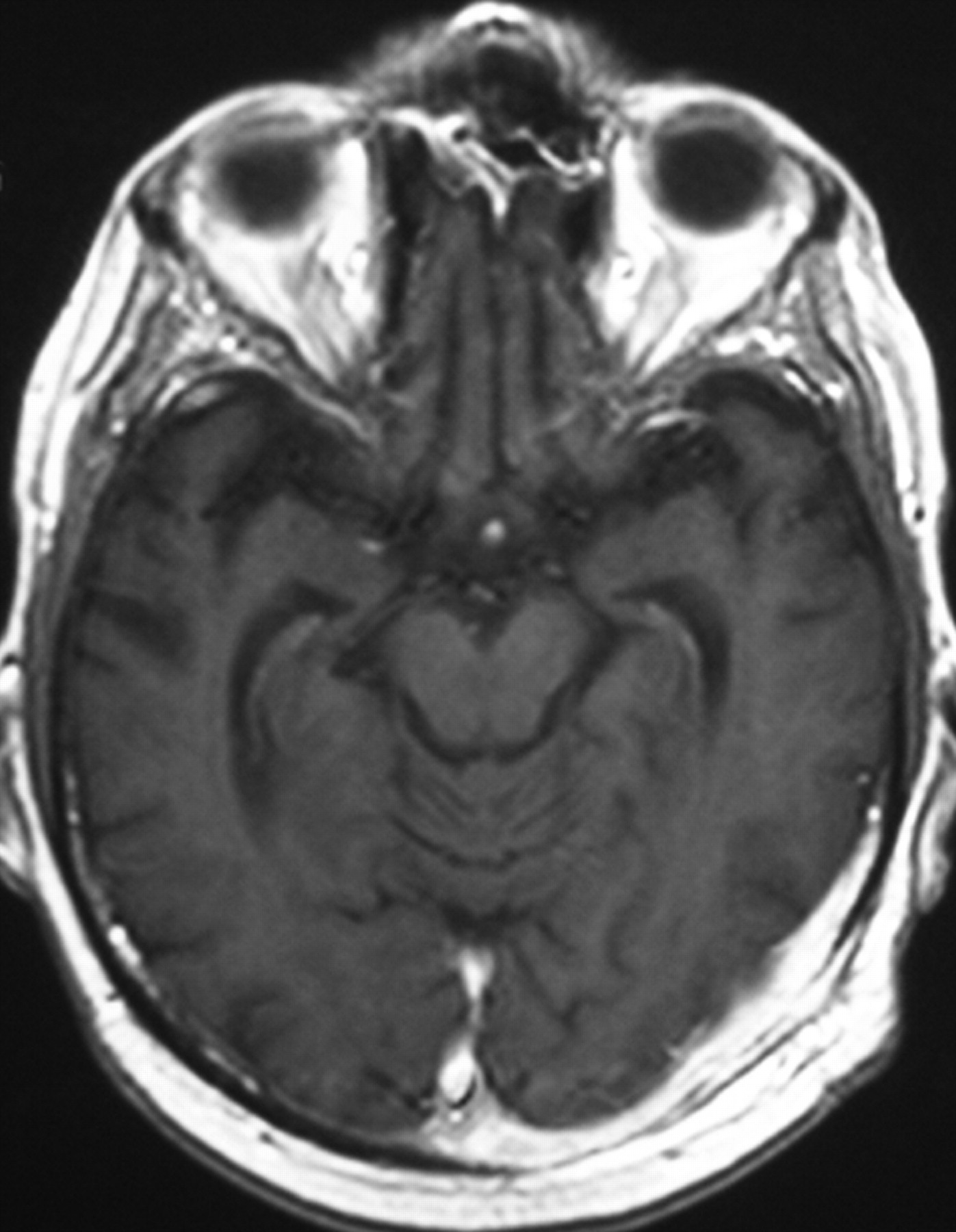
The patient was placed on chemotherapy (rituximab, cyclophosphamide [Cytoxan], doxorubicin hydrochloride [Adriamycin], vincristine, and prednisone) and radiation therapy regimens, after which the mass markedly decreased in size, with only 2-cm dural thickness on a follow-up MR imaging study, but with persistent occlusion of the left transverse sinus and lymphomatous involvement of the left mastoid bone (Fig 4). The patient died 3 months after the initial presentation to the emergency department for the cranial mass.

Fig 4.
  • Download figure
  • Open in new tab
  • Download powerpoint
Fig 4.

Post-therapy gadolinium-enhanced axial fast spin-echo T1-weighted image (340/14/1 [TR/TE/excitations]) reveals marked reduction of the mass size with continued abnormal enhancement along the dura.

Discussion

The occurrence of diffuse large cell non-Hodgkin lymphoma in the setting of CLL was first described by Richter in 19289 and appointed as “Richter syndrome” in 1964.10 Richter transformation is an unusual and serious complication of CLL, with an incidence occurring between 1% and 10% of CLL cases, a 5–6 month median survival duration despite multiagent therapy,3,8,11,12 and a significantly higher incidence in patients younger than 55 years of age.13 Hodgkin disease in the setting of CLL has also been described as a variant of Richter transformation.3 Diffuse large cell non-Hodgkin lymphoma has been demonstrated in 6% of patients with Waldenstrom macroglobulinemia.14 Reports have documented the presence of 2 types of Richter syndrome: the “classical” type as a terminal event of CLL and the “variant” type as the first clinical presentation of previously undetected subclinical CLL.15,16 There has been controversy about whether the transformation represents the emergence of a more malignant clone or the chance occurrence of 2 independent neoplasms.3,4,8

The incidence of Richter transformation in the study by Robertson et al8 was similar in patients who had and who had not received prior fludarabine therapy, implying that therapy does not reverse a component of CLL-associated immunodeficiency leading to the development of Richter transformation.3 Follow-up has been suggested to evaluate whether increased immunosuppression correlates with an increased incidence of Richter transformation.3

Only an awareness of the patient’s pre-existing condition and the possibility of transformation of CLL to non-Hodgkin lymphoma can lead to the inclusion of Richter transformation. The mass in this patient was dural-based, and because previously reported cases of central nervous system Richter transformation4,5,8 do not specify a dural mass, this case may be even more atypical.

Acknowledgments

We thank Negar Imani, who assisted in preparing the images for this case report.

References

  1. ↵
    Agard G, Hamidou M, Leautez S, et al. Localisation neuroméningée d’um syndrome de Richter. Rev Méd Interne 1999;20:64–67
    PubMed
  2. Bayliss KM, Kueck BD, Hanson CA, et al. Richter’s syndrome presenting as primary central nervous system lymphoma: transformation of an identical clone. Am J Clin Pathol 1990;93:117–23
    PubMed
  3. ↵
    Giles FJ, O’Brien SM, Keating MJ. Chronic lymphocytic leukemia in (Richter’s) transformation. Semin Oncol 1998;25:117–25
    PubMed
  4. ↵
    Lane PK, Townsend RM, Beckstead JH, et al. Central nervous system involvement in a patient with chronic lymphocytic leukemia and non-Hodgkin’s lymphoma (Richter’s syndrome), with concordant cell surface immunoglobulin isotypic and immunophenotypic markers. Am J Clin Pathol 1988;89:254–59
    PubMed
  5. ↵
    Mahé B, Moreau P, Bonnemain B, et al. Isolated Richter’s syndrome of the brain: two recent cases. Nouv Rev Fr Hematol 1994;36:383–85
    PubMed
  6. O’Neill BP, Habermann TM, Banks PM, et al. Primary central nervous system lymphoma as a variant of Richter’s syndrome in two patients with chronic lymphocytic leukemia. Cancer 1989;15:1296–300
  7. ↵
    Resende LS, Bacchi CE, Resende LA, et al. Isolated Richter’s syndrome in central nervous system: case report [in Portuguese]. Arq Neuropsiquiatr 2005;63(2B):530–31. Epub 2005 Jul 25
  8. ↵
    Robertson LE, Pugh W, O’Brien S, et al. Richter’s syndrome: a report on 39 patients. J Clin Oncol 1993;11:1985–89
    Abstract/FREE Full Text
  9. ↵
    Richter MN. Generalized reticular cell sarcoma of lymph nodes associated with lymphatic leukemia. Am J Pathol 1928;6:285–99
  10. ↵
    Lortholary P, Boiron M, Ripault J, et al. Leucémie lymphoïde chronique secondairement associée à une réticulopathie maligne (syndrome de Richter). Nouv Rev Fr Hematol 1964;4:621–44
  11. ↵
    Rai KR, Keating MJ. Staging and prognosis of chronic lymphocytic leukemia. Up-to-date May 10, 2005. Available at http://patients.uptodate.com/topic.asp?file=leukemia/12607
  12. ↵
    Rai KR, Dohner H, Keating MJ, et al. Chronic lymphocytic leukemia: case-based session. Hematology Am Soc Hematol Educ Program 2001 :140–56
  13. ↵
    Mauro FR, Foa R, Giannarelli D, et al. Clinical characteristics and outcome of young chronic lymphocytic leukemia patients: a single institution study of 204 cases. Blood 1999;94:448–54
    Abstract/FREE Full Text
  14. ↵
    Tardif S, de Kerviler E, Chaibi P, et al. CT and MR patterns of spinal involvement in Richter syndrome. J Comput Assist Tomogr 1995;19:146–49
    PubMed
  15. ↵
    Jelic S, Jovanovic V, Milanovic N, et al. Richter syndrome with emphasis on large-cell non-Hodgkin lymphoma in previously unrecognized subclinical chronic lymphocytic leukemia. Neoplasma 1997;44:63–68
    PubMed
  16. ↵
    Strauchen JA, May MM, Crown J. Large cell transformation of subclinical small lymphocytic leukemia/lymphoma: a variant of Richter’s syndrome. Hematol Oncol 1987;5:167–74
    PubMed
  • Received November 23, 2005.
  • Accepted after revision January 3, 2006.
  • Copyright © American Society of Neuroradiology
View Abstract
PreviousNext
Back to top

In this issue

American Journal of Neuroradiology: 28 (2)
American Journal of Neuroradiology
Vol. 28, Issue 2
February 2007
  • Table of Contents
  • Index by author
Advertisement
Print
Download PDF
Email Article

Thank you for your interest in spreading the word on American Journal of Neuroradiology.

NOTE: We only request your email address so that the person you are recommending the page to knows that you wanted them to see it, and that it is not junk mail. We do not capture any email address.

Enter multiple addresses on separate lines or separate them with commas.
Richter Transformation of Chronic Lymphocytic Leukemia Presenting as a Dural-Based Non-Hodgkin Lymphoma Mass
(Your Name) has sent you a message from American Journal of Neuroradiology
(Your Name) thought you would like to see the American Journal of Neuroradiology web site.
CAPTCHA
This question is for testing whether or not you are a human visitor and to prevent automated spam submissions.
Cite this article
M. Ghofrani, B. Tantiwongkosi, A.S. Smith, D.A. Wasdahl
Richter Transformation of Chronic Lymphocytic Leukemia Presenting as a Dural-Based Non-Hodgkin Lymphoma Mass
American Journal of Neuroradiology Feb 2007, 28 (2) 318-320;

Citation Manager Formats

  • BibTeX
  • Bookends
  • EasyBib
  • EndNote (tagged)
  • EndNote 8 (xml)
  • Medlars
  • Mendeley
  • Papers
  • RefWorks Tagged
  • Ref Manager
  • RIS
  • Zotero
0 Responses
Respond to this article
Share
Bookmark this article
Richter Transformation of Chronic Lymphocytic Leukemia Presenting as a Dural-Based Non-Hodgkin Lymphoma Mass
M. Ghofrani, B. Tantiwongkosi, A.S. Smith, D.A. Wasdahl
American Journal of Neuroradiology Feb 2007, 28 (2) 318-320;
del.icio.us logo Twitter logo Facebook logo Mendeley logo
  • Tweet Widget
  • Facebook Like
  • Google Plus One

Jump to section

  • Article
    • Abstract
    • Case Report
    • Discussion
    • Acknowledgments
    • References
  • Figures & Data
  • Info & Metrics
  • Responses
  • References
  • PDF

Related Articles

  • No related articles found.
  • PubMed
  • Google Scholar

Cited By...

  • No citing articles found.
  • Crossref
  • Google Scholar

This article has not yet been cited by articles in journals that are participating in Crossref Cited-by Linking.

More in this TOC Section

  • Optimal MRI Sequence for Identifying Occlusion Location in Acute Stroke: Which Value of Time-Resolved Contrast-Enhanced MRA?
  • Evaluating the Effects of White Matter Multiple Sclerosis Lesions on the Volume Estimation of 6 Brain Tissue Segmentation Methods
  • Quiet PROPELLER MRI Techniques Match the Quality of Conventional PROPELLER Brain Imaging Techniques
Show more BRAIN

Similar Articles

Advertisement

Indexed Content

  • Current Issue
  • Accepted Manuscripts
  • Article Preview
  • Past Issues
  • Editorials
  • Editor's Choice
  • Fellows' Journal Club
  • Letters to the Editor
  • Video Articles

Cases

  • Case Collection
  • Archive - Case of the Week
  • Archive - Case of the Month
  • Archive - Classic Case

Special Collections

  • AJNR Awards
  • ASNR Foundation Special Collection
  • Most Impactful AJNR Articles
  • Photon-Counting CT
  • Spinal CSF Leak Articles (Jan 2020-June 2024)

More from AJNR

  • Trainee Corner
  • Imaging Protocols
  • MRI Safety Corner

Multimedia

  • AJNR Podcasts
  • AJNR Scantastics

Resources

  • Turnaround Time
  • Submit a Manuscript
  • Submit a Video Article
  • Submit an eLetter to the Editor/Response
  • Manuscript Submission Guidelines
  • Statistical Tips
  • Fast Publishing of Accepted Manuscripts
  • Graphical Abstract Preparation
  • Imaging Protocol Submission
  • Evidence-Based Medicine Level Guide
  • Publishing Checklists
  • Author Policies
  • Become a Reviewer/Academy of Reviewers
  • News and Updates

About Us

  • About AJNR
  • Editorial Board
  • Editorial Board Alumni
  • Alerts
  • Permissions
  • Not an AJNR Subscriber? Join Now
  • Advertise with Us
  • Librarian Resources
  • Feedback
  • Terms and Conditions
  • AJNR Editorial Board Alumni

American Society of Neuroradiology

  • Not an ASNR Member? Join Now

© 2025 by the American Society of Neuroradiology All rights, including for text and data mining, AI training, and similar technologies, are reserved.
Print ISSN: 0195-6108 Online ISSN: 1936-959X

Powered by HighWire