Advertisement

Main menu

  • Home
  • Content
    • Current Issue
    • Accepted Manuscripts
    • Article Preview
    • Past Issue Archive
    • Video Articles
    • AJNR Case Collection
    • Case of the Week Archive
    • Case of the Month Archive
    • Classic Case Archive
  • Special Collections
    • AJNR Awards
    • ASNR Foundation Special Collection
    • Most Impactful AJNR Articles
    • Photon-Counting CT
    • Spinal CSF Leak Articles (Jan 2020-June 2024)
  • Multimedia
    • AJNR Podcasts
    • AJNR SCANtastic
    • Trainee Corner
    • MRI Safety Corner
    • Imaging Protocols
  • For Authors
    • Submit a Manuscript
    • Submit a Video Article
    • Submit an eLetter to the Editor/Response
    • Manuscript Submission Guidelines
    • Statistical Tips
    • Fast Publishing of Accepted Manuscripts
    • Graphical Abstract Preparation
    • Imaging Protocol Submission
    • Author Policies
  • About Us
    • About AJNR
    • Editorial Board
    • Editorial Board Alumni
  • More
    • Become a Reviewer/Academy of Reviewers
    • Subscribers
    • Permissions
    • Alerts
    • Feedback
    • Advertisers
    • ASNR Home

User menu

  • Alerts
  • Log in

Search

  • Advanced search
American Journal of Neuroradiology

American Journal of Neuroradiology

ASHNR American Society of Functional Neuroradiology ASHNR American Society of Pediatric Neuroradiology ASSR
  • Alerts
  • Log in

Advanced Search

  • Home
  • Content
    • Current Issue
    • Accepted Manuscripts
    • Article Preview
    • Past Issue Archive
    • Video Articles
    • AJNR Case Collection
    • Case of the Week Archive
    • Case of the Month Archive
    • Classic Case Archive
  • Special Collections
    • AJNR Awards
    • ASNR Foundation Special Collection
    • Most Impactful AJNR Articles
    • Photon-Counting CT
    • Spinal CSF Leak Articles (Jan 2020-June 2024)
  • Multimedia
    • AJNR Podcasts
    • AJNR SCANtastic
    • Trainee Corner
    • MRI Safety Corner
    • Imaging Protocols
  • For Authors
    • Submit a Manuscript
    • Submit a Video Article
    • Submit an eLetter to the Editor/Response
    • Manuscript Submission Guidelines
    • Statistical Tips
    • Fast Publishing of Accepted Manuscripts
    • Graphical Abstract Preparation
    • Imaging Protocol Submission
    • Author Policies
  • About Us
    • About AJNR
    • Editorial Board
    • Editorial Board Alumni
  • More
    • Become a Reviewer/Academy of Reviewers
    • Subscribers
    • Permissions
    • Alerts
    • Feedback
    • Advertisers
    • ASNR Home
  • Follow AJNR on Twitter
  • Visit AJNR on Facebook
  • Follow AJNR on Instagram
  • Join AJNR on LinkedIn
  • RSS Feeds

AJNR is seeking candidates for the AJNR Podcast Editor. Read the position description.

Article CommentaryCOMMENTARY

Controversies: Is There a Role for Positron-Emission Tomographic CT in the Initial Staging of Head and Neck Squamous Cell Carcinoma?

Suresh K. Mukherji and Carol R. Bradford
American Journal of Neuroradiology February 2006, 27 (2) 243-245;
Suresh K. Mukherji
  • Find this author on Google Scholar
  • Find this author on PubMed
  • Search for this author on this site
Carol R. Bradford
  • Find this author on Google Scholar
  • Find this author on PubMed
  • Search for this author on this site
  • Article
  • Figures & Data
  • Info & Metrics
  • Responses
  • References
  • PDF
Loading

Positron-emission tomographic CT (PET-CT) is gaining greater acceptance in a wide variety of oncologic indications in numerous organ systems (head and neck, central nervous system, breast, gynecologic, pulmonary, lymphoma). The dual-technique capability of PET-CT, which permits direct image fusion and improves the ability to anatomically localize foci of fluorodeoxyglucose (FDG) uptake, is replacing stand-alone PET systems. There are numerous potential clinical applications for PET-CT to evaluate malignancies of the head and neck, specifically squamous cell carcinoma (HNSCCA). Potential clinical applications include pretreatment staging, treatment monitoring and evaluation of the previously treated patients.

The current literature suggests that most primary site HNSCCA with volumes >1 mL will be FDG avid. These correspond to lesions that are moderately sized T1 or greater. Tumors with volumes <1 mL may be detected with FDG, however, the sensitivity decreases with decreasing size. PET also has the ability to detect metastatic cervical lymph nodes, which may be both clinically occult and not detected by CT or MR. In light of these potential benefits, there is debate as to how to use PET-CT for the initial staging of HNSCCA. The current consensus does not support the use of CT-PET for routine staging of all newly diagnosed squamous cell carcinomas. The intent of this manuscript is to propose potential indications for performing PET-CT for initial staging of HNSCCA before treatment.

One potential application is to perform PET-CT in advanced stage HNSCCA to evaluate for occult distant metastases to the lungs or bones. The presence of pulmonary metastases upstages a patient from M0 to M1 and alters treatment intent (Fig 1). The likelihood of pulmonary metastases is low in patients with early-stage disease and the routine imaging work-up for pulmonary metastases is conventional radiography of the chest at most institutions. An argument can be made to perform chest CT in all patients with advanced stage disease; however, if a solitary nodule is identified, it is often unclear whether this is metastasis or a granuloma. PET may help in this evaluation as a FDG-positive nodule will likely require biopsy, whereas an FDG-negative nodule (>8 mm) likely indicates a granuloma, and a biopsy may be avoided.

Various studies have been performed to evaluate the diagnostic accuracy of PET-FDG for detecting metastatic cervical lymph nodes. The consensus of the current literature suggests that sensitivity ranges of 70%–90%, whereas the specificity is slightly higher (80%–95%). The negative predictive value (NPV) is approximately 90%. This is because >40% of metastatic lymph nodes are <7 mm in diameter. As a result, PET-CT has not gained widespread acceptance to be used to exclude the presence of metastases in the clinically N0 neck. In fact, there is currently no imaging study that has a negative predictive value that has been shown to be consistently >95%. It is conceivable that future advances in CT detector technology in PET-CT units will permit diagnostic CT (<2.5 mm and gantry angulation) to be performed. If so, PET-CT may have greater impact on management of the N0 neck as the NPV of this technique will increase if both studies (PET and CT) are of diagnostic quality and both yield normal results.

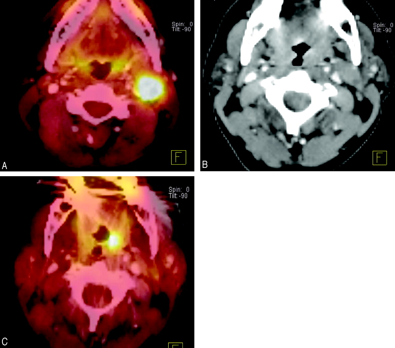
Numerous investigators have documented the ability of PET-CT to detect unknown primary tumors of the upper arodigestive tract. The current literature suggests that PET can detect HNSCCA in 30%–50% of patients presenting with an unknown primary tumor (Fig 2). At most institutions, PET-CT is performed after confirming the presence of metastatic HNSCCA and following a negative endoscopy. PET is usually performed before endoscopic biopsies to help improve the yield of the speculative tissue sampling. The diagnostic yield will likely increase with PET-CT because this technique improves accurate anatomic localization of areas of abnormal FDG uptake.

An area of potential utilization of PET-CT currently under investigation is in determining response to nonsurgical treatment modalities, either chemotherapy and/or radiation. Comparison of pretreatment standard uptake values to SUVs 2 weeks into treatment can allow measurement of the speed of response and the sensitivity of the tumor to the treatment technique. Poorly responsive tumors can then be treated to higher effective tumor doses of radiation, for example, or surgery can be performed. Furthermore, initial results suggest that PET-CT can be used to assist in defining primary site and nodal tumor targets for intensity-modulated radiation therapy approaches.

There are numerous professional and financial issues surrounding PET-CT that will require further discussion. Important topics that will need to be addressed include

  1. Who should interpret PET-CT? Should these be interpreted by a nuclear medicine physician (PET-CT), the subspecialist who would usually interpret the CT (CT-PET), or some form of joint interpretation?

  2. Should intravenous contrast routinely be given for the CT portion of the CT-PET?

  3. How should the CT component of the PET be interpreted? Will this only be used as an “anatomic localizer,” or will all PET-CT studies need to be interpreted for unsuspected findings, which would be akin to “screening CT”?

  4. How will we bill for PET-CT? The CT technology of earlier versions of CT-PET consisted of 2 or 4 detector rows, which were unable to obtain images to obtain thin sections (<5 mm). Newer versions of PET-CT now integrate state-of-the-art CT 16- and 64-row detector configurations, so it is possible that the PET-CT will provide diagnostic quality CT studies. Will we have one code for a combined PET-CT study; will we bill for a PET study with a modifier for the CT component, or will we independently bill for both the CT and PET components? How will this affect states that have certificate of need requirements that regulate the number of CT scanners that an institution may have at any one time?

At our institution, we are now routinely administering intravenous contrast for all PET-CT performed of the extracranial head and neck. The studies are jointly interpreted by faculty members of the divisions of neuroradiology and nuclear medicine. It is our belief that PET-CT is a useful adjunct to initial clinical staging of HNSCCA for specific indications and utilization of pretreatment PET-CT will continue to increase with advances in PET-CT technology.

Fig 1.
  • Download figure
  • Open in new tab
  • Download powerpoint
Fig 1.

T3N2 pyriform sinus carcinoma.

A, Axial PET-CT demonstrates avid FDG uptake in a right pyriform sinus carcinoma and a metastatic right cervical lymph node.

B, PET-CT of the chest shows a mediastinal mass with focal increased uptake. This was not detected on conventional radiography of the chest.

Fig 2.
  • Download figure
  • Open in new tab
  • Download powerpoint
Fig 2.

Unknown primary detected with PET-CT.

A, Axial PET demonstrates and enlarged left-sided lymph node with avid FDG uptake. Biopsy revealed squamous cell carcinoma but there was no mass seen at endoscopy.

B, Contrast-enhanced CT shows no evidence of an aggressive focal mass.

C, PET-CT demonstrates abnormal focal uptake in the left tonsil. The patient underwent a left tonsillectomy and the pathology revealed squamous cell carcinoma.

References

  1. Bruschini P, Giorgetti A, Bruschini L, et al. Positron emission tomography (PET) in the staging of head neck cancer: comparison between PET and CT. Acta Otorhinolaryngol Ital 2003;23:446–53
    PubMed
  2. McGuirt WF, Greven K, Williams D 3rd, Keyes JW Jr, et al. PET scanning in head and neck oncology: a review. Head Neck 1998;20:208–15
    CrossRefPubMed
  3. Di Martino E, Nowak B, Hassan HA, et al. Diagnosis and staging of head and neck cancer: a comparison of modern imaging modalities (positron emission tomography, computed tomography, color-coded duplex sonography) with panendoscopic and histopathologic findings. Arch Otolaryngol Head Neck Surg 2000;126:1457–61
    PubMed
  4. Benchaou M, Lehmann W, Slosman DO, et al. The role of FDG-PET in the preoperative assessment of n-staging in head and neck cancer. Acta Otolaryngol 1996;116:332–35
    CrossRefPubMed
  5. Hubner KF, Thie JA, Smith GT, et al. Clinical utility of FDG-PET in detecting head and neck tumors: a comparison of diagnostic methods and modalities. Clin Positron Imaging 2000;3:7–16
    PubMed
  6. Dammann F, Horger M, Mueller-Berg M, et al. Rational diagnosis of squamous cell carcinoma of the head and neck region: comparative evaluation of CT, MRI, and 18FDG PET. AJR Am J Roentgenol 2005;184:1326–31
    CrossRefPubMed
  7. Halfpenny W, Hain SF, Biassoni L, et al. FDG-PET: a possible prognostic factor in head and neck cancer. Br J Cancer 2002;86:512–16
    CrossRefPubMed
  8. Haberkorn U, Strauss LG, Dimitrakopoulou A, et al. Fluorodeoxyglucose imaging of advanced head and neck cancer after chemotherapy. J Nucl Med 1993;34:12–17
    Abstract/FREE Full Text
  9. Mukherji SK, Drane WE, Mancuso AA, et al. Occult primary tumors of the head and neck: detection with 2-[F-18] fluoro-2-deoxy-D-glucose SPECT. Radiology 1996;199:761–66
    PubMed
  10. Bruschini P, Giorgetti A, Bruschini L, et al. Positron emission tomography (PET) in the staging of head neck cancer: comparison between PET and CT. Acta Otorhinolaryngol Ital 2003;23:446–53
  11. Wong WL, Chevretton EB, McGurk M, et al. A prospective study of PET-FDG imaging for the assessment of head and neck squamous cell carcinoma. Clin Otolaryngol Allied Sci 1997;22:209–14
  12. Lowe VJ, Dunphy FR, Varvares M, et al. Evaluation of chemotherapy response in patients with advanced head and neck cancer using [F-18]fluorodeoxyglucose positron emission tomography. Head Neck 1997;19:666–74
    CrossRefPubMed
  13. Delgado-Bolton RC, Fernandez-Perez C, Gonzalez-Mate A, et al. Meta-analysis of the performance of 18F-FDG PET in primary tumor detection in unknown primary tumors. J Nucl Med 2003;44:1301–14
    Abstract/FREE Full Text
  14. Sheikholeslam-Zadeh R, Choufani G, Goldman S, et al. Unknown primary detected by FDG-PET: a review of the present indications of FDG-PET in head and neck cancers. Acta Otorhinolaryngol Belg 2002;56:77–82
    PubMed
  15. Paulino AC, Koshy M, Howell R, et al. Comparison of CT- and FDG-PET-defined gross tumor volume in intensity-modulated radiotherapy for head-and-neck cancer. Int J Radiat Oncol Biol Phys 2005;61:1385–92
    CrossRefPubMed
  16. Schwartz DL, Rajendran J, Yueh B, et al. FDG-PET prediction of head and neck squamous cell cancer outcomes. Arch Otolaryngol Head Neck Surg 2004;130:1361–67
    CrossRefPubMed
  17. Yao M, Buatti JM, Dornfeld KJ, et al. Can post-RT FDG PET accurately predict the pathologic status in neck dissection after radiation for locally advanced head and neck cancer? In regard to Rogers, et al. Int J Radiat Oncol Biol Phys 2004;58:694–97; Int J Radiat Oncol Biol Phys 2005;61:306–307; author reply 307
  18. Koshy M, Paulino AC, Howell R, et al. F-18 FDG PET-CT fusion in radiotherapy treatment planning for head and neck cancer. Head Neck 2005;27:494–502
    CrossRefPubMed
  19. Nishiyama Y, Yamamoto Y, Yokoe K, et al. FDG PET as a procedure for detecting simultaneous tumours in head and neck cancer patients. Nucl Med Commun 2005;26:239–44
    CrossRefPubMed
  20. Branstetter BF 4th, Blodgett TM, Zimmer LA, et al. Head and neck malignancy: is PET/CT more accurate than PET or CT alone? Radiology 2005;235:580–86
    PubMed
  21. Gutzeit A, Antoch G, Kuhl H, et al. Unknown primary tumors: detection with dual-modality PET/CT: initial experience. Radiology 2005;234:227–34
    PubMed
  • Copyright © American Society of Neuroradiology
PreviousNext
Back to top

In this issue

American Journal of Neuroradiology: 27 (2)
American Journal of Neuroradiology
Vol. 27, Issue 2
February, 2006
  • Table of Contents
  • Index by author
Advertisement
Print
Download PDF
Email Article

Thank you for your interest in spreading the word on American Journal of Neuroradiology.

NOTE: We only request your email address so that the person you are recommending the page to knows that you wanted them to see it, and that it is not junk mail. We do not capture any email address.

Enter multiple addresses on separate lines or separate them with commas.
Controversies: Is There a Role for Positron-Emission Tomographic CT in the Initial Staging of Head and Neck Squamous Cell Carcinoma?
(Your Name) has sent you a message from American Journal of Neuroradiology
(Your Name) thought you would like to see the American Journal of Neuroradiology web site.
CAPTCHA
This question is for testing whether or not you are a human visitor and to prevent automated spam submissions.
Cite this article
Suresh K. Mukherji, Carol R. Bradford
Controversies: Is There a Role for Positron-Emission Tomographic CT in the Initial Staging of Head and Neck Squamous Cell Carcinoma?
American Journal of Neuroradiology Feb 2006, 27 (2) 243-245;

Citation Manager Formats

  • BibTeX
  • Bookends
  • EasyBib
  • EndNote (tagged)
  • EndNote 8 (xml)
  • Medlars
  • Mendeley
  • Papers
  • RefWorks Tagged
  • Ref Manager
  • RIS
  • Zotero
0 Responses
Respond to this article
Share
Bookmark this article
Controversies: Is There a Role for Positron-Emission Tomographic CT in the Initial Staging of Head and Neck Squamous Cell Carcinoma?
Suresh K. Mukherji, Carol R. Bradford
American Journal of Neuroradiology Feb 2006, 27 (2) 243-245;
del.icio.us logo Twitter logo Facebook logo Mendeley logo
  • Tweet Widget
  • Facebook Like
  • Google Plus One

Jump to section

  • Article
    • References
  • Figures & Data
  • Info & Metrics
  • Responses
  • References
  • PDF

Related Articles

  • No related articles found.
  • PubMed
  • Google Scholar

Cited By...

  • Do Radiologists Report the TNM Staging in Radiology Reports for Head and Neck Cancers? A National Survey Study
  • Crossref
  • Google Scholar

This article has not yet been cited by articles in journals that are participating in Crossref Cited-by Linking.

More in this TOC Section

  • Evidence for a Link of COVID-19-Associated Long-Term Neurologic Symptoms and Altered Brain Integrity?
  • Neonatal Intracranial Bleeds Around Birth
  • Cerebral Veins: A New “New Frontier”
Show more Commentary

Similar Articles

Advertisement

Indexed Content

  • Current Issue
  • Accepted Manuscripts
  • Article Preview
  • Past Issues
  • Editorials
  • Editor's Choice
  • Fellows' Journal Club
  • Letters to the Editor
  • Video Articles

Cases

  • Case Collection
  • Archive - Case of the Week
  • Archive - Case of the Month
  • Archive - Classic Case

Special Collections

  • AJNR Awards
  • ASNR Foundation Special Collection
  • Most Impactful AJNR Articles
  • Photon-Counting CT
  • Spinal CSF Leak Articles (Jan 2020-June 2024)

More from AJNR

  • Trainee Corner
  • Imaging Protocols
  • MRI Safety Corner

Multimedia

  • AJNR Podcasts
  • AJNR Scantastics

Resources

  • Turnaround Time
  • Submit a Manuscript
  • Submit a Video Article
  • Submit an eLetter to the Editor/Response
  • Manuscript Submission Guidelines
  • Statistical Tips
  • Fast Publishing of Accepted Manuscripts
  • Graphical Abstract Preparation
  • Imaging Protocol Submission
  • Evidence-Based Medicine Level Guide
  • Publishing Checklists
  • Author Policies
  • Become a Reviewer/Academy of Reviewers
  • News and Updates

About Us

  • About AJNR
  • Editorial Board
  • Editorial Board Alumni
  • Alerts
  • Permissions
  • Not an AJNR Subscriber? Join Now
  • Advertise with Us
  • Librarian Resources
  • Feedback
  • Terms and Conditions
  • AJNR Editorial Board Alumni

American Society of Neuroradiology

  • Not an ASNR Member? Join Now

© 2025 by the American Society of Neuroradiology All rights, including for text and data mining, AI training, and similar technologies, are reserved.
Print ISSN: 0195-6108 Online ISSN: 1936-959X

Powered by HighWire