Advertisement

Main menu

  • Home
  • Content
    • Current Issue
    • Accepted Manuscripts
    • Article Preview
    • Past Issue Archive
    • Video Articles
    • AJNR Case Collection
    • Case of the Week Archive
    • Case of the Month Archive
    • Classic Case Archive
  • Special Collections
    • AJNR Awards
    • ASNR Foundation Special Collection
    • Most Impactful AJNR Articles
    • Photon-Counting CT
    • Spinal CSF Leak Articles (Jan 2020-June 2024)
  • Multimedia
    • AJNR Podcasts
    • AJNR SCANtastic
    • Trainee Corner
    • MRI Safety Corner
    • Imaging Protocols
  • For Authors
    • Submit a Manuscript
    • Submit a Video Article
    • Submit an eLetter to the Editor/Response
    • Manuscript Submission Guidelines
    • Statistical Tips
    • Fast Publishing of Accepted Manuscripts
    • Graphical Abstract Preparation
    • Imaging Protocol Submission
    • Author Policies
  • About Us
    • About AJNR
    • Editorial Board
    • Editorial Board Alumni
  • More
    • Become a Reviewer/Academy of Reviewers
    • Subscribers
    • Permissions
    • Alerts
    • Feedback
    • Advertisers
    • ASNR Home

User menu

  • Alerts
  • Log in

Search

  • Advanced search
American Journal of Neuroradiology

American Journal of Neuroradiology

ASHNR American Society of Functional Neuroradiology ASHNR American Society of Pediatric Neuroradiology ASSR
  • Alerts
  • Log in

Advanced Search

  • Home
  • Content
    • Current Issue
    • Accepted Manuscripts
    • Article Preview
    • Past Issue Archive
    • Video Articles
    • AJNR Case Collection
    • Case of the Week Archive
    • Case of the Month Archive
    • Classic Case Archive
  • Special Collections
    • AJNR Awards
    • ASNR Foundation Special Collection
    • Most Impactful AJNR Articles
    • Photon-Counting CT
    • Spinal CSF Leak Articles (Jan 2020-June 2024)
  • Multimedia
    • AJNR Podcasts
    • AJNR SCANtastic
    • Trainee Corner
    • MRI Safety Corner
    • Imaging Protocols
  • For Authors
    • Submit a Manuscript
    • Submit a Video Article
    • Submit an eLetter to the Editor/Response
    • Manuscript Submission Guidelines
    • Statistical Tips
    • Fast Publishing of Accepted Manuscripts
    • Graphical Abstract Preparation
    • Imaging Protocol Submission
    • Author Policies
  • About Us
    • About AJNR
    • Editorial Board
    • Editorial Board Alumni
  • More
    • Become a Reviewer/Academy of Reviewers
    • Subscribers
    • Permissions
    • Alerts
    • Feedback
    • Advertisers
    • ASNR Home
  • Follow AJNR on Twitter
  • Visit AJNR on Facebook
  • Follow AJNR on Instagram
  • Join AJNR on LinkedIn
  • RSS Feeds

AJNR is seeking candidates for the AJNR Podcast Editor. Read the position description.

OtherBRAIN

Relatively Decreased 11C-Methionine Uptake within the Anaplastic Component of a Mixed-Grade Oligodendroglioma

K. Miwa, J. Shinoda, H. Yano and T. Iwama
American Journal of Neuroradiology November 2007, 28 (10) 2005-2007; DOI: https://doi.org/10.3174/ajnr.A0695
K. Miwa
  • Find this author on Google Scholar
  • Find this author on PubMed
  • Search for this author on this site
J. Shinoda
  • Find this author on Google Scholar
  • Find this author on PubMed
  • Search for this author on this site
H. Yano
  • Find this author on Google Scholar
  • Find this author on PubMed
  • Search for this author on this site
T. Iwama
  • Find this author on Google Scholar
  • Find this author on PubMed
  • Search for this author on this site
  • Article
  • Figures & Data
  • Info & Metrics
  • Responses
  • References
  • PDF
Loading

Abstract

SUMMARY: A 56-year-old woman presented with a mixed-grade oligodendroglioma. On 11C-methionine [MET]-positron-emission tomography images, heterogeneous uptake of MET was demonstrated in the mass lesion. The part of the lesion with higher MET uptake was identified as an ordinary oligodendroglioma, whereas the part of the lesion with lower MET uptake was an anaplastic component of oligodendroglioma. With oligodendrogliomas, we should be aware of the possibility that MET uptake decreases paradoxically with an increased anaplastic component of oligodendroglioma cells.

Positron-emission tomography (PET) after injection of 11C-methionine (MET) shows that gliomas accumulate this amino-acid tracer.1,2 Some articles have reported a correlation between MET uptake and histologic grade in gliomas.3–5 However, no one to date has found a correlation between MET uptake and distinct features within a tumor that has several histologic components. We present a case of mixed-grade oligodendroglioma that had a combination of low- and high-grade malignant features and examine the histologic findings in the context of images obtained with MET-PET, MR imaging, and CT.

Case Report

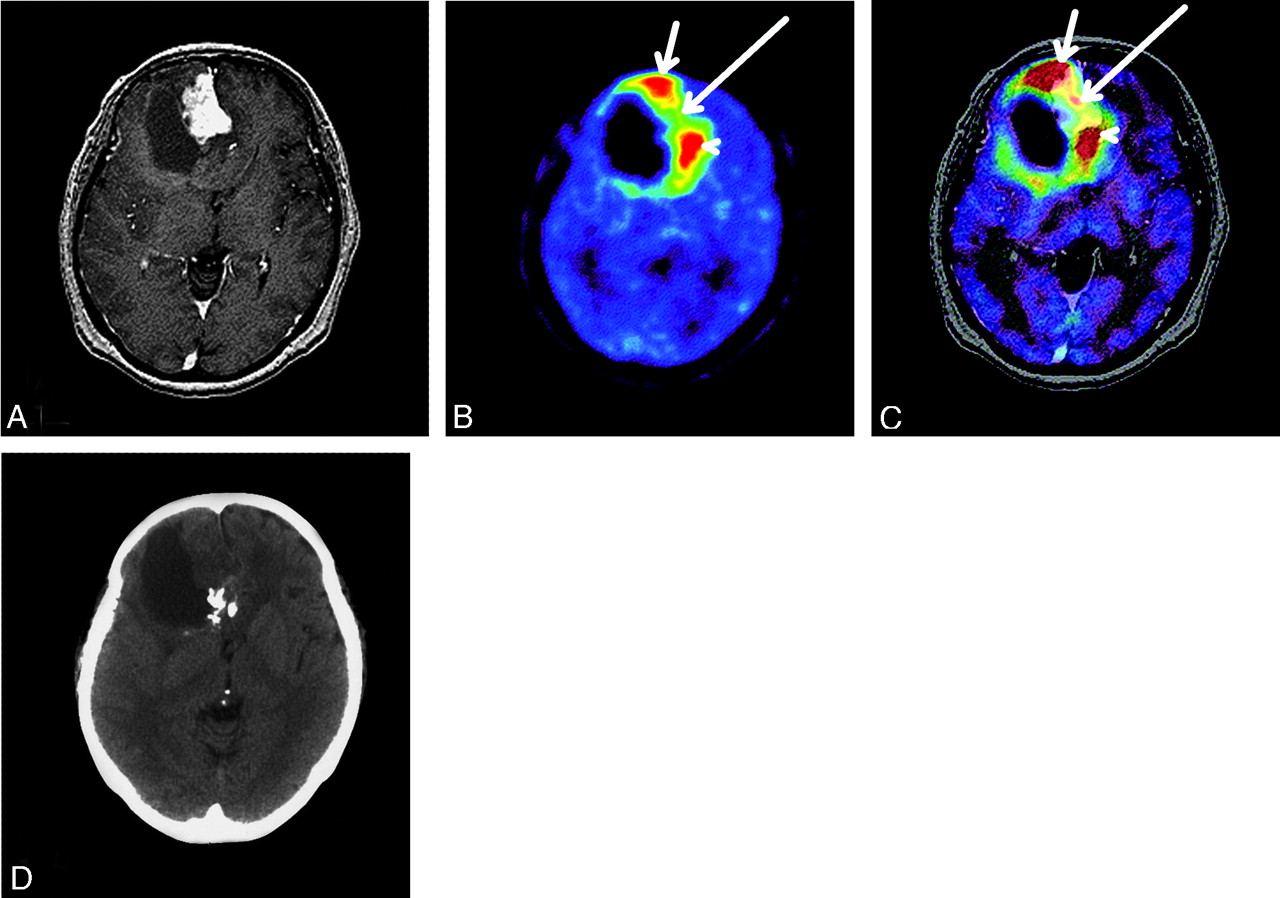
A 56-year-old woman was admitted to our hospital with a 2-month history of headache. CT demonstrated a cystic right frontal mass with calcification. A 1.5T MR imaging system (Signa Horizon LX; GE Healthcare, Waukesha, Wis) was used to obtain transaxial T1-weighted images. For contrast enhancement studies, 0.1 mmol/kg of body weight of gadolinium was injected intravenously. On MR imaging with gadolinium, an enhanced mass lesion with a large cyst was demonstrated in the right frontal lobe (Fig 1A). PET was carried out with an Advance NXi Imaging System (GE Medical Systems, Tokyo, Japan). After a 7-minute transmission scan was obtained, a dose of 370 MBq of MET was intravenously injected into a cubital vein within 1 minute. A 10-minute static PET scan was begun 20 minutes after MET injection. With MET-PET, high uptake of MET was demonstrated in the anterior right frontal lobe (Fig 1B, tumor area A: short arrow), moderate uptake of MET was demonstrated in the anteromedial right frontal lobe (Fig 1B, tumor area B: long arrow), and high uptake of MET was demonstrated in the posteromedial right frontal lobe (Fig 1B, tumor area C: arrowhead). MET uptake ratio was defined as the ratio of radioisotope count of MET in tumor to that in normal gray matter. MET uptake ratio was 3.9 for tumor area A, 2.1 for tumor area B, and 4.1 for tumor area C.

Fig 1.
  • Download figure
  • Open in new tab
  • Download powerpoint
Fig 1.

Imaging findings at initial imaging. A, Gadolinium-enhanced T1-weighted MR imaging demonstrates an enhanced mass lesion with a large cyst in the right frontal lobe. B, MET-PET image demonstrates intense high uptake of MET in the anterior right frontal lobe (tumor area A: short arrow), moderate uptake of MET in the anteromedial right frontal lobe (tumor area B: long arrow), and intense uptake of MET in the posteromedial right frontal lobe (tumor area C: arrowhead). C, MET-PET image coregistered with gadolinium-enhanced MR imaging. On coregistered images, tumor area B was intensively enhanced with gadolinium (long arrow), but tumor areas A and C had no gadolinium enhancement (short arrow and arrowhead, respectively). D, CT scans demonstrate a cystic right frontal mass with calcification. Tumor area C almost completely corresponded with the calcified lesion.

PET and MR imaging datasets were transferred to a SUN workstation (SPARC MP20; Sun Microsystems, Mountain View, Calif). Coregistration of MET-PET and MR imaging data used a commercial software package (Dr. View; Asahi Kasei Joho System, Tokyo, Japan). On coregistered images, tumor area B demonstrated intensive gadolinium enhancement; however, there was no enhancement in areas A or C (Fig 1C). Tumor area C corresponded largely with the calcified lesion seen on CT scans (Fig 1D).

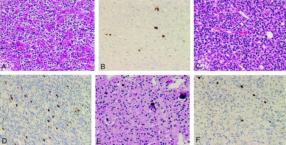
With reference to coregistered images, the tumor was totally removed with a neuronavigated surgical system (Brain LAB, Munich, Germany). Histologic examination was done with hematoxylin-eosin (H&E) staining. Within each tumor area, the degree of proliferative activity (Ki-67 index) was estimated. Tumor area A was a low-grade oligodendroglioma (Fig 2A). In tumor area B, histologic features consistent with high-grade (anaplastic) oligodendroglioma were evident (Fig 2C). Tumor area C was a low-grade oligodendroglioma with abundant calcification (Fig 2E). The Ki-67 index was 1.8% for area A, 7.6% for area B, and 2.2% for area C (Fig 2B, D, F). Histologic and neuroradiologic features for all of the areas are shown in the Table.

Fig 2.
  • Download figure
  • Open in new tab
  • Download powerpoint
Fig 2.

Histologic findings of tumor area A (A and B), tumor area B (C and D), and tumor area C (E and F). (H&E, ×200 [A, C, and E], MIB-1 immunostaining, hematoxylin counterstaining, ×200 [B, D, and F].) A, Tumor area A is moderately cellular and composed of cells with rounded, homogeneous nuclei and a swollen, clear cytoplasm (honeycomb appearance), consistent with low-grade oligodendroglioma. B, Tumor area A has MIB-1 positive cells and an MIB-1 labeling index of 1.8%. C, Tumor area B shows increased cellularity, marked cytological atypia, and high percentage of mitotic activity, consistent with high-grade (anaplastic) oligodendroglioma. D, Tumor area B shows frequent MIB-1 positive cells and an MIB-1 labeling index of 7.6%. E, Tumor area C is moderately cellular and composed of cells with rounded, homogeneous nuclei with calcification, consistent with low-grade oligodendroglioma. F, Tumor area C shows MIB-1 positive cells and an MIB-1 labeling index of 2.2%.

View this table:
  • View inline
  • View popup

Histologic and neuroradiologic features of tumor areas A, B, and C

Discussion

MET uptake by tumor tissue was first described in 1976 by Comar et al.6 Previous reports have shown that MET uptake by gliomas depends on histologic grade.1,3,7,8 Among glial tumors, oligodendrogliomas show high MET uptake.9 It has been suggested that oligodendroglial tumors have a high rate of cell turnover,10 which could enhance amino acid uptake and metabolism. High-grade oligodendrogliomas have been shown to have significantly higher MET uptake than low-grade tumors.9

However, no sophisticated correlation has been demonstrated between MET uptake and a histologic feature within an individual tumor that has combined low- and high-grade oligodendroglioma. In our patient, an exact local comparison of MET uptake and a histologic feature was performed using the neuronavigation system with reference to coregistered images. The oligodendroglioma in our case was separated in 3 tumor areas defined by different histologic features. We found a paradoxical result between MET uptake and histologic grade compared with previous studies. The lesion with higher MET uptake ratio (tumor areas A and C) had normal oligodendroglial cells, whereas the lesion with lower MET uptake ratio (tumor area B) was composed of anaplastic oligodendroglial cells. The increased Ki-67 index of tumor area B was consistent with the malignant features seen with H&E staining.

The reason for such a paradoxical result remains unclear, but we speculate that in a high-grade tumor, the cell is so deranged that its amino acid uptake mechanism no longer works and, hence, no MET uptake. On the other hand, anaplastic area was demonstrated with gadolinium-enhancement on MR imaging, which reflected disruption of the blood-brain barrier. We also suspect that MET uptake ratio does not always correlate with disruption of the blood-brain barrier. Only tumor area C demonstrated remarkable calcification independent of MET uptake ratio.

Conclusions

We demonstrated the case of relatively decreased MET uptake within the anaplastic component of a mixed-grade oligodendroglioma. Our exceptional case is interesting because of its value in elucidating the histologic and metabolic value of oligodendroglioma.

References

  1. ↵
    Bustany P, Chatel M, Derlon JM, et al. Brain tumor protein synthesis and histological grades: a study by positron emission tomography (PET) with C11-L-Methionine. J Neurooncol 1986;3:397–404
    CrossRefPubMed
  2. ↵
    Mineura K, Sasajima T, Suda Y, et al. Amino acid study of cerebral gliomas using positron emission tomography-analysis of (11C-methyl)-L-methionine uptake index. Neutrol Med Chir 1990;30:997–1002
  3. ↵
    Kaschten B, Stevenaert A, Sadzot B, et al. Preoperative evaluation of 54 gliomas by PET with fluorine-18-fluorodeoxyglucose and/or carbon-11-methionine. J Nucl Med 1998;39:778–85
    Abstract/FREE Full Text
  4. Ogawa T, Shishido F, Kanno I, et al. Cerebral glioma: evaluation with methionine PET. Radiology 1993;186:45–53
    PubMed
  5. ↵
    Ogawa T, Inugami A, Hatazawa J, et al. Clinical positron emission tomography for brain tumors: comparison of fludeoxyglucose F 18 and L-methyl-11C-methionine. AJNR Am J Neuroradiol 1996;17:345–53
    Abstract
  6. ↵
    Comar D, Cartron J, Mazière M, et al. Labeling and metabolism of methionine-methyl-11C. Eur J Nucl Med 1976;1:11–14
    PubMed
  7. ↵
    Derlon JM, Bourdet C, Bustany P, et al. [11C]L-methionine uptake in gliomas. Neurosurgery 1989;25:720–28
    CrossRefPubMed
  8. ↵
    Olivier DW, Ilan G, David W, et al. Positron emission tomography with injection of methionine as a prognostic factor of glioma. J Neurosurg 2001;95:746–50
    CrossRefPubMed
  9. ↵
    Derlon JM, Chapon F, Noël MH, et al. Non-invasive grading of oligodendrogliomas: correlation between in vivo metabolic pattern and histopathology. Eur J Nucl Med 2000;27:778–87
    CrossRefPubMed
  10. ↵
    Daumas-Duport C. Histological grading of gliomas. Curr Opin Neurol Neurosurg 1992;5:924–31
    PubMed
  • Received February 10, 2007.
  • Accepted after revision April 30, 2007.
  • Copyright © American Society of Neuroradiology
View Abstract
PreviousNext
Back to top

In this issue

American Journal of Neuroradiology: 28 (10)
American Journal of Neuroradiology
Vol. 28, Issue 10
November 2007
  • Table of Contents
  • Index by author
Advertisement
Print
Download PDF
Email Article

Thank you for your interest in spreading the word on American Journal of Neuroradiology.

NOTE: We only request your email address so that the person you are recommending the page to knows that you wanted them to see it, and that it is not junk mail. We do not capture any email address.

Enter multiple addresses on separate lines or separate them with commas.
Relatively Decreased 11C-Methionine Uptake within the Anaplastic Component of a Mixed-Grade Oligodendroglioma
(Your Name) has sent you a message from American Journal of Neuroradiology
(Your Name) thought you would like to see the American Journal of Neuroradiology web site.
CAPTCHA
This question is for testing whether or not you are a human visitor and to prevent automated spam submissions.
Cite this article
K. Miwa, J. Shinoda, H. Yano, T. Iwama
Relatively Decreased 11C-Methionine Uptake within the Anaplastic Component of a Mixed-Grade Oligodendroglioma
American Journal of Neuroradiology Nov 2007, 28 (10) 2005-2007; DOI: 10.3174/ajnr.A0695

Citation Manager Formats

  • BibTeX
  • Bookends
  • EasyBib
  • EndNote (tagged)
  • EndNote 8 (xml)
  • Medlars
  • Mendeley
  • Papers
  • RefWorks Tagged
  • Ref Manager
  • RIS
  • Zotero
0 Responses
Respond to this article
Share
Bookmark this article
Relatively Decreased 11C-Methionine Uptake within the Anaplastic Component of a Mixed-Grade Oligodendroglioma
K. Miwa, J. Shinoda, H. Yano, T. Iwama
American Journal of Neuroradiology Nov 2007, 28 (10) 2005-2007; DOI: 10.3174/ajnr.A0695
del.icio.us logo Twitter logo Facebook logo Mendeley logo
  • Tweet Widget
  • Facebook Like
  • Google Plus One
Purchase

Jump to section

  • Article
    • Abstract
    • Case Report
    • Discussion
    • Conclusions
    • References
  • Figures & Data
  • Info & Metrics
  • Responses
  • References
  • PDF

Related Articles

  • No related articles found.
  • PubMed
  • Google Scholar

Cited By...

  • No citing articles found.
  • Crossref (4)
  • Google Scholar

This article has been cited by the following articles in journals that are participating in Crossref Cited-by Linking.

  • Contributions of biological tumor parameters to the incorporation rate of l-[methyl-11C] methionine into astrocytomas and oligodendrogliomas
    Takeko Nojiri, Tadashi Nariai, Masaru Aoyagi, Michio Senda, Kenji Ishii, Kiichi Ishiwata, Kikuo Ohno
    Journal of Neuro-Oncology 2009 93 2
  • Detection of histological anaplasia in gliomas with oligodendroglial components using positron emission tomography with 18F-FDG and 11C-methionine: report of two cases
    Shigeru Yamaguchi, Hiroyuki Kobayashi, Kenji Hirata, Tohru Shiga, Shinya Tanaka, Junichi Murata, Shunsuke Terasaka
    Journal of Neuro-Oncology 2011 101 2
  • Diagnostic Imaging: Brain
    2016
  • Imaging in Neurology
    2016

More in this TOC Section

  • Usefulness of Quantitative Susceptibility Mapping for the Diagnosis of Parkinson Disease
  • White Matter Alterations in the Brains of Patients with Active, Remitted, and Cured Cushing Syndrome: A DTI Study
  • Qualitative and Quantitative Analysis of MR Imaging Findings in Patients with Middle Cerebral Artery Stroke Implanted with Mesenchymal Stem Cells
Show more BRAIN

Similar Articles

Advertisement

Indexed Content

  • Current Issue
  • Accepted Manuscripts
  • Article Preview
  • Past Issues
  • Editorials
  • Editor's Choice
  • Fellows' Journal Club
  • Letters to the Editor
  • Video Articles

Cases

  • Case Collection
  • Archive - Case of the Week
  • Archive - Case of the Month
  • Archive - Classic Case

Special Collections

  • AJNR Awards
  • ASNR Foundation Special Collection
  • Most Impactful AJNR Articles
  • Photon-Counting CT
  • Spinal CSF Leak Articles (Jan 2020-June 2024)

More from AJNR

  • Trainee Corner
  • Imaging Protocols
  • MRI Safety Corner

Multimedia

  • AJNR Podcasts
  • AJNR Scantastics

Resources

  • Turnaround Time
  • Submit a Manuscript
  • Submit a Video Article
  • Submit an eLetter to the Editor/Response
  • Manuscript Submission Guidelines
  • Statistical Tips
  • Fast Publishing of Accepted Manuscripts
  • Graphical Abstract Preparation
  • Imaging Protocol Submission
  • Evidence-Based Medicine Level Guide
  • Publishing Checklists
  • Author Policies
  • Become a Reviewer/Academy of Reviewers
  • News and Updates

About Us

  • About AJNR
  • Editorial Board
  • Editorial Board Alumni
  • Alerts
  • Permissions
  • Not an AJNR Subscriber? Join Now
  • Advertise with Us
  • Librarian Resources
  • Feedback
  • Terms and Conditions
  • AJNR Editorial Board Alumni

American Society of Neuroradiology

  • Not an ASNR Member? Join Now

© 2025 by the American Society of Neuroradiology All rights, including for text and data mining, AI training, and similar technologies, are reserved.
Print ISSN: 0195-6108 Online ISSN: 1936-959X

Powered by HighWire