Advertisement

Main menu

  • Home
  • Content
    • Current Issue
    • Accepted Manuscripts
    • Article Preview
    • Past Issue Archive
    • Video Articles
    • AJNR Case Collection
    • Case of the Week Archive
    • Case of the Month Archive
    • Classic Case Archive
  • Special Collections
    • AJNR Awards
    • ASNR Foundation Special Collection
    • Most Impactful AJNR Articles
    • Photon-Counting CT
    • Spinal CSF Leak Articles (Jan 2020-June 2024)
  • Multimedia
    • AJNR Podcasts
    • AJNR SCANtastic
    • Trainee Corner
    • MRI Safety Corner
    • Imaging Protocols
  • For Authors
    • Submit a Manuscript
    • Submit a Video Article
    • Submit an eLetter to the Editor/Response
    • Manuscript Submission Guidelines
    • Statistical Tips
    • Fast Publishing of Accepted Manuscripts
    • Graphical Abstract Preparation
    • Imaging Protocol Submission
    • Author Policies
  • About Us
    • About AJNR
    • Editorial Board
    • Editorial Board Alumni
  • More
    • Become a Reviewer/Academy of Reviewers
    • Subscribers
    • Permissions
    • Alerts
    • Feedback
    • Advertisers
    • ASNR Home

User menu

  • Alerts
  • Log in

Search

  • Advanced search
American Journal of Neuroradiology

American Journal of Neuroradiology

ASHNR American Society of Functional Neuroradiology ASHNR American Society of Pediatric Neuroradiology ASSR
  • Alerts
  • Log in

Advanced Search

  • Home
  • Content
    • Current Issue
    • Accepted Manuscripts
    • Article Preview
    • Past Issue Archive
    • Video Articles
    • AJNR Case Collection
    • Case of the Week Archive
    • Case of the Month Archive
    • Classic Case Archive
  • Special Collections
    • AJNR Awards
    • ASNR Foundation Special Collection
    • Most Impactful AJNR Articles
    • Photon-Counting CT
    • Spinal CSF Leak Articles (Jan 2020-June 2024)
  • Multimedia
    • AJNR Podcasts
    • AJNR SCANtastic
    • Trainee Corner
    • MRI Safety Corner
    • Imaging Protocols
  • For Authors
    • Submit a Manuscript
    • Submit a Video Article
    • Submit an eLetter to the Editor/Response
    • Manuscript Submission Guidelines
    • Statistical Tips
    • Fast Publishing of Accepted Manuscripts
    • Graphical Abstract Preparation
    • Imaging Protocol Submission
    • Author Policies
  • About Us
    • About AJNR
    • Editorial Board
    • Editorial Board Alumni
  • More
    • Become a Reviewer/Academy of Reviewers
    • Subscribers
    • Permissions
    • Alerts
    • Feedback
    • Advertisers
    • ASNR Home
  • Follow AJNR on Twitter
  • Visit AJNR on Facebook
  • Follow AJNR on Instagram
  • Join AJNR on LinkedIn
  • RSS Feeds

AJNR is seeking candidates for the AJNR Podcast Editor. Read the position description.

OtherPEDIATRICS

MR Imaging Features of Brain Stem Hypoplasia in Familial Horizontal Gaze Palsy and Scoliosis

A.V. dos Santos, S. Matias, P. Saraiva and A. Goulão
American Journal of Neuroradiology June 2006, 27 (6) 1382-1383;
A.V. dos Santos
  • Find this author on Google Scholar
  • Find this author on PubMed
  • Search for this author on this site
S. Matias
  • Find this author on Google Scholar
  • Find this author on PubMed
  • Search for this author on this site
P. Saraiva
  • Find this author on Google Scholar
  • Find this author on PubMed
  • Search for this author on this site
A. Goulão
  • Find this author on Google Scholar
  • Find this author on PubMed
  • Search for this author on this site
  • Article
  • Figures & Data
  • Info & Metrics
  • Responses
  • References
  • PDF
Loading

Abstract

Summary: We report the case of a child with horizontal gaze palsy, pendular nystagmus, and discrete thoracolumbar scoliosis. MR imaging of the brain depicted pons hypoplasia with an absence of the facial colliculi, hypoplasia, butterfly configuration of the medulla, and the presence of a deep midline pontine cleft (split pons sign). These MR imaging findings suggest familial horizontal gaze palsy with progressive kyphoscoliosis, a rare congenital disorder. To the best of our knowledge, MR imaging findings of only 4 similar cases, with or without progressive idiopathic scoliosis, have been reported. We discuss the pathogenesis substratum of this entity. Early recognition of this rare entity is important if supportive therapeutic measures in progressive scoliosis are to be applied.

Isolated malformations involving the pons and medulla oblongata are extremely rare. They usually occur associated with disorders of the cerebellum. Horizontal gaze palsy with progressive scoliosis (HGPPS) is a rare congenital disorder with autosomal recessive inherence, believed to result from cranial nuclear maldevelopment and characterized by absence of conjugate horizontal eye movements, preservation of vertical gaze and convergence, progressive scoliosis developing in childhood and adolescence, midline pontine cleft, butterfly configuration of the medulla, brain stem hypoplasia, and absence of facial colliculi.1 Although several clinical cases of brain stem hypoplasia with familial HGPPS have been published, Pieh et al and Rossi et al were the only authors to report on MR imaging findings of HGPPS.2,3 Congenital cleavage of the dorsal pons and medulla oblongata associated with horizontal gaze palsy without scoliosis in 2 children have also been reported.4,5

We report the clinical and imaging findings of one child with horizontal gaze palsy and congenital midsagittal cleft in the dorsal pons, discuss the pathogenesis of this rare entity, and hypothesize that early recognition of these clinical and imaging findings could help prevent severe scoliosis by enabling application of supportive therapeutic measures at an early phase of the disease.

Case Report

A 20-month-old boy born at term to healthy nonconsanguineous parents presented with hypotonia and abnormal eye movements since the neonatal period. Neurologic and ophthalmologic examinations revealed pendular nystagmus and total absence of horizontal gaze consistent with involvement of the abducens nucleus, without pseudobulbar or long tracts signs. Convergence and pupillary reflexes were normal. Discrete thoracolumbar scoliosis and slight mental retardation were also present. MR imaging findings revealed a hypoplastic pons with a midsagittal cleft extending ventrally from the fourth ventricular floor, generating a split axial pons sign on axial images and hypoplasia of the medulla oblongata with butterfly configuration (Figs 1 and 2). The fourth ventricular floor was tent shaped, and the facial colliculi were absent (Fig 3). The cerebral hemispheres, corpus callosum, and cerebellum were normal (Fig 4).

Fig 1.
  • Download figure
  • Open in new tab
  • Download powerpoint
Fig 1.

Sagittal T1-weighted image. Hypoplasia of the pons and medulla oblongata. Normal corpus callosum.

Fig 2.
  • Download figure
  • Open in new tab
  • Download powerpoint
Fig 2.

Axial T2-weighted image. Deep midsagittal cleft extends ventrally from the fourth ventricular floor originating the split pons sign.

Fig 3.
  • Download figure
  • Open in new tab
  • Download powerpoint
Fig 3.

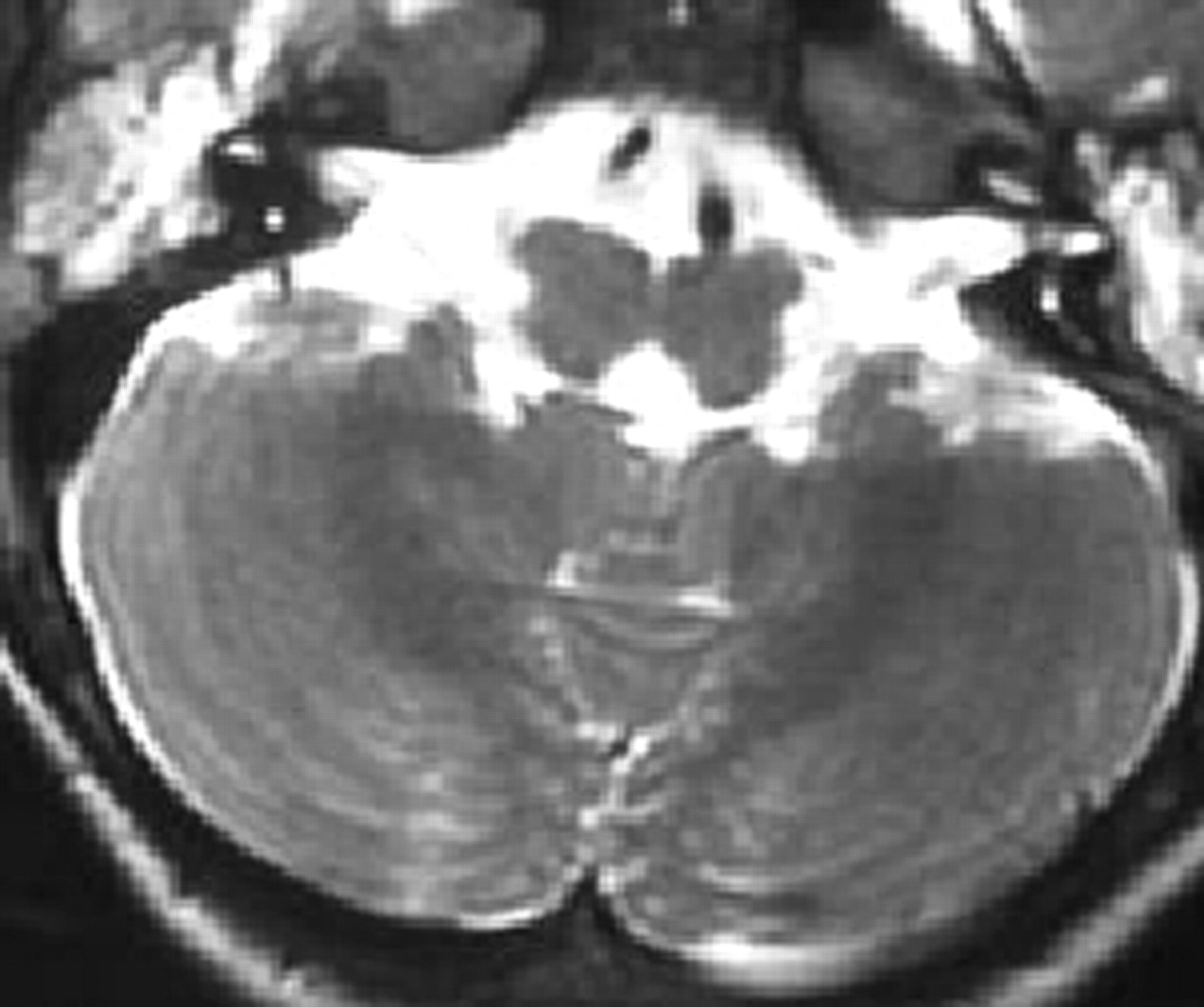
Axial T2-weighted image. Hypoplasia of the medulla oblongata with butterfly morphology.

Fig 4.
  • Download figure
  • Open in new tab
  • Download powerpoint
Fig 4.

Axial T2-weighted image. At the level of the pons, the floor of the fourth ventricle is flattened and has a tent shape. Absence of the facial colliculi is notable.

Discussion

Smooth horizontal gaze is a coordinated activity controlled by the abducens nucleus. This nucleus, located in the lower part of the pontine tegmentum at the level of the fourth ventricular floor, contains 2 types of neurons, one directly innervating the ipsilateral lateral rectus muscle and the other consisting of internuclear neurons that project through the medial longitudinal fasciculus (MLF) to the contralateral oculomotor nucleus, promoting the coordination of eye movement.

Facial colliculi, a paired prominence at the floor of the fourth ventricle, result from axons of the motor nuclei of the facial nerves passing dorsally to the abducens nuclei.

Absence of conjugate horizontal eye movement and facial colliculi not visible on axial MR imaging with intact facial, vestibulocochlear, and oculomotor nerves suggest selective agenesis of the abducens nuclei.

The split pons sign found on the axial MR imaging was similar to those previously reported by Rossi et al, Squirrel et al and by Nuri Sener.3–5 This could be due to the abnormal development of the abducens nuclei and medial longitudinal fasciculus occurring between gestational weeks 5 and 8 as the ventral furrow that indents the posterior metencephalon progressively disappears while the dorsomedial nuclei and tracts develop.

Progressive idiopathic scoliosis has been described in childhood. Although its pathogenesis is still poorly understood, a primary neurologic dysfunction involving the proprioceptive inputs mediated by the posterior column pathways of the spinal cord and medial lemniscus, the postural equilibrium, and the labyrinthine function mediated by the vestibular nuclei and the interplay of visual and vestibular reflexes mediated by the vestibular nuclei and the medial longitudinal fasciculus have been described as a possible explanation.3 In our case, the discrete scoliosis could be explained by the child’s age or by this being a genetic syndrome with variable expression.

Conditions such as Möbius syndrome and Duane retraction syndrome have similar clinical findings and should be included in the differential diagnosis. Abnormal development of the abducens nucleus plays a crucial role in the pathogenesis of both of these entities.

Möbius syndrome, a congenital disorder characterized by partial or complete facial diplegia and horizontal gaze palsy, with frequent involvement of other cranial nerves, results from agenesis or destruction of both the abducens and facial nuclei, possibly accompanied by abnormalities of other cranial nerve nuclei.5 Duane retraction syndrome is characterized by absent abduction, insufficient adduction, globe retraction, and palpebral narrowing during fixation,6 caused by selective absence of direct abducens nerve motoneurons with preservation of internuclear neurons visible.1 Aplasia of abducens nerve in Duane retraction syndrome and hypoplasia of both abducens and facial nuclei associated with hypoplasia of the caudal of the brain stem and absence of hypoglossal eminence in Möbius syndrome have been rarely reported as some of the imaging features of Duane retraction and Möbius syndromes.5,7 There are no reports of split pons sign associated with these syndromes. If this is the case, it can also help in the differential diagnosis of genetic eye motility disorders.

Conclusion

MR imaging may play a critical role on the precocious diagnosis of HGPPS by showing the maldevelopment of the dorsomedial brain stem structures and absence of the facial colliculi, believed to result from agenesis of the abducens nerves.

Early diagnosis of this congenital entity may be important to apply supportive therapies to prevent rapid progression of idiopathic scoliosis.

References

  1. 1.↵
    Engle EC, Leigh RJ. Genes, brainstem developmental, and eye movements. Neurology 2002;59:304–05
    FREE Full Text
  2. 2.↵
    Pieh C, Lengyel D, Neff A, et al. Brainstem hypoplasia in familial horizontal gaze palsy and scoliosis. Neurology 2002;59:462–63
    FREE Full Text
  3. 3.↵
    Rossi A, Catala M, Biancheri R, et al. MR imaging of brain-stem hypoplasia in horizontal gaze palsy with progressive scoliosis. AJNR Am J Neuroradiol 2004;25:1046–48
    Abstract/FREE Full Text
  4. 4.↵
    Nuri Sener R. Congenital cleft in the pontomedullary junction: case report. J Comp Assist Tomogr27:544–46
  5. 5.↵
    Squirrel DM, Griffiths PD, Burke JP. Congenital cleavage of dorsal pons and medulla. J Pediatr Ophtalmol Strabismus 2001;38:308–10
    PubMed
  6. 5.
    Pedraza S, Gamez J, Rovira A, et al. MRI findings in Mobius syndrome: correlation with clinical features. Neurology 2000;55:1058–60
    Abstract/FREE Full Text
  7. 6.↵
    Verzijl HTFM, van der Zwaag B, Cruysberg JRM, et al. Mobius syndrome redefined: a syndrome of rhombencephalic maldevelopmental. Neurology 2003;61:327–33
    Abstract/FREE Full Text
  8. 7.↵
    Ozkurt H, Basak M, Oral Y, et al. Magnetic resonance imaging in Duane’s retraction syndrome. J Pediatr Ophthalmol Strabismus 2003;40:19–22
    PubMed
  • Received February 16, 2005.
  • Accepted after revision April 23, 2005.
  • Copyright © American Society of Neuroradiology
View Abstract
PreviousNext
Back to top

In this issue

American Journal of Neuroradiology: 27 (6)
American Journal of Neuroradiology
Vol. 27, Issue 6
June 2006
  • Table of Contents
  • Index by author
Advertisement
Print
Download PDF
Email Article

Thank you for your interest in spreading the word on American Journal of Neuroradiology.

NOTE: We only request your email address so that the person you are recommending the page to knows that you wanted them to see it, and that it is not junk mail. We do not capture any email address.

Enter multiple addresses on separate lines or separate them with commas.
MR Imaging Features of Brain Stem Hypoplasia in Familial Horizontal Gaze Palsy and Scoliosis
(Your Name) has sent you a message from American Journal of Neuroradiology
(Your Name) thought you would like to see the American Journal of Neuroradiology web site.
CAPTCHA
This question is for testing whether or not you are a human visitor and to prevent automated spam submissions.
Cite this article
A.V. dos Santos, S. Matias, P. Saraiva, A. Goulão
MR Imaging Features of Brain Stem Hypoplasia in Familial Horizontal Gaze Palsy and Scoliosis
American Journal of Neuroradiology Jun 2006, 27 (6) 1382-1383;

Citation Manager Formats

  • BibTeX
  • Bookends
  • EasyBib
  • EndNote (tagged)
  • EndNote 8 (xml)
  • Medlars
  • Mendeley
  • Papers
  • RefWorks Tagged
  • Ref Manager
  • RIS
  • Zotero
0 Responses
Respond to this article
Share
Bookmark this article
MR Imaging Features of Brain Stem Hypoplasia in Familial Horizontal Gaze Palsy and Scoliosis
A.V. dos Santos, S. Matias, P. Saraiva, A. Goulão
American Journal of Neuroradiology Jun 2006, 27 (6) 1382-1383;
del.icio.us logo Twitter logo Facebook logo Mendeley logo
  • Tweet Widget
  • Facebook Like
  • Google Plus One

Jump to section

  • Article
    • Abstract
    • Case Report
    • Discussion
    • Conclusion
    • References
  • Figures & Data
  • Info & Metrics
  • Responses
  • References
  • PDF

Related Articles

  • No related articles found.
  • PubMed
  • Google Scholar

Cited By...

  • No citing articles found.
  • Crossref
  • Google Scholar

This article has not yet been cited by articles in journals that are participating in Crossref Cited-by Linking.

More in this TOC Section

  • SyMRI & MR Fingerprinting in Brainstem Myelination
  • Comparison of Image Quality and Radiation Dose in Pediatric Temporal Bone CT Using Photon-Counting Detector CT and Energy-Integrating Detector CT
  • Neonatal Hypocalcemia: Cerebral MRI Abnormalities
Show more Pediatrics

Similar Articles

Advertisement

Indexed Content

  • Current Issue
  • Accepted Manuscripts
  • Article Preview
  • Past Issues
  • Editorials
  • Editor's Choice
  • Fellows' Journal Club
  • Letters to the Editor
  • Video Articles

Cases

  • Case Collection
  • Archive - Case of the Week
  • Archive - Case of the Month
  • Archive - Classic Case

Special Collections

  • AJNR Awards
  • ASNR Foundation Special Collection
  • Most Impactful AJNR Articles
  • Photon-Counting CT
  • Spinal CSF Leak Articles (Jan 2020-June 2024)

More from AJNR

  • Trainee Corner
  • Imaging Protocols
  • MRI Safety Corner

Multimedia

  • AJNR Podcasts
  • AJNR Scantastics

Resources

  • Turnaround Time
  • Submit a Manuscript
  • Submit a Video Article
  • Submit an eLetter to the Editor/Response
  • Manuscript Submission Guidelines
  • Statistical Tips
  • Fast Publishing of Accepted Manuscripts
  • Graphical Abstract Preparation
  • Imaging Protocol Submission
  • Evidence-Based Medicine Level Guide
  • Publishing Checklists
  • Author Policies
  • Become a Reviewer/Academy of Reviewers
  • News and Updates

About Us

  • About AJNR
  • Editorial Board
  • Editorial Board Alumni
  • Alerts
  • Permissions
  • Not an AJNR Subscriber? Join Now
  • Advertise with Us
  • Librarian Resources
  • Feedback
  • Terms and Conditions
  • AJNR Editorial Board Alumni

American Society of Neuroradiology

  • Not an ASNR Member? Join Now

© 2025 by the American Society of Neuroradiology All rights, including for text and data mining, AI training, and similar technologies, are reserved.
Print ISSN: 0195-6108 Online ISSN: 1936-959X

Powered by HighWire