Advertisement

Main menu

  • Home
  • Content
    • Current Issue
    • Accepted Manuscripts
    • Article Preview
    • Past Issue Archive
    • Video Articles
    • AJNR Case Collection
    • Case of the Week Archive
    • Case of the Month Archive
    • Classic Case Archive
  • Special Collections
    • AJNR Awards
    • ASNR Foundation Special Collection
    • Most Impactful AJNR Articles
    • Photon-Counting CT
    • Spinal CSF Leak Articles (Jan 2020-June 2024)
  • Multimedia
    • AJNR Podcasts
    • AJNR SCANtastic
    • Trainee Corner
    • MRI Safety Corner
    • Imaging Protocols
  • For Authors
    • Submit a Manuscript
    • Submit a Video Article
    • Submit an eLetter to the Editor/Response
    • Manuscript Submission Guidelines
    • Statistical Tips
    • Fast Publishing of Accepted Manuscripts
    • Graphical Abstract Preparation
    • Imaging Protocol Submission
    • Author Policies
  • About Us
    • About AJNR
    • Editorial Board
    • Editorial Board Alumni
  • More
    • Become a Reviewer/Academy of Reviewers
    • Subscribers
    • Permissions
    • Alerts
    • Feedback
    • Advertisers
    • ASNR Home

User menu

  • Alerts
  • Log in

Search

  • Advanced search
American Journal of Neuroradiology

American Journal of Neuroradiology

ASHNR American Society of Functional Neuroradiology ASHNR American Society of Pediatric Neuroradiology ASSR
  • Alerts
  • Log in

Advanced Search

  • Home
  • Content
    • Current Issue
    • Accepted Manuscripts
    • Article Preview
    • Past Issue Archive
    • Video Articles
    • AJNR Case Collection
    • Case of the Week Archive
    • Case of the Month Archive
    • Classic Case Archive
  • Special Collections
    • AJNR Awards
    • ASNR Foundation Special Collection
    • Most Impactful AJNR Articles
    • Photon-Counting CT
    • Spinal CSF Leak Articles (Jan 2020-June 2024)
  • Multimedia
    • AJNR Podcasts
    • AJNR SCANtastic
    • Trainee Corner
    • MRI Safety Corner
    • Imaging Protocols
  • For Authors
    • Submit a Manuscript
    • Submit a Video Article
    • Submit an eLetter to the Editor/Response
    • Manuscript Submission Guidelines
    • Statistical Tips
    • Fast Publishing of Accepted Manuscripts
    • Graphical Abstract Preparation
    • Imaging Protocol Submission
    • Author Policies
  • About Us
    • About AJNR
    • Editorial Board
    • Editorial Board Alumni
  • More
    • Become a Reviewer/Academy of Reviewers
    • Subscribers
    • Permissions
    • Alerts
    • Feedback
    • Advertisers
    • ASNR Home
  • Follow AJNR on Twitter
  • Visit AJNR on Facebook
  • Follow AJNR on Instagram
  • Join AJNR on LinkedIn
  • RSS Feeds

AJNR is seeking candidates for the AJNR Podcast Editor. Read the position description.

OtherPEDIATRICS

Klippel-Feil Syndrome and Sprengel Deformity Combined with an Intraspinal Course of the Left Subclavian Artery and a Bovine Aortic Arch Variant

F. Floemer, O. Magerkurth, C. Jauckus, J. Lütschg and J.F. Schneider
American Journal of Neuroradiology February 2008, 29 (2) 306-307; DOI: https://doi.org/10.3174/ajnr.A0878
F. Floemer
  • Find this author on Google Scholar
  • Find this author on PubMed
  • Search for this author on this site
O. Magerkurth
  • Find this author on Google Scholar
  • Find this author on PubMed
  • Search for this author on this site
C. Jauckus
  • Find this author on Google Scholar
  • Find this author on PubMed
  • Search for this author on this site
J. Lütschg
  • Find this author on Google Scholar
  • Find this author on PubMed
  • Search for this author on this site
J.F. Schneider
  • Find this author on Google Scholar
  • Find this author on PubMed
  • Search for this author on this site
  • Article
  • Figures & Data
  • Info & Metrics
  • Responses
  • References
  • PDF
Loading

Abstract

SUMMARY: We present a case of Klippel-Feil syndrome and Sprengel deformity with a bovine aortic arch and an aberrant course of the left subclavian artery in a 14-year-old boy. CT and MR imaging of the neck and upper thorax demonstrated a cervical osseous segmentation anomaly, a left common carotid artery originating from the innominate artery, and a left subclavian artery coursing through the intraspinal space at the C6 through T1 level. Possible embryonic mechanisms and clinical significance of this variant are reviewed.

Klippel-Feil syndrome (KFS) is a heterogeneous entity that includes a large vascular variability. Exact evaluation of supra-aortic vessels before orthopedic correction has, therefore, been strongly recommended. Sprengel deformity (SD) has been readily associated with KFS, and both anomalies share many developmental and embryologic steps. In this setting, abnormal development of the fetal aortic arch and the vertebral or subclavian artery or both has been implicated as a causative factor in both syndromes. Nevertheless, an association between KFS, SD, and an aberrant intraspinal course of the left subclavian artery has, to the best of our knowledge, never been described. This variation, if not duly recognized, may have devastating consequences during surgery. Chronic intraspinal pulsation of this vessel, on the other hand, may eventually lead to myelopathic changes due to its proximity to the cervical spinal cord.

Case Report

A 14-year-old boy with known KFS clinically presented with repetitive episodes of severe occipital headache, vertigo, and nausea combined with neck and shoulder discomfort, which lasted for weeks. Clinical examination showed a markedly reduced vertebral range of motion. An SD and fusion of the left ribs 1–3 were known. Sonography demonstrated a prominent right vertebral artery without stenosis. Blood pressure was normal. Contrast-enhanced CT of the head, neck, and upper thorax was performed to exclude cranial abnormalities and cervical fracture.

Findings of cranial CT were normal. Fusion of the left side of the odontoid process and the lateral mass of C2 with the lateral mass of C1 and a bifid anterior and posterior C1 arch were noted. Multisegmental left-sided hemivertebral fusions were present at the levels C5 through T3 with a bifid spinous process, which led to a right-sided cervicothoracic angulation and scoliosis.

Vessel status revealed a so-called bovine aortic arch variant. The first branch of the aortic arch was a common trunk, comprising the innominate artery and the left common carotid artery (Fig 1A). The second branch was the left subclavian artery. The first segment of this vessel showed a posterior course, entering the spinal canal at T1 (Fig 1A, -B) with a horizontal course through the left epidural space, exiting posteriorly through an osseous defect of the left posterior arch at the same level (Fig 2A, -B) before resuming its normal trajectory within the supraclavicular space. The left vertebral artery originated from the left subclavian artery just before its intraspinal penetrance and showed a normal cranial continuation after entering the left transverse foramen at the level of C6. The U-shaped loop of the subclavian artery due to its intraspinal course was clearly depicted. There was no spinal canal stenosis or displacement of the cervical cord (Fig 2B). On a follow-up MR imaging, vascular anomalies were confirmed but the spinal cord itself revealed no signal-intensity anomaly, hemorrhagic changes, or evidence of myelopathy.

Fig 1.
  • Download figure
  • Open in new tab
  • Download powerpoint
Fig 1.

A and B, Volume rendering reconstructions with anteroposterior view (A) and left-lateral view (B) showing a bovine aortic arch variant (white arrow) and a U-shaped dorsal running loop of the left subclavian artery. The posterior course of the proximal part of the left subclavian artery entering the intraspinal space through the left-sided fused vertebrae at the T1 level with the same-level dorsal exiting is clearly seen.

Fig 2.
  • Download figure
  • Open in new tab
  • Download powerpoint
Fig 2.

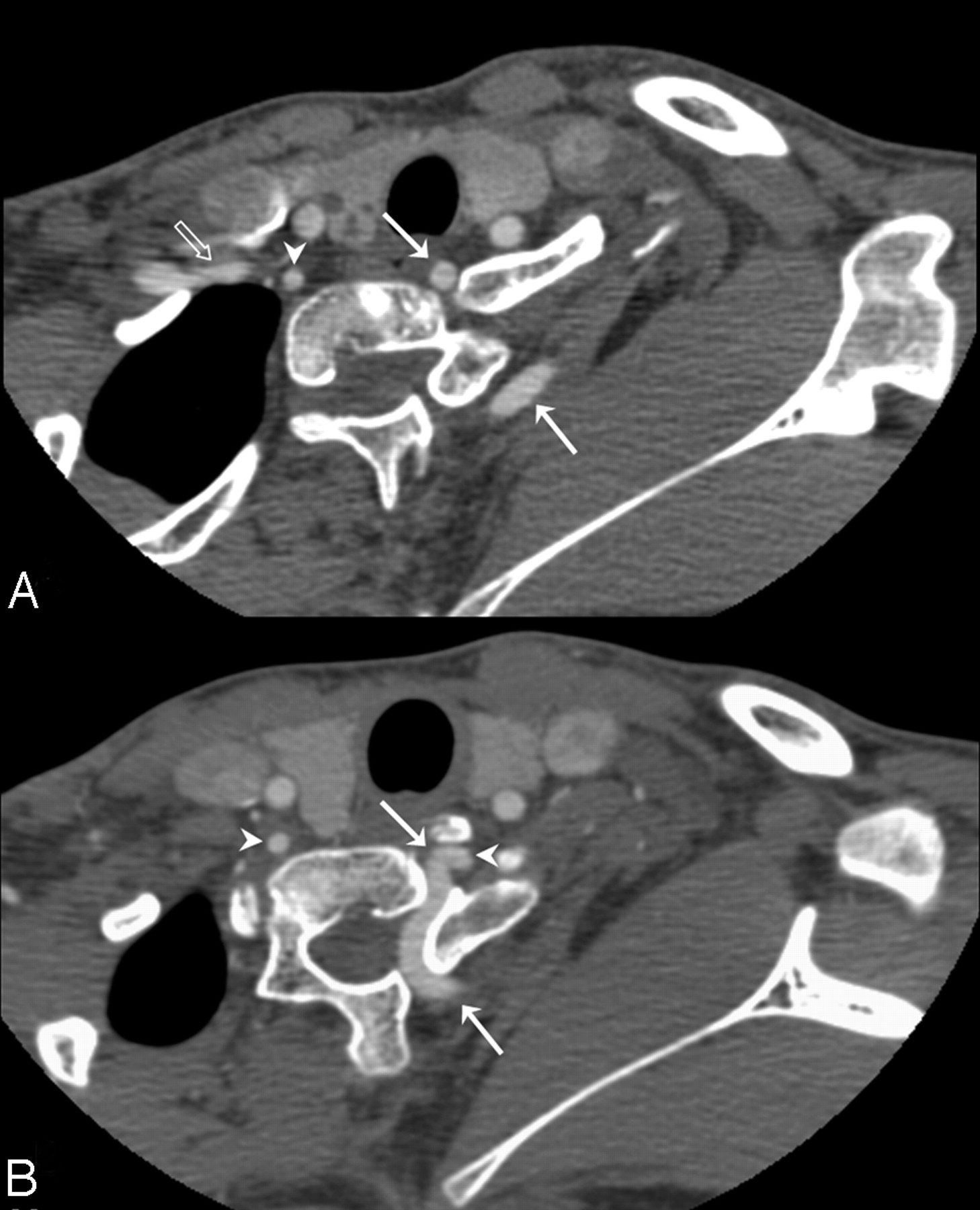
A and B, Axial multisection CT reference images. Arrowheads point to the vertebral artery of the right and left side. The transparent arrow shows the right subclavian artery. A, Anterior and posterior part of the left subclavian artery (white arrows). B, Origin of the left vertebral artery (white arrowheads) contralateral to the normal course of the right vertebral artery.

Discussion

In 1912, Maurice Klippel and Andre Feil first described a patient presenting an association of short neck, low posterior hairline, and limited range of motion. The complete clinical triad is seen in <50% of the cases, and many patients with KFS demonstrate additional developmental anomalies. Therefore, KFS is diagnosed by the presence of 2 or more nonsegmented cervical vertebrae alone.1

KFS is reported to be present in 1 of 42,000 individuals, and 57%–70% of all patients are female. Various classification schemes were developed. KFS was classically categorized into 3 groups, depending on the level and extent of vertebral fusion. Genetic analysis revealed an autosomal recessive trait for types I and III and an autosomal dominant trait for type II. Subsequent studies incorporated the mode of inheritance with the location of the most rostral vertebral fusion. This system includes 4 groups with different autosomal dominant or recessive or X-linked traits.

Anomalies of the aortic arch, carotid arteries, subclavian artery, vertebral artery, and a persistent trigeminal artery are known in association with KFS.2–6

SD has been found in 7%–42% of the patients with KFS. The scapula develops from the paraxial mesoderm and is, therefore, closely associated with the development of the cervical spine. Patients with KFS and SD show more congenital fused segments, especially at the C6 through C7 level.7

Cervical spine development starts with gastrulation in the fourteenth week by formation of the 3 germ cell layers and the notochord. Between days 20–30, the paraxial mesoderm subdivides into somites, which divide into sclerotomes and dermomyotomes. Sclerotomes resegment into a cranial and a caudal area, and in between forms the intervertebral disk. Fusion of the caudal section from a rostral somite with the cranial section of the corresponding caudal somite forms a vertebral body. Errors in segmentation may result in KFS. Vascular system development runs parallel to the development of the spinal cord. At days 21–29, thirty pairs of dorsal intersegmental arteries arise from the dorsal aortae and supply their corresponding somites. Between days 32–42, vertical and right-angle anastomoses develop between them and cross the vertebral bodies. The anastomoses serve as the origin of the vertebral arteries. During gestational days 37–42, the first 6 cervical intersegmental arteries regress, and only the seventh intersegmental arteries persist. The entire left and the greater part of the right subclavian artery originate from this seventh intersegmental artery.

Fetal vascular disruption disorders have been found to be responsible for various congenital anomalies, depending on the extent and timing of the disruptive event. A subclavian artery supply disruption sequence has been hypothesized to result in KFS on the basis of the theory that vascular disruption of the vertebral artery leads to ischemia during morphogenesis. Subsequent structural anomalies will involve not only the definitive vascular pattern by itself but also the vasculature-dependent soft-tissue territories.8,9 Due to the heterogeneity in KFS, the exact influence of vascular and genetic factors remains unclear. Because of the complex vascular variants in this group of patients, prior imaging of the aortic arch and supra-aortic vessels is recommended.10

References

  1. ↵
    Tracy MR, Dormans JP, Kusumi K. Klippel-Feil Syndrome. Clin Orthop Relat Res 2004;424:183–90
    CrossRefPubMed
  2. ↵
    Abbas J, Nazzal M, Serrano P, et al. Aortic arch abnormality in a patient with Klippel-Feil syndrome. Vascular 2006;14:43–46
    Abstract/FREE Full Text
  3. Layton KF, Kallmes DF, Cloft HJ, et al. Bovine aortic arch variant in humans: clarification of a common misnomer. AJNR Am J Neuroradiol 2006;27:1541–42
    Abstract/FREE Full Text
  4. Gailloud P, Murphy KJ, Rigamonti D. Bilateral thoracic bifurcation of the common carotid artery associated with Klippel-Feil anomaly. AJNR Am J Neuroradiol 2000;21:941–44
    Abstract/FREE Full Text
  5. Hasan I, Wapnick S, Kutscher ML, et al. Vertebral artery dissection associated with Klippel-Feil syndrome in a child. Child’s Nerv Syst 2002;18:67–70
    CrossRefPubMed
  6. ↵
    Paksoy Y, Seker M, Kalkan E. Klippel-Feil syndrome associated with persistent trigeminal artery. Spine 2004;29:193–99
    CrossRefPubMed
  7. ↵
    Samartzis D, Herman J, Lubicky JP, et al. Sprengel’s deformity in Klippel-Feil syndrome. Spine 2007;32:512–16
    CrossRefPubMed
  8. ↵
    Bavinck JN, Weaver DD. Subclavian artery supply disruption sequence: hypothesis of a vascular etiology for Poland, Klippel-Feil, and Möbius anomalies. Am J Med Genet 1986;23:903–18
    CrossRefPubMed
  9. ↵
    Brill CB, Peyster RG, Keller MS, et al. Isolation of the right subclavian artery with subclavian steal in a child with Klippel-Feil anomaly: an example of the subclavian artery supply disruption sequence. Am J Med Genet 1987;26:933–40
    CrossRefPubMed
  10. ↵
    Paul I, Badmanaban B, Campalani G. Patients with Klippel-Feil syndrome should have imaging of the subclavian artery and its branches prior to coronary artery bypass grafting. Interact Cardiovasc Thorac Surg 2007;6:403–04. Epub 2007 Feb 8
    Abstract/FREE Full Text
  • Received October 3, 2007.
  • Accepted after revision October 12, 2007.
  • Copyright © American Society of Neuroradiology
View Abstract
PreviousNext
Back to top

In this issue

American Journal of Neuroradiology: 29 (2)
American Journal of Neuroradiology
Vol. 29, Issue 2
February 2008
  • Table of Contents
  • Index by author
Advertisement
Print
Download PDF
Email Article

Thank you for your interest in spreading the word on American Journal of Neuroradiology.

NOTE: We only request your email address so that the person you are recommending the page to knows that you wanted them to see it, and that it is not junk mail. We do not capture any email address.

Enter multiple addresses on separate lines or separate them with commas.
Klippel-Feil Syndrome and Sprengel Deformity Combined with an Intraspinal Course of the Left Subclavian Artery and a Bovine Aortic Arch Variant
(Your Name) has sent you a message from American Journal of Neuroradiology
(Your Name) thought you would like to see the American Journal of Neuroradiology web site.
CAPTCHA
This question is for testing whether or not you are a human visitor and to prevent automated spam submissions.
Cite this article
F. Floemer, O. Magerkurth, C. Jauckus, J. Lütschg, J.F. Schneider
Klippel-Feil Syndrome and Sprengel Deformity Combined with an Intraspinal Course of the Left Subclavian Artery and a Bovine Aortic Arch Variant
American Journal of Neuroradiology Feb 2008, 29 (2) 306-307; DOI: 10.3174/ajnr.A0878

Citation Manager Formats

  • BibTeX
  • Bookends
  • EasyBib
  • EndNote (tagged)
  • EndNote 8 (xml)
  • Medlars
  • Mendeley
  • Papers
  • RefWorks Tagged
  • Ref Manager
  • RIS
  • Zotero
0 Responses
Respond to this article
Share
Bookmark this article
Klippel-Feil Syndrome and Sprengel Deformity Combined with an Intraspinal Course of the Left Subclavian Artery and a Bovine Aortic Arch Variant
F. Floemer, O. Magerkurth, C. Jauckus, J. Lütschg, J.F. Schneider
American Journal of Neuroradiology Feb 2008, 29 (2) 306-307; DOI: 10.3174/ajnr.A0878
del.icio.us logo Twitter logo Facebook logo Mendeley logo
  • Tweet Widget
  • Facebook Like
  • Google Plus One
Purchase

Jump to section

  • Article
    • Abstract
    • Case Report
    • Discussion
    • References
  • Figures & Data
  • Info & Metrics
  • Responses
  • References
  • PDF

Related Articles

  • No related articles found.
  • PubMed
  • Google Scholar

Cited By...

  • No citing articles found.
  • Crossref (19)
  • Google Scholar

This article has been cited by the following articles in journals that are participating in Crossref Cited-by Linking.

  • Congenital high scapula (Sprengel&#039;s deformity): Four cases
    R. Guillaume, E. Nectoux, J. Bigot, L. Vandenbussche, D. Fron, A. Mézel, B. Herbaux, N. Boutry
    Diagnostic and Interventional Imaging 2012 93 11
  • Bilateral Multilevel Cervical Rib and Bilateral Omovertebra in Klippel-Feil Syndrome
    Serap Satış, Nur Alparslan, Mustafa Tuna, Osman Dere, Alparslan Yetişgin
    World Neurosurgery 2020 136
  • Severe Sprengel deformity associated with Klippel–Feil syndrome and a complex vascular abnormality that determined the corrective surgery technique
    Cesar G. Fontecha, Ester Navarro Cano, Francisco Soldado, Ignasi Barber
    Journal of Pediatric Orthopaedics B 2014 23 6
  • Surgical management of Sprengel’s deformity by a modification of Green’s procedure
    Yu Jiang, Yuan Guo, Zhenghua Zhu, Yukun Wang
    Der Orthopäde 2020 49 3
  • Functional improvement in patients with Sprengel’s deformity following Modified Green’s procedure and simplified clavicle osteotomy—a study of forty cases
    Premal Naik, Hitesh Chauhan
    International Orthopaedics 2020 44 12
  • Juvenile Bow Hunter&#039;s Stroke without Hemodynamic Changes
    Kozue Saito, Makito Hirano, Toshiaki Taoka, Hiroyuki Nakagawa, Takanori Kitauchi, Masanori Ikeda, Emi Tanizawa, Kimihiko Kichikawa, Satoshi Ueno
    Clinical Medicine Insights: Case Reports 2010 3
  • Congenital deformation of the posterior arch of the atlas: Subluxation of the atlanto-axial joint with temporary quadriplegia
    Tobias M Ballhause, Mirko Velickovic, Darius M Thiesen, Marc Dreimann
    SAGE Open Medical Case Reports 2019 7
  • A case of coexistence of multiple vascular anomalies including the absence of a left internal carotid artery, a left vertebral artery arising from the subclavian artery in a high cervical location, and a bovine arch associated with vertebral fusion
    Tamaki Ichikawa, Shuichi Kawada, Takashi Okazaki, Kento Yokoyama, Makiko Kobayashi, Hiroyuki Katoh, Masahiko Watanabe, Jun Hashimoto
    Radiology Case Reports 2024 19 11
  • Anomalous vasculature in the neck diagnosed by ultrasound
    Sarah Constantine, Denise Roach
    Australasian Journal of Ultrasound in Medicine 2020 23 4
  • Die Scapula
    Lars-J. Lehmann
    2015

More in this TOC Section

  • SyMRI & MR Fingerprinting in Brainstem Myelination
  • Comparison of Image Quality and Radiation Dose in Pediatric Temporal Bone CT Using Photon-Counting Detector CT and Energy-Integrating Detector CT
  • Neonatal Hypocalcemia: Cerebral MRI Abnormalities
Show more Pediatrics

Similar Articles

Advertisement

Indexed Content

  • Current Issue
  • Accepted Manuscripts
  • Article Preview
  • Past Issues
  • Editorials
  • Editor's Choice
  • Fellows' Journal Club
  • Letters to the Editor
  • Video Articles

Cases

  • Case Collection
  • Archive - Case of the Week
  • Archive - Case of the Month
  • Archive - Classic Case

Special Collections

  • AJNR Awards
  • ASNR Foundation Special Collection
  • Most Impactful AJNR Articles
  • Photon-Counting CT
  • Spinal CSF Leak Articles (Jan 2020-June 2024)

More from AJNR

  • Trainee Corner
  • Imaging Protocols
  • MRI Safety Corner

Multimedia

  • AJNR Podcasts
  • AJNR Scantastics

Resources

  • Turnaround Time
  • Submit a Manuscript
  • Submit a Video Article
  • Submit an eLetter to the Editor/Response
  • Manuscript Submission Guidelines
  • Statistical Tips
  • Fast Publishing of Accepted Manuscripts
  • Graphical Abstract Preparation
  • Imaging Protocol Submission
  • Evidence-Based Medicine Level Guide
  • Publishing Checklists
  • Author Policies
  • Become a Reviewer/Academy of Reviewers
  • News and Updates

About Us

  • About AJNR
  • Editorial Board
  • Editorial Board Alumni
  • Alerts
  • Permissions
  • Not an AJNR Subscriber? Join Now
  • Advertise with Us
  • Librarian Resources
  • Feedback
  • Terms and Conditions
  • AJNR Editorial Board Alumni

American Society of Neuroradiology

  • Not an ASNR Member? Join Now

© 2025 by the American Society of Neuroradiology All rights, including for text and data mining, AI training, and similar technologies, are reserved.
Print ISSN: 0195-6108 Online ISSN: 1936-959X

Powered by HighWire