Advertisement

Main menu

  • Home
  • Content
    • Current Issue
    • Accepted Manuscripts
    • Article Preview
    • Past Issue Archive
    • Video Articles
    • AJNR Case Collection
    • Case of the Week Archive
    • Case of the Month Archive
    • Classic Case Archive
  • Special Collections
    • AJNR Awards
    • ASNR Foundation Special Collection
    • Most Impactful AJNR Articles
    • Photon-Counting CT
    • Spinal CSF Leak Articles (Jan 2020-June 2024)
  • Multimedia
    • AJNR Podcasts
    • AJNR SCANtastic
    • Trainee Corner
    • MRI Safety Corner
    • Imaging Protocols
  • For Authors
    • Submit a Manuscript
    • Submit a Video Article
    • Submit an eLetter to the Editor/Response
    • Manuscript Submission Guidelines
    • Statistical Tips
    • Fast Publishing of Accepted Manuscripts
    • Graphical Abstract Preparation
    • Imaging Protocol Submission
    • Author Policies
  • About Us
    • About AJNR
    • Editorial Board
    • Editorial Board Alumni
  • More
    • Become a Reviewer/Academy of Reviewers
    • Subscribers
    • Permissions
    • Alerts
    • Feedback
    • Advertisers
    • ASNR Home

User menu

  • Alerts
  • Log in

Search

  • Advanced search
American Journal of Neuroradiology

American Journal of Neuroradiology

ASHNR American Society of Functional Neuroradiology ASHNR American Society of Pediatric Neuroradiology ASSR
  • Alerts
  • Log in

Advanced Search

  • Home
  • Content
    • Current Issue
    • Accepted Manuscripts
    • Article Preview
    • Past Issue Archive
    • Video Articles
    • AJNR Case Collection
    • Case of the Week Archive
    • Case of the Month Archive
    • Classic Case Archive
  • Special Collections
    • AJNR Awards
    • ASNR Foundation Special Collection
    • Most Impactful AJNR Articles
    • Photon-Counting CT
    • Spinal CSF Leak Articles (Jan 2020-June 2024)
  • Multimedia
    • AJNR Podcasts
    • AJNR SCANtastic
    • Trainee Corner
    • MRI Safety Corner
    • Imaging Protocols
  • For Authors
    • Submit a Manuscript
    • Submit a Video Article
    • Submit an eLetter to the Editor/Response
    • Manuscript Submission Guidelines
    • Statistical Tips
    • Fast Publishing of Accepted Manuscripts
    • Graphical Abstract Preparation
    • Imaging Protocol Submission
    • Author Policies
  • About Us
    • About AJNR
    • Editorial Board
    • Editorial Board Alumni
  • More
    • Become a Reviewer/Academy of Reviewers
    • Subscribers
    • Permissions
    • Alerts
    • Feedback
    • Advertisers
    • ASNR Home
  • Follow AJNR on Twitter
  • Visit AJNR on Facebook
  • Follow AJNR on Instagram
  • Join AJNR on LinkedIn
  • RSS Feeds

AJNR is seeking candidates for the AJNR Podcast Editor. Read the position description.

OtherHEAD & NECK

Atypical Cystic Meningioma of the Trigeminal Nerve in a Pediatric Patient

K. Obeng, Z. Rumboldt, G. Tuite, C.T. Welsh, S. Patel and M.V. Spampinato
American Journal of Neuroradiology February 2008, 29 (2) 398-399; DOI: https://doi.org/10.3174/ajnr.A0876
K. Obeng
  • Find this author on Google Scholar
  • Find this author on PubMed
  • Search for this author on this site
Z. Rumboldt
  • Find this author on Google Scholar
  • Find this author on PubMed
  • Search for this author on this site
G. Tuite
  • Find this author on Google Scholar
  • Find this author on PubMed
  • Search for this author on this site
C.T. Welsh
  • Find this author on Google Scholar
  • Find this author on PubMed
  • Search for this author on this site
S. Patel
  • Find this author on Google Scholar
  • Find this author on PubMed
  • Search for this author on this site
M.V. Spampinato
  • Find this author on Google Scholar
  • Find this author on PubMed
  • Search for this author on this site
  • Article
  • Figures & Data
  • Info & Metrics
  • Responses
  • References
  • PDF
Loading

Abstract

SUMMARY: We report an unusual case of atypical cystic meningioma of the trigeminal nerve proved by histology in a 15-year-old white girl. A review of the literature showed that this is only the second reported case of a meningioma of the trigeminal nerve without dural attachment and the first occurrence in a pediatric patient.

Primary tumors of the trigeminal nerve and Meckel cave represent less than 0.5% of intracranial tumors.1 We report the case of a dumbbell-shaped atypical meningioma of the trigeminal nerve without dural attachment, involving the Meckel cave and the posterior fossa in an adolescent.

Case Report

A 15-year-old white girl presented with a 2-month history of left-sided facial numbness and loss of sensation of the left hemitongue. Physical examination showed hypoesthesia in the left V1, V2, and V3 dermatome distribution and absent left corneal reflex. She had no family history of neurofibromatosis, and physical examination revealed no evidence of café au lait spots or freckling in the axillary or inguinal area. Aside from the absence of a left corneal reflex, the findings of the ophthalmologic examination were unremarkable, with no evidence of lens abnormalities.

A brain MR imaging revealed an extra-axial dumbbell-shaped cystic mass, without obvious “dural tail” or dural attachment, demonstrating peripheral rim enhancement (Fig 1), with a cystic component in the posterior fossa at the root entry zone of the left trigeminal nerve, and a second cystic lesion with a fluid-fluid level at the level of the left Meckel cave. The mass was thought to be consistent with a cystic schwannoma of the left trigeminal nerve. There were no masses along the course of the 8 cranial nerves.

Fig 1.
  • Download figure
  • Open in new tab
  • Download powerpoint
Fig 1.

A–C, Axial T2-weighted (A and B) and axial postcontrast T1-weighted (C) images demonstrate a dumbbell-shaped extra-axial mass in the Meckel cave (short arrows) and in the left prepontine cistern (long arrows).

A 1-stage surgical excision of the middle and posterior fossa components of the mass was planned. The patient underwent a left-sided skull base craniotomy, with a left temporal craniotomy and zygomatic osteotomy, by using an extradural approach. At surgery, it was noted that the mass arose from the left trigeminal nerve, did not have a dural attachment, and was heavily intermixed with the surrounding trigeminal nerve. Surgical resection of the mass was thought to be complete.

The resected gross specimen consisted of multiple fragments of brown-red variegated tissue. Histology (Fig 2) showed concentric structures typical of meningioma, hypercellularity, mitotically active foci, and focal necrosis, making it atypical. Embedded nerve fascicles of the excised trigeminal nerve appeared infiltrated by tumor. Reticulin stain showed chiefly perivascular staining in the tumor, highlighting its vascularity. A final diagnosis of atypical meningioma was made.

Fig 2.
  • Download figure
  • Open in new tab
  • Download powerpoint
Fig 2.

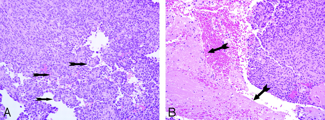
A, The concentric structures (arrows) typical of a meningioma are depicted. Small round-to-oval nuclei with inconspicuous nucleoli are present (hematoxylin-eosin [HE] stain, original magnification ×20). B, Necrosis (arrows) is also a prominent feature (HE stain, original magnification ×20).

Given the histology and the location of the neoplasm, we planned to treat residual or recurrent tumor with stereotactic radiosurgery. Since surgery, a postoperative MR imaging and subsequent follow-up MR imaging performed every 2 months in the last 4 months did not demonstrate evidence of macroscopic residual or recurrent tumor. Findings of an MR imaging of the cervical spine performed 2 months after surgery were normal, with no evidence of intra- or extradural masses.

Discussion

Approximately one third of all primary tumors of the Meckel cave are schwannomas, with the remainder consisting of meningiomas, epidermoids, dermoids, lipomas, metastases, hemangiomas, and lymphomas.1,2 Although very few pediatric cases of trigeminal schwannomas have been reported in the literature,3,4 to our knowledge, this is the first case of an atypical meningioma along the trigeminal nerve in a pediatric patient.

Meningiomas of the Meckel cave are rare and account for approximately 1% of all intracranial meningiomas.5,6 The Meckel cave is a dural recess in the posterior medial portion of the middle cranial fossa, containing the trigeminal ganglion. Meningiomas of the Meckel cave typically originate from the dura mater, compress the trigeminal ganglion, and can extend toward the middle cranial fossa or through the porus trigeminus into the posterior fossa in a dumbbell-shaped configuration.2

Meningiomas generally derive from dural-based arachnoid cap cells; however, intracranial meningiomas without dural attachment occur in the ventricles, Sylvian fissure, pineal region, and even in the brain parenchyma.7 The average age at diagnosis of patients affected by meningiomas without dural attachment is 26.5 years, approximately 20 years younger than the average age at diagnosis of meningiomas in general, with a male-to-female ratio of meningiomas without dural attachment of 1.39:1.7 The diagnosis of meningioma at an early age should raise the possibility of neurofibromatosis type 2. Our patient did not fulfill the diagnostic criteria for neurofibromatosis type 1 or 2.8

Atypical meningiomas (World Health Organization grade II/IV) exhibit a more aggressive behavior and an increased risk of recurrence after treatment.9 Most of the previously reported atypical meningiomas without dural attachment were intraventricular, with the exception of a single case of intraparenchymal atypical meningioma in a 16-year-old boy.7 A review of the literature yielded a single documented case of trigeminal nerve meningioma without attachment to the dura in a 69-year-old woman, but this lesion had no atypical features.10

In our paitent and in the case previously described by Fujimoto et al,10 the tumor most likely originated from arachnoid cap cells within the left trigeminal nerve sheath or from the perineural cells of the nerve and not from the dura of the Meckel cave.10 In both cases, the trigeminal nerve meningiomas had a cystic appearance and involved the cisternal portion of the nerve and Meckel cave. Therefore, meningiomas of the trigeminal nerve, though rare, should be included in the differential diagnosis of cystic dumbbell-shaped trigeminal nerve tumors. To our knowledge, this is the first reported atypical meningioma originating from the trigeminal nerve in a pediatric patient and the second documented case of a meningioma without dural attachment arising from the trigeminal nerve.

References

  1. ↵
    Yuh WT, Wright DC, Barloon TJ, et al. MR imaging of primary tumors of trigeminal nerve and Meckel cave. AJR Am J Roentgenol 1988;151:577–82
    PubMed
  2. ↵
    Kapila A, Chakeres DW, Blanco E. The Meckel cave: computed tomographic study. Part I. Normal anatomy. Part II. Pathology. Radiology 1984;152:425–33
    PubMed
  3. ↵
    Ross DL, Tew JM Jr, Benton C, et al. Trigeminal schwannoma in a child. Neurosurgery 1984;15:108–10
    PubMed
  4. ↵
    Verstappen CC, Beems T, Erasmus CE, et al. Dumbbell trigeminal schwannoma in a child: complete removal by a one-stage pterional surgical approach. Childs Nerv Syst 2005;21:1008–11. Epub 2005 Mar 16
    CrossRefPubMed
  5. ↵
    Butti G, Gaetani P, Giordana MT, et al. Meningiomas of Meckel cave. Surg Neurol 1983;20:305–09
    CrossRefPubMed
  6. ↵
    Delfini R, Innocenzi G, Ciappetta P, et al. Meningiomas of Meckel cave. Neurosurgery 1992;31:1000–06, discussion 1006–07
    CrossRefPubMed
  7. ↵
    Zhang J, Chi LY, Meng B, et al. Meningioma without dural attachment: case report, classification, and review of the literature. Surg Neurol 2007;67:535–39
    CrossRefPubMed
  8. ↵
    Neurofibromatosis. Natl Inst Health Consens Dev Conf Consens Statement 1987;6:1–7
    PubMed
  9. ↵
    Louis D, Scheithauer B, Budka H, et al. Pathology and Genetics of Tumours of the Nervous System. Lyon, France: IARC Press;2000 :175–95
  10. ↵
    Fujimoto Y, Kato A, Taniguchi M, et al. Meningioma arising from the trigeminal nerve: a case report and literature review. J Neurooncol 2004;68:185–87
    CrossRefPubMed
  • Received July 26, 2007.
  • Accepted after revision October 2, 2007.
  • Copyright © American Society of Neuroradiology
View Abstract
PreviousNext
Back to top

In this issue

American Journal of Neuroradiology: 29 (2)
American Journal of Neuroradiology
Vol. 29, Issue 2
February 2008
  • Table of Contents
  • Index by author
Advertisement
Print
Download PDF
Email Article

Thank you for your interest in spreading the word on American Journal of Neuroradiology.

NOTE: We only request your email address so that the person you are recommending the page to knows that you wanted them to see it, and that it is not junk mail. We do not capture any email address.

Enter multiple addresses on separate lines or separate them with commas.
Atypical Cystic Meningioma of the Trigeminal Nerve in a Pediatric Patient
(Your Name) has sent you a message from American Journal of Neuroradiology
(Your Name) thought you would like to see the American Journal of Neuroradiology web site.
CAPTCHA
This question is for testing whether or not you are a human visitor and to prevent automated spam submissions.
Cite this article
K. Obeng, Z. Rumboldt, G. Tuite, C.T. Welsh, S. Patel, M.V. Spampinato
Atypical Cystic Meningioma of the Trigeminal Nerve in a Pediatric Patient
American Journal of Neuroradiology Feb 2008, 29 (2) 398-399; DOI: 10.3174/ajnr.A0876

Citation Manager Formats

  • BibTeX
  • Bookends
  • EasyBib
  • EndNote (tagged)
  • EndNote 8 (xml)
  • Medlars
  • Mendeley
  • Papers
  • RefWorks Tagged
  • Ref Manager
  • RIS
  • Zotero
0 Responses
Respond to this article
Share
Bookmark this article
Atypical Cystic Meningioma of the Trigeminal Nerve in a Pediatric Patient
K. Obeng, Z. Rumboldt, G. Tuite, C.T. Welsh, S. Patel, M.V. Spampinato
American Journal of Neuroradiology Feb 2008, 29 (2) 398-399; DOI: 10.3174/ajnr.A0876
del.icio.us logo Twitter logo Facebook logo Mendeley logo
  • Tweet Widget
  • Facebook Like
  • Google Plus One
Purchase

Jump to section

  • Article
    • Abstract
    • Case Report
    • Discussion
    • References
  • Figures & Data
  • Info & Metrics
  • Responses
  • References
  • PDF

Related Articles

  • No related articles found.
  • PubMed
  • Google Scholar

Cited By...

  • No citing articles found.
  • Crossref (6)
  • Google Scholar

This article has been cited by the following articles in journals that are participating in Crossref Cited-by Linking.

  • Atypical histopathologic type of cystic meningioma
    Sheng Li Hu, Fei Li, Rong Hu, GaoYu Cui, Hui Meng, Hua Feng
    Acta Neurochirurgica 2010 152 1
  • Extradural Middle Fossa Approach to a Clear Cell Meningioma in a Child
    James King, Michael Cusimano, Cynthia Hawkins, Peter Dirks
    Canadian Journal of Neurological Sciences / Journal Canadien des Sciences Neurologiques 2009 36 2
  • Idiopathic trigeminal neuropathy with trigeminal mass lesion on MRI: Neoplasm or not?
    Keita Sakurai, Masaki Hara, Kenji Okita, Shoji Kawashima, Takemori Yamawaki, Yuta Shibamoto
    Cephalalgia 2010 30 8
  • Peritumoral cystic meningioma: A report of two cases and review of the literature
    PENGFEI WANG, SONG HAN, NING LIU, CHUNJIANG YU, XUELING QI, MINGWANG ZHU, XIANGQIAN ZHANG, LI WANG, CHANGXIANG YAN
    Experimental and Therapeutic Medicine 2016 11 3
  • Atypical cystic meningiomas arising from the trigeminal nerve: Surgical and neuroradiological consideration
    Michele Rotondo, Raffaele D’Avanzo, Massimo Natale, Annamaria Porto, Rossella Ferrara, Assunta Scuotto
    Clinical Neurology and Neurosurgery 2012 114 2
  • Evaluation of magnetic resonance imaging criteria for Meckel's cave lesion: logistic regression analysis and correlation with surgical findings
    Zuohua Tang, Xiaoyuan Feng, Wen Qian, Jichang Song
    Clinical Imaging 2011 35 5

More in this TOC Section

  • Chondrosarcoma vs Synovial Chondromatosis: Imaging
  • WHO Classification Update: Nasal&Skull Base Tumors
  • Peritumoral Signal in Vestibular Schwannomas
Show more HEAD & NECK

Similar Articles

Advertisement

Indexed Content

  • Current Issue
  • Accepted Manuscripts
  • Article Preview
  • Past Issues
  • Editorials
  • Editor's Choice
  • Fellows' Journal Club
  • Letters to the Editor
  • Video Articles

Cases

  • Case Collection
  • Archive - Case of the Week
  • Archive - Case of the Month
  • Archive - Classic Case

Special Collections

  • AJNR Awards
  • ASNR Foundation Special Collection
  • Most Impactful AJNR Articles
  • Photon-Counting CT
  • Spinal CSF Leak Articles (Jan 2020-June 2024)

More from AJNR

  • Trainee Corner
  • Imaging Protocols
  • MRI Safety Corner

Multimedia

  • AJNR Podcasts
  • AJNR Scantastics

Resources

  • Turnaround Time
  • Submit a Manuscript
  • Submit a Video Article
  • Submit an eLetter to the Editor/Response
  • Manuscript Submission Guidelines
  • Statistical Tips
  • Fast Publishing of Accepted Manuscripts
  • Graphical Abstract Preparation
  • Imaging Protocol Submission
  • Evidence-Based Medicine Level Guide
  • Publishing Checklists
  • Author Policies
  • Become a Reviewer/Academy of Reviewers
  • News and Updates

About Us

  • About AJNR
  • Editorial Board
  • Editorial Board Alumni
  • Alerts
  • Permissions
  • Not an AJNR Subscriber? Join Now
  • Advertise with Us
  • Librarian Resources
  • Feedback
  • Terms and Conditions
  • AJNR Editorial Board Alumni

American Society of Neuroradiology

  • Not an ASNR Member? Join Now

© 2025 by the American Society of Neuroradiology All rights, including for text and data mining, AI training, and similar technologies, are reserved.
Print ISSN: 0195-6108 Online ISSN: 1936-959X

Powered by HighWire