Advertisement

Main menu

  • Home
  • Content
    • Current Issue
    • Accepted Manuscripts
    • Article Preview
    • Past Issue Archive
    • Video Articles
    • AJNR Case Collection
    • Case of the Week Archive
    • Case of the Month Archive
    • Classic Case Archive
  • Special Collections
    • AJNR Awards
    • ASNR Foundation Special Collection
    • Most Impactful AJNR Articles
    • Photon-Counting CT
    • Spinal CSF Leak Articles (Jan 2020-June 2024)
  • Multimedia
    • AJNR Podcasts
    • AJNR SCANtastic
    • Trainee Corner
    • MRI Safety Corner
    • Imaging Protocols
  • For Authors
    • Submit a Manuscript
    • Submit a Video Article
    • Submit an eLetter to the Editor/Response
    • Manuscript Submission Guidelines
    • Statistical Tips
    • Fast Publishing of Accepted Manuscripts
    • Graphical Abstract Preparation
    • Imaging Protocol Submission
    • Author Policies
  • About Us
    • About AJNR
    • Editorial Board
    • Editorial Board Alumni
  • More
    • Become a Reviewer/Academy of Reviewers
    • Subscribers
    • Permissions
    • Alerts
    • Feedback
    • Advertisers
    • ASNR Home

User menu

  • Alerts
  • Log in

Search

  • Advanced search
American Journal of Neuroradiology

American Journal of Neuroradiology

ASHNR American Society of Functional Neuroradiology ASHNR American Society of Pediatric Neuroradiology ASSR
  • Alerts
  • Log in

Advanced Search

  • Home
  • Content
    • Current Issue
    • Accepted Manuscripts
    • Article Preview
    • Past Issue Archive
    • Video Articles
    • AJNR Case Collection
    • Case of the Week Archive
    • Case of the Month Archive
    • Classic Case Archive
  • Special Collections
    • AJNR Awards
    • ASNR Foundation Special Collection
    • Most Impactful AJNR Articles
    • Photon-Counting CT
    • Spinal CSF Leak Articles (Jan 2020-June 2024)
  • Multimedia
    • AJNR Podcasts
    • AJNR SCANtastic
    • Trainee Corner
    • MRI Safety Corner
    • Imaging Protocols
  • For Authors
    • Submit a Manuscript
    • Submit a Video Article
    • Submit an eLetter to the Editor/Response
    • Manuscript Submission Guidelines
    • Statistical Tips
    • Fast Publishing of Accepted Manuscripts
    • Graphical Abstract Preparation
    • Imaging Protocol Submission
    • Author Policies
  • About Us
    • About AJNR
    • Editorial Board
    • Editorial Board Alumni
  • More
    • Become a Reviewer/Academy of Reviewers
    • Subscribers
    • Permissions
    • Alerts
    • Feedback
    • Advertisers
    • ASNR Home
  • Follow AJNR on Twitter
  • Visit AJNR on Facebook
  • Follow AJNR on Instagram
  • Join AJNR on LinkedIn
  • RSS Feeds

AJNR is seeking candidates for the AJNR Podcast Editor. Read the position description.

OtherBRAIN
Open Access

Frontal Bone Window Improves the Ability of Transcranial Color-Coded Sonography to Visualize the Anterior Cerebral Artery of Asian Patients with Stroke

S. Yoshimura, M. Koga, K. Toyoda, T. Mukai, B.-H. Hyun, M. Naganuma, K. Nagatsuka and K. Minematsu
American Journal of Neuroradiology June 2009, 30 (6) 1268-1269; DOI: https://doi.org/10.3174/ajnr.A1452
S. Yoshimura
  • Find this author on Google Scholar
  • Find this author on PubMed
  • Search for this author on this site
M. Koga
  • Find this author on Google Scholar
  • Find this author on PubMed
  • Search for this author on this site
K. Toyoda
  • Find this author on Google Scholar
  • Find this author on PubMed
  • Search for this author on this site
T. Mukai
  • Find this author on Google Scholar
  • Find this author on PubMed
  • Search for this author on this site
B.-H. Hyun
  • Find this author on Google Scholar
  • Find this author on PubMed
  • Search for this author on this site
M. Naganuma
  • Find this author on Google Scholar
  • Find this author on PubMed
  • Search for this author on this site
K. Nagatsuka
  • Find this author on Google Scholar
  • Find this author on PubMed
  • Search for this author on this site
K. Minematsu
  • Find this author on Google Scholar
  • Find this author on PubMed
  • Search for this author on this site
  • Article
  • Figures & Data
  • Supplemental
  • Info & Metrics
  • Responses
  • References
  • PDF
Loading

Abstract

SUMMARY: The frontal bone window (FBW) is a promising approach in evaluating the anterior cerebral artery. The goal of the present study was to determine the rates of detection of the basal cerebral arteries by using the FBW alone and a combination of the FBW with the temporal bone window (TBW) in 163 patients. The combined application improved detection rates of A1 (58.6% versus 46.0%, P = .001) and A2 (43.6% versus 6.7%, P < .001) compared with the TBW alone.

Transcranial color-coded sonography (TCCS) is an established tool for evaluating intracranial cerebral arteries because it can identify and focus a sonographic beam on occluded sites. Although a proper bone window for TCCS is essential for detecting intracranial vascular lesions, the acoustic property of the standard approach via the temporal bone window (TBW) is limited in elderly and female patients.1,2 In particular, an optimal TBW is difficult to apply in Asians.3,4

Improving the detection rate of the anterior cerebral artery (ACA) by TCCS will increase the opportunity for such patients to undergo noninvasive and real-time evaluations at the bedside in general practice. In particular, Japanese patients will receive considerable benefits from an improved TCCS examination because pathologic changes often arise in the intracranial arteries, including the ACA.5,6 However, the ACA is more difficult to detect than the middle cerebral artery (MCA) through the TBW.7 Studies of a small cohort have indicated that this limitation could be overcome by an approach through the frontal bone window (FBW) to evaluate the postcommunicating segment (A2) of the ACA from a single center.8 However, the utility of the FBW for Asian patients with stroke has not been determined. For Asians, TCCS via the FBW and the TBW might be equally disadvantageous.

The present study determined the rate of detecting the basal cerebral arteries, including the ACA via the FBW alone and via the FBW combined with the TBW in Japanese patients with stroke.

Technique

A total of 163 consecutive Japanese patients (46 women; mean age, 69.5 ± 11.4 years) with cerebrovascular disease were recruited. The detection rates of the main trunk of the MCA (M1), the precommunicating segment (A1) and A2 of the ACA, the precommunicating (P1) and the postcommunicating (P2) segments of the posterior cerebral artery (PCA) by TCCS through the FBW and TBW were evaluated by using a Sonos 5500 equipped with an S3 transducer (1.0–3.0 MHz; Philips Medical Systems, Tokyo, Japan) or an iE33 with an S5–1 transducer (1.0–5.0 MHz; Philips Medical Systems). Patients were randomly examined with 1 examination from each scanner. The TBW is located upward and forward of the earhole. The FBW consists of the lateral FBW located above the lateral aspect of the eyebrow and the paramedian FBW slightly lateral to the midline of the forehead (Fig 1). The most prominent vascular structure identified via the lateral FBW is A2 with blood flowing toward the probe. The ipsilateral A1 is color-coded with flow moving away from the probe, and M1 is coded with flow moving toward the probe. P1 and P2 are coded with flow moving toward and away, respectively, from the transducer in the lateral frontal insonation plane. Via the paramedian FBW, A1 and A2 are coded with flow toward the probe, and the distal ACA and cortical branches are coded with flow away from the probe.

  Fig 1.
  • Download figure
  • Open in new tab
  • Download powerpoint
Fig 1.

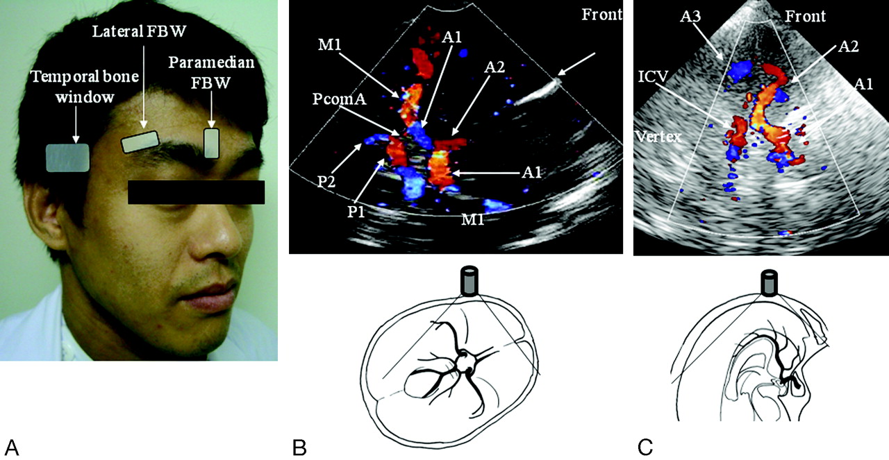
The lateral and paramedian FBW.8 A, The lateral FBW is above the lateral aspect the eyebrow. The paramedian FBW is slightly lateral to the midline of the forehead. B, TCCS image obtained via the lateral FBW and the schema. M1 and A2 are color-coded with flow moving toward probe. The ipsilateral A1 is coded with flow moving away from probe. P1 and P2 are coded with flow moving toward and away, respectively, from the transducer in the lateral frontal insonation plane. C, TCCS image obtained via the paramedian FBW and the schema. A1 and A2 are color-coded with flow moving toward the probe; and the distal ACA and A3 are color-coded with flow moving away from probe. PcomA indicates posterior communicating artery; ICV, internal cerebral veins.

Because contralateral intracranial arteries were not often detectable via the TBW and FBW in Japanese patients with stroke, only ipsilateral arteries were assessed. The detection of vascular insonation was defined on the basis of the consensus of 2 or more vascular neurologists. According to detection on color-flow imaging and a sufficiently measurable Doppler spectrum, each arterial site was judged as visible. The rates of visualizing a target artery were calculated for both bone windows. Sonographic contrast-enhancing agents were not applied. Categorized variables were compared by using the χ2 tests or Fisher exact test as appropriate. A value of P < .05 was considered statistically significant.

On-line Table 1 summarizes the rates of visualizing a target artery via the FBW, categorized by age and sex. Detection rates for M1 (P < .001), A1 (P < .001), A2 (P < .001), P1 (P < .001), and P2 (P = .047) were always higher in men than in women. The detection rates of A1 (P < .001) and M1 (P = .016) via the lateral FBW were significantly higher in patients 60 years of age or younger than those older than 60 years of age. On-line Table 2 shows the detection rates of target arteries with TCCS examinations via the TBW and the combination of both windows. The detection rate of A2 was higher via the FBW than via the TBW (40.2% versus 6.7%, P < .001). The combination of the TBW and FBW significantly improved the detection rates of A1 and A2 to 58.6% (P = .001) and 43.6% (P < .001) compared with only the TBW (46.0% and 6.7%, respectively). Only via the FBW, 36.9% of A2, 12.6% of A1, 4.9% of M1, 6.1% of P1, and 2.3% of P2 were detected.

Discussion

This is the first report to describe the clinical utility of TCCS examinations via the FBW in Asian patients with stroke. The rates of detecting all examined arteries were higher in male than in female patients. Approximately 50% of male and 20% of female patients with stroke had a proper FBW when the ACA and M1 were visualized. Although the detection rates via the FBW were not better than those via the TBW in all arteries except A2, the combination of the FBW and TBW improved the rate of detecting the ACA. Notably, 36.9% of A2 and 12.6% of A1 arteries were detected only via the FBW. The lateral FBW was more useful for the patients without a proper TBW for detecting ACA segments compared with the paramedian FBW.

An improper bone window via the TBW in elderly and female patients is a critical limitation of TCCS examinations.3,4 A demonstration of improved rates of ACA visualization by using a combination of the TBW and FBW is important because the insonation quality of Japanese patients with stroke via the TBW is inferior, especially in the ACA territory. Sonographic contrast may be useful for the FBW, to improve detection rates as demonstrated in the TBW.

The present study has some limitations. First, the rates determined in such a small number of female and younger patients with stroke were not statistically reliable. Second, a way to affix the transcranial probe for the FBW for long-term monitoring does not presently exist. Third, the utility of sonographic contrast agents via the FBW was not assessed.

In conclusion, TCCS examination via the FBW combined with the TBW improved the rate of detecting the ACA in Japanese patients with stroke. The compensatory technique appears to be useful for patients with an inadequate TBW.

Footnotes

  • This study was supported in part by grants-in-aid (H20-Junkanki-Ippan-019) and a Research Grant for Cardiovascular Disease 18C-5 from the Ministry of Health, Labor and Welfare of Japan.

  • indicates article with supplemental on-line tables.

References

  1. ↵
    Comerota AJ, Katz ML, Hosking JD, et al. Is transcranial Doppler a worthwhile addition to screening tests for cerebrovascular disease? J Vasc Surg 1995;21:90–95, discussion 95–97
    CrossRefPubMed
  2. ↵
    Marinoni M, Ginanneschi A, Forleo P, et al. Technical limits in transcranial Doppler recording: inadequate acoustic windows. Ultrasound Med Biol 1997;23:1275–77
    CrossRefPubMed
  3. ↵
    Itoh T, Matsumoto M, Handa N, et al. Rate of successful recording of blood flow signals in the middle cerebral artery using transcranial Doppler sonography. Stroke 1993;24:1192–95
    Abstract/FREE Full Text
  4. ↵
    Yagita Y, Etani H, Handa N, et al. Effect of transcranial Doppler intensity on successful recording in Japanese patients. Ultrasound Med Biol 1996;22:701–05
    CrossRefPubMed
  5. ↵
    Kazui S, Sawada T, Naritomi H, et al. Angiographic evaluation of brain infarction limited to the anterior cerebral artery territory. Stroke 1993;24:549–53
    Abstract/FREE Full Text
  6. ↵
    Kameyama M, Okinaka SH. Collateral circulation of the brain with special reference to atherosclerosis of the major cervical and cerebral arteries. Neurology 1963;13:279–86
    FREE Full Text
  7. ↵
    Krejza J, Mariak Z, Walecki J, et al. Transcranial color Doppler sonography of basal cerebral arteries in 182 healthy subjects: age and sex variability and normal reference values for blood flow parameters. AJR Am J Roentgenol 1999;172:213–18
    CrossRefPubMed
  8. ↵
    Stolz E, Kaps M, Kern A, et al. Frontal bone windows for transcranial color-coded duplex sonography. Stroke 1999;30:814–20
    Abstract/FREE Full Text
  • Received August 7, 2008.
  • Accepted after revision November 11, 2008.
  • Copyright © American Society of Neuroradiology
View Abstract
PreviousNext
Back to top

In this issue

American Journal of Neuroradiology: 30 (6)
American Journal of Neuroradiology
Vol. 30, Issue 6
June 2009
  • Table of Contents
  • Index by author
Advertisement
Print
Download PDF
Email Article

Thank you for your interest in spreading the word on American Journal of Neuroradiology.

NOTE: We only request your email address so that the person you are recommending the page to knows that you wanted them to see it, and that it is not junk mail. We do not capture any email address.

Enter multiple addresses on separate lines or separate them with commas.
Frontal Bone Window Improves the Ability of Transcranial Color-Coded Sonography to Visualize the Anterior Cerebral Artery of Asian Patients with Stroke
(Your Name) has sent you a message from American Journal of Neuroradiology
(Your Name) thought you would like to see the American Journal of Neuroradiology web site.
CAPTCHA
This question is for testing whether or not you are a human visitor and to prevent automated spam submissions.
Cite this article
S. Yoshimura, M. Koga, K. Toyoda, T. Mukai, B.-H. Hyun, M. Naganuma, K. Nagatsuka, K. Minematsu
Frontal Bone Window Improves the Ability of Transcranial Color-Coded Sonography to Visualize the Anterior Cerebral Artery of Asian Patients with Stroke
American Journal of Neuroradiology Jun 2009, 30 (6) 1268-1269; DOI: 10.3174/ajnr.A1452

Citation Manager Formats

  • BibTeX
  • Bookends
  • EasyBib
  • EndNote (tagged)
  • EndNote 8 (xml)
  • Medlars
  • Mendeley
  • Papers
  • RefWorks Tagged
  • Ref Manager
  • RIS
  • Zotero
0 Responses
Respond to this article
Share
Bookmark this article
Frontal Bone Window Improves the Ability of Transcranial Color-Coded Sonography to Visualize the Anterior Cerebral Artery of Asian Patients with Stroke
S. Yoshimura, M. Koga, K. Toyoda, T. Mukai, B.-H. Hyun, M. Naganuma, K. Nagatsuka, K. Minematsu
American Journal of Neuroradiology Jun 2009, 30 (6) 1268-1269; DOI: 10.3174/ajnr.A1452
del.icio.us logo Twitter logo Facebook logo Mendeley logo
  • Tweet Widget
  • Facebook Like
  • Google Plus One
Purchase

Jump to section

  • Article
    • Abstract
    • Technique
    • Discussion
    • Footnotes
    • References
  • Figures & Data
  • Supplemental
  • Info & Metrics
  • Responses
  • References
  • PDF

Related Articles

  • PubMed
  • Google Scholar

Cited By...

  • No citing articles found.
  • Crossref (9)
  • Google Scholar

This article has been cited by the following articles in journals that are participating in Crossref Cited-by Linking.

  • Echography in brain imaging in intensive care unit: State of the art
    Anselmo Caricato
    World Journal of Radiology 2014 6 9
  • Neuro-ultrasonography
    Ryan Hakimi, Andrei V. Alexandrov, Zsolt Garami
    Neurologic Clinics 2020 38 1
  • The Frontal Bone Window for Transcranial Doppler Ultrasonography in Critically Ill Patients: Validation of a New Approach in the ICU
    Pierre Sentenac, Jonathan Charbit, Camille Maury, Paul Bory, Geoffrey Dagod, Frédéric Greco, Xavier Capdevila, Pierre-François Perrigault
    Neurocritical Care 2020 33 1
  • Non-invasive Detection of Diffuse Intracranial Vertebrobasilar Artery Stenosis: A Prospective Comparison with Digital Subtraction Angiography
    Jie Zhang, Lijuan Wang, Ying Chen, Sibo Wang, Li Cui
    Ultrasound in Medicine & Biology 2022 48 3
  • Cryptogenic ischemic stroke and embolic stroke of undetermined source: clinical implications and importance for detection of covert atrial fibrillation in Japan
    Kazunori Toyoda, Ken Okumura, Yoichiro Hashimoto, Takanori Ikeda, Takashi Komatsu, Teruyuki Hirano, Haruhisa Fukuda, Kazuo Matsumoto, Masahiro Yasaka
    Nosotchu 2016 38 2
  • Neurosonology in Critical Care
    Jorge Carrizosa
    2022
  • Neurosonology in Critical Care
    Camilo N. Rodríguez, Deborah Pugin
    2022
  • Transcranial Doppler and transcranial color-coded sonography in patients with hyperacute ischemic stroke
    Masatoshi KOGA
    Choonpa Igaku 2010 37 3
  • Évaluation et validation de la fenêtre frontale en doppler transcrânien chez les patients neurolésés hospitalisés en réanimation : une étude prospective bicentrique
    Pierre Sentenac, Camille Maury, Geoffrey Dagod, Jonathan Charbit, Frédéric Greco, Sophie Bringuier, Pierre François Perrigault, Xavier Capdevila
    Anesthésie & Réanimation 2015 1

More in this TOC Section

  • Usefulness of Quantitative Susceptibility Mapping for the Diagnosis of Parkinson Disease
  • White Matter Alterations in the Brains of Patients with Active, Remitted, and Cured Cushing Syndrome: A DTI Study
  • Qualitative and Quantitative Analysis of MR Imaging Findings in Patients with Middle Cerebral Artery Stroke Implanted with Mesenchymal Stem Cells
Show more BRAIN

Similar Articles

Advertisement

Indexed Content

  • Current Issue
  • Accepted Manuscripts
  • Article Preview
  • Past Issues
  • Editorials
  • Editor's Choice
  • Fellows' Journal Club
  • Letters to the Editor
  • Video Articles

Cases

  • Case Collection
  • Archive - Case of the Week
  • Archive - Case of the Month
  • Archive - Classic Case

Special Collections

  • AJNR Awards
  • ASNR Foundation Special Collection
  • Most Impactful AJNR Articles
  • Photon-Counting CT
  • Spinal CSF Leak Articles (Jan 2020-June 2024)

More from AJNR

  • Trainee Corner
  • Imaging Protocols
  • MRI Safety Corner

Multimedia

  • AJNR Podcasts
  • AJNR Scantastics

Resources

  • Turnaround Time
  • Submit a Manuscript
  • Submit a Video Article
  • Submit an eLetter to the Editor/Response
  • Manuscript Submission Guidelines
  • Statistical Tips
  • Fast Publishing of Accepted Manuscripts
  • Graphical Abstract Preparation
  • Imaging Protocol Submission
  • Evidence-Based Medicine Level Guide
  • Publishing Checklists
  • Author Policies
  • Become a Reviewer/Academy of Reviewers
  • News and Updates

About Us

  • About AJNR
  • Editorial Board
  • Editorial Board Alumni
  • Alerts
  • Permissions
  • Not an AJNR Subscriber? Join Now
  • Advertise with Us
  • Librarian Resources
  • Feedback
  • Terms and Conditions
  • AJNR Editorial Board Alumni

American Society of Neuroradiology

  • Not an ASNR Member? Join Now

© 2025 by the American Society of Neuroradiology All rights, including for text and data mining, AI training, and similar technologies, are reserved.
Print ISSN: 0195-6108 Online ISSN: 1936-959X

Powered by HighWire