
Abstract
BACKGROUND AND PURPOSE: Anterior scalene block is a helpful diagnostic test for NTOS and a good predictor of surgical outcome. The purpose of this study was to describe the technique, success rate, and complications associated with CT-guided anesthetic and botulinum toxin injection of the ASM/MSM in patients with NTOS symptoms.
MATERIALS AND METHODS: One hundred six participants (mean age, 41.5 ± 10 years; 80 women) were identified via a retrospective review of medical records for CT-guided scalene blocks. The procedure was evaluated regarding the technical success, defined as satisfactory detection of the ASM/MSM; intramuscular needle placement; intramuscular injection of contrast; appropriate delivery of medication; and frequency of unintended BP block or other complications. We also determined the outcome of patients who underwent surgery following the block.
RESULTS: Study participants underwent 146 scalene injections, 83 blocks, and 63 chemodenervations, which were included in this investigation. In all cases, detection of the ASM/MSM and intramuscular needle placement was satisfactory. Postprocedural complications included 5 (3.4%) temporary BP blocks, 1 patient with (0.7%) Horner sign, 7 (4.8%) needle-induced pain reports, 1 (0.7%) case of dysphagia, and 2 (1.4%) instances of muscle weakness. There were no major complications reported. The rate of good outcome following surgery was the same in patients with positive versus negative blocks, 30/43 (70%) versus 5/7 (71%), respectively.
CONCLUSIONS: CT guidance is a useful adjunct in performing accurate ASM/MSM blocks with a low rate of minor complications.
Abbreviations
- ASM
- anterior scalene muscle
- BP
- brachial plexus
- CR
- cervical rib
- EMG
- electromyography
- EMR
- electronic medical record
- IJV
- internal jugular vein
- MSM
- middle scalene muscle
- NTOS
- neurogenic thoracic outlet syndrome
- TOS
- thoracic outlet syndrome
Estimates of TOS incidence vary widely, with rates ranging from 3 to 80 cases per 1000 people.1,2 TOS is caused by compression of the neurovascular structures passing through the superior thoracic outlet, and it results from neck trauma (often from auto crashes), repetitive stress injury, congenital abnormalities (eg, cervical ribs), or a combinations of these.3,4 There are 3 types of TOS: venous, arterial, and neurogenic. NTOS comprises almost 95% of TOS cases5–7 and is mostly seen in the third and fourth decades of life, with a female/male ratio of 3:5–4:1.2,6–8 Symptoms of NTOS include paresthesias; pain on the affected side of the neck, shoulder, or arm; discoloration and cold intolerance of fingers in cases involving sympathetic fibers; and, less commonly, weakness of the upper extremity. Venous TOS commonly presents with acute effort thrombosis of the subclavian vein, also known as Paget-Schroetter syndrome.2,6 Patients usually are athletes who present with an acutely swollen discolored upper extremity; pain due to subclavian vein obstruction, with or without thrombosis; and visible subcutaneous veins over the involved shoulder and chest wall.2,3,5,6 Arterial TOS is associated with hand ischemia caused by either external compression of the subclavian artery or from emboli arising from a subclavian artery aneurysm.3,5,6 The symptoms of arterial TOS include digital ischemia, claudication, pallor, coldness, paresthesia, and pain in the hand but seldom in the shoulder or neck. Occasionally, the neurologic and vascular components may coexist in the same patient.2
If ergonomic modifications, exercise, and physiotherapy do not improve the symptoms, anesthetic injection of the anterior scalene muscle, as a diagnostic test, is often performed.9 The anesthetic injection allows the first rib to descend by relaxing the scalene muscle and thereby decompressing the BP.6,9–11 The anterior scalene block has been the most helpful test to confirm the diagnosis of NTOS5,6,11–15 and to predict the response to surgery, because it may mimic the results of first-rib resection and anterior scalenectomy.6,10–12,14,16 However, to be predictive, the injection should avoid anesthesia of the BP and sympathetic chain, and the patient should be both medically and psychologically stable.13
Different methods have been used to guide the scalene injection, including the use of anatomic landmarks,13,17 EMG,12 sonography,11 combinations of EMG and sonography,18 EMG and fluoroscopy,18 and CT most recently.9,10
CT-guided injection of the scalene muscle is a novel technique,9 which has not been previously evaluated in the radiology literature, to our knowledge. The purpose of this study was to describe the technique, findings, and complications associated with CT-guided injection of the ASM/MSM in patients being evaluated or treated for NTOS. We also assessed the predictive value of the CT-guided scalene block by determining the outcome of patients who underwent surgical decompression following the block.
Materials and Methods
Study Design
Our institutional review board approved the retrospective review of patient data for this study. Informed consent was waived by the institutional review board, and the study was compliant with the Health Insurance Portability and Accountability Act.
We identified patients via a retrospective review of the EMR for CT-guided scalene injections. Unavailable images excluded patients from the study. Patients displayed different distributions of pain in the neck, shoulder, and upper extremity; paresthesia of the hand; tenderness of the scalene muscles; positive findings on the arm stress test; or difficulty doing overhead activities and those requiring repetitive movements. Patients were evaluated for NTOS by a single vascular surgeon. They were then referred for CT-guided anesthetic injections of the ASM alone or ASM/MSM, which were performed by 3 different anesthesiologists from 2005 to 2009.
The CT images of the neck were investigated independently and separately by a board-certified subspecialty-certified neuroradiologist with 13 years' experience and a neuroradiology research fellow with 1 year of neuroimaging research experience. Any discordant readings were reviewed by consensus and with a subspecialty certified neuroradiologist with 22 years' experience. We evaluated the satisfactory detection of the scalene muscles, intramuscular needle placement, and the location of the contrast injected. We assessed if, in any image, there was intravascular needle placement or contrast accumulation within arterial or venous structures of the neck.
Twenty-three patients had images limited to 1 cervical vertebra at the level of injection, usually C6 or C7. In this group of patients, findings other than the ones related to the scalene injection procedure could not be consistently reported. Typically the scanning ranged from C4 to T1. The operator then selected the level that best demonstrated the anterior scalene muscle. That level was usually around C6-C7.
In addition, we reported imaging findings that may account for the patient's symptoms, either as a predisposing factor or as a sign of other diseases that may mimic NTOS symptoms. They included cervical ribs, an elongated C7 transverse process (defined as the C7 transverse processes extended beyond the T1 transverse process), a bulky callus of the first rib or clavicle, degenerative joint diseases, and apophysomegaly of the facet and transverse process of the cervical vertebrae. We also described incidental lesions in the cervical and upper thoracic areas, like thyroid nodules or calcified atherosclerotic plaques.
After investigating the CT images, we inspected patient histories via the EMR for the frequency of unintended BP block, sympathectomy, hematoma, infection, abnormal bleeding, pneumothorax, vascular uptake, dysphagia, neck weakness, phonation problems, or any other complications. We also assessed the incidence of positive scalene injections.
Forty-three patients with a positive block and 7 with a negative block underwent thoracic outlet decompression surgery. This was not a randomized controlled trial, but a biased subset evaluated clinically. The outcome of surgery was evaluated in both groups via review of postoperative records. The surgical outcome was assessed clinically on the basis of patient histories and physical examinations by the thoracic surgeon who performed the procedure. Notes as to the result with respect to pain relief and upper extremity function were reviewed in the EMR.
Good outcome was defined as significant pain relief and improvement in function and range of motion of the affected upper extremity at follow-up. Among the 7 patients with a negative block who underwent surgery, 4 had clinical signs of arterial involvement, like pulse obliteration and bruit onset with arm hyperabduction; 1 of these 4 also had vascular studies indicating collapse of the subclavian vein with arm abduction. The fifth patient had a history of multiple surgeries on the shoulder and exacerbation of her pain after those surgeries. The sixth patient had a history of recurrent effort thrombosis. Finally, the last patient underwent surgery despite ambiguity associated with the result of the scalene block.
Technique of Scalene Block
Written informed consent was obtained from all participants before the procedure. Scanning was performed on the axial plane by using a 16-section multidetector row CT scanner (Aquilon 16; Toshiba Medical Systems, Tokyo, Japan). Patients were positioned supine, and a strip of radiopaque markers was placed on the neck. A scout film was obtained from the C4 to T1 levels by using 3-mm sections. We identified the ASM/MSM. The neck was prepared in the usual sterile manner, and a 1.5-inch 22- or 25-ga needle was inserted into the muscle belly under CT guidance. A focal area of the neck was rescanned on 1–2 more occasions for evaluation and adjustment of needle position. Then 0.1- to 0.3-mL radiographic contrast (iohexol, Omnipaque 180; GE Healthcare, Milwaukee, Wisconsin) was injected into the ASM/MSM to verify both needle placement and spread of material within the muscle.9
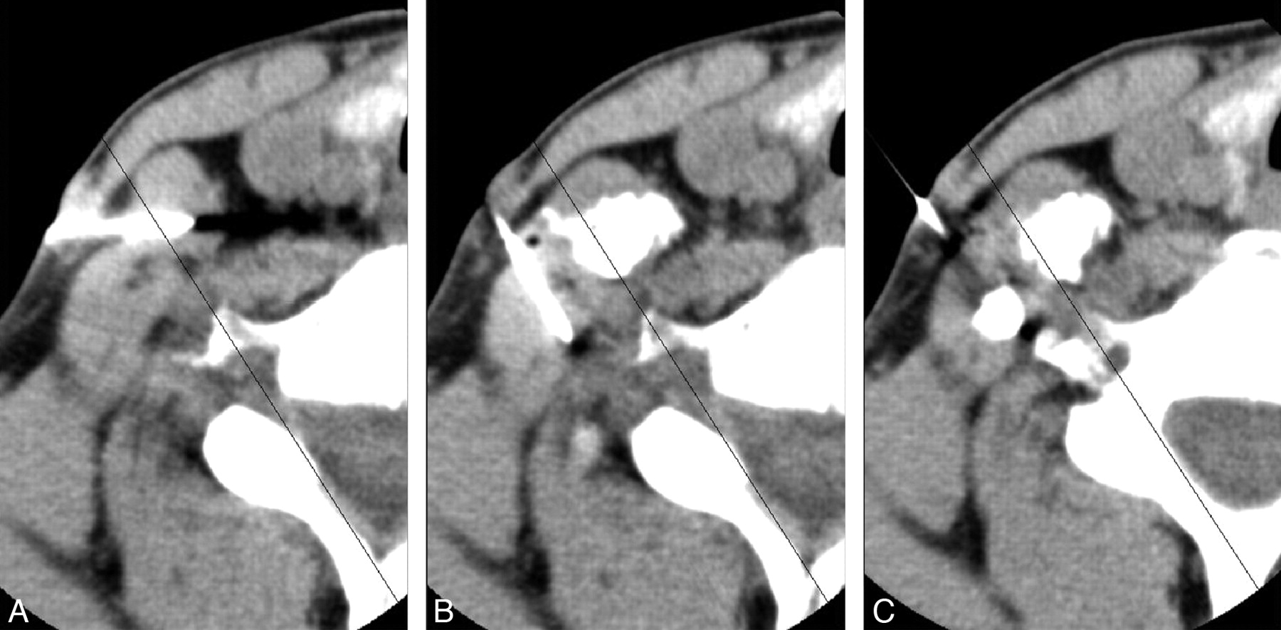
For the scalene block, 1–2 mL of bupivacaine 0.25% alone was injected or the bupivacaine was followed by either 20–40 mg of methylprednisolone (Depo-Medrol) or 10 mg of triamcinolone (Kenalog) (Fig 1). For scalene chemodenervation, either 1 mL of bupivacaine 0.25% or 0.5% followed by 16–20 U of botulinum toxin was injected, or 16–20 U of botulinum toxin alone was injected after negative findings on aspiration. Then the patient was rescanned to confirm selective injection into the ASM/MSM.9 The entire procedure took approximately 10 minutes of scanner time in most patients. A “positive” scalene injection was characterized as a ≥50% reduction in numeric pain scores during elevated arm stress test immediately after the scalene block. For chemodenervation with botulinum toxin, a positive scalene injection was defined as significant pain relief and improvement in function and range of motion of the affected upper extremity.

Stages of injection into the scalene muscle by CT guidance. A, The ASM is identified, and the distance from the skin surface to the belly of the ASM is measured. B, The needle is inserted into the ASM. C, Contrast is injected into the ASM. D, More spread of contrast inside the ASM after botulinum injection confirms selective injection into the ASM.
Pain scores were assessed by using a standard visual analog scale by the pain management specialist.
Results
We identified 106 patients with a mean age of 41.5 ± 10 years (range, 20–67 years); 80 (75%) were women. Among 106 patients, 44 (41%) had a history of neck trauma; whiplash injury (61%) was the most common type of trauma, followed by repetitive injury (14%), which included heavy lifting and typing on a computer keyboard for long hours.
Although we studied 106 patients, we examined 146 scalene injections because some patients underwent both a scalene block and a chemodenervation later. Therefore, in these patients, the number of scalene injections exceeded 1. Of 146 scalene injections, 83 were scalene blocks and 63 were scalene chemodenervations (Table 1). In all cases, detection of the ASM/MSM and intramuscular needle placement were satisfactory on the basis of CT guidance. Postinjection, there were no instances of hematoma; infection; abnormal bleeding; pneumothorax; or vascular uptake of the contrast, anesthetic, or botulinum toxin. Details about the outcomes and the complications (Figs 2⇓–4) of the injections are described in the On-line Table.
Scalene muscles injected
Injection of bupivcaine and botulinum into the right ASM and MSM. A, Needle placed into the ASM. B, Contrast injected into the right ASM; needle now in the right MSM. C, Contrast injected into the right MSM.

Incorrect needle placement. A, Axial CT image of the ASM anteriorly, the MSM posteriorly, and the BP passing through them. B, The needle is initially inserted into BP. C, It is then reinserted into the ASM.

Spread of contrast in the BP. A, An axial view of the left ASM, MSM, and BP. B, Contrast injected into the ASM spreads out posteriorly. C, After injection of bupivacaine, more spread of contrast is observed around the BP and into the ASM, which results in transient paresthesia.
Of 43 patients with positive blocks (39 injections into the ASM alone, 4 into both the ASM and MSM) who then underwent first-rib resection and scalenectomy, 30 (70%) experienced a good outcome. The mean follow-up time was 8 ± 6.5 months. Eleven (26%) patients did not improve after surgical decompression, and 2 patients did not have follow-up. Seven7 patients with negative blocks also underwent surgery; 5 (71%) experienced a good outcome at a follow-up of 8 ± 5.5 months and 2 had persistent symptoms following the surgery.
Imaging findings that could be contributory factors for the patients' symptoms were discovered on the accompanying CT scans. Cervical ribs (Fig 5) were seen in 4 (3.8%) patients; 3 were bilateral. There were 6 (5.6%) elongated C7 transverse processes (Fig 6), and 4 were bilateral. Facet/uncovertebral joint hypertrophy associated with neural foraminal narrowing and disk herniations was discovered in 46 (43%) and 12 (11%) patients, respectively.

Unilateral cervical rib (C7) compressing the BP in a 42-year-old woman.

Right C7 elongated transverse process compressing the BP in a 52-year-old woman.
Discussion
NTOS is induced by compression of the BP in any of 3 parts of the superior thoracic outlet: the interscalene triangle, costoclavicular space, or retropectoralis minor space.7 Paresthesia is the most common symptom,3 mostly evident in the ring and little finger.5,7 Pain is usually distributed in the shoulder region and radiates along the inner aspect of the arm. In severe cases, there may be atrophy of the thenar eminence, ulnar intrinsic hand musculature, and forearm muscles.10 Patients may present with symptoms of upper plexus compression (C5-C7), like pain on the ipsilateral aspect of the neck or mastoid area or even occipital headaches.6,7 Pain may also radiate to the deltoid region, trapezius, and the rhomboid muscles; the upper pectoral region; and down the lateral aspect of the arm. In most cases, however, patients present with involvement of the lower plexus (C8-T1). Symptoms are usually aggravated by continuous elevation of the arm.5,7,13 However, because 99% of patients with NTOS lack objective symptoms,5,12 the diagnosis and treatment of NTOS are highly disputed.
Anatomic variations often represent asymptomatic predisposing factors until trauma is superimposed.5,6 Variations can include cervical ribs, an elongated C7 transverse process, insertion variations of the scalene muscles, redundant muscles like the scalene minimus, a bulky callus or tumor of the first rib or clavicle, and fibrous bands. Cervical ribs occur in less than 1% of the general population.5–7 Fifty percent are bilateral,6 and 70% occur in women.5 These have been reported in 5%–9% of patients with TOS.7 Most cervical ribs are asymptomatic but can produce the neurogenic form of TOS more frequently than the vascular form.5 The C7 transverse process is considered elongated if it extends beyond the T1 transverse process, but this variant is less common than the cervical rib.7 Among our 106 patients, we found almost the same frequency of cervical ribs (4%) as in previous studies, but the elongated C7 transverse processes were more frequent (5.6%).7 They were mainly bilateral and were only found in female patients. Furthermore, there were different degrees of facet degenerative joint disease in 43% of our patients, which could contribute to their specific symptoms.
Posttraumatic or postoperative fibrosis (in the neck/shoulder area) often precipitates symptoms of NTOS.5,7 Trauma may induce fibrosis or spasm of the scalene muscles, leading to elevation of the first rib and narrowing of the thoracic outlet.6,7 The most common type of trauma leading to NTOS is whiplash injury, followed by repetitive injury such as typing on a computer keyboard for long hours or heavy lifting.5–7 The frequency of neck trauma as the etiology of NTOS has been reported from 20% to 80%.5
Two previous studies illustrate that a higher percentage of patients with a positive scalene block respond favorably to surgical decompression.12,16 If ergonomic modifications, exercise, and physiotherapy do not improve the symptoms, surgery may be an option. In our study, the rate of successful surgical outcome did not vary according to positive or negative anesthetic injections into the ASM/MSM, 30/43 (70%) versus 5/7 (71%), respectively. However, the results may not be comparable because the sample size of the patient population with negative tests who had surgery was very small compared with those who underwent surgery with positive scalene injections. In addition, those patients who had surgical decompression despite a negative scalene block displayed compelling signs and symptoms of vascular involvement that required surgery. Therefore, a successful surgical outcome in this group may result from vascular rather than neurogenic TOS.
Localization techniques for scalene injections include palpation and external visible anatomic landmarks,13,17 EMG guidance,12 sonographic guidance,11,18,19 and fluoroscopic guidance.18,20 Success rates by these means are expected to be very operator-dependent based on experience, whereas CT guidance lends a simplicity and accuracy that would allow a shorter learning curve. With no imaging guidance, there is a high possibility of a false-positive result because of accidental BP anesthesia (≤10% of cases)12 or a false-negative result due to inaccurate targeting of the scalene muscles or inadvertent puncture of important vascular structures.12,13 When blocks are performed by means of fluoroscopy and EMG, there may be a 1.2%–7% rate of dysphagia and a 0.6% rate of undesired muscle weakness.18,20 Moreover, frequent paresthesia during needle placement has been reported.18 Torriani et al11 performed 29 sonographically guided anesthetic injections of the ASM and experienced temporary BP blocks in 10/29 (34%) injections.
CT-guided local anesthetic or botulinum toxin injection of the scalene muscle is a novel technique,9,10 and we evaluated the outcomes and complications of this procedure in a large group of patients. In our study, 68/83 (82%) of the anesthetic injections into the ASM alone or ASM/MSM together resulted in a positive scalene block, which was higher than that in studies using needle guidance by sonography (11/29, 38%),11 EMG and fluoroscopy (4/22, 18%),20 or EMG alone (93/122, 72%).12 Although placebo effects and coexisting spinal pathology may challenge the results of our study, the high rate of improvement (70%) after surgery retrospectively verifies cases of true NTOS.
Christo et al9 reported minor neck weakness and no significant dysphagia, phonation disturbance, or aspiration after CT-guided ASM injection with botulinum toxin (Botox). Mauro et al10 incurred no complications other than a few instances of needle-induced pain and 2/14 patients reporting hand numbness after CT-guided ASM injection with local anesthetic.
Compared with the use of superficial anatomic landmarks,12,13 sonographic guidance,11 or fluoroscopic and EMG guidance,18,20 CT guidance minimized the occurrence of Horner sign, dysphonia, BP block, and dysphagia and produced a comparably low rate of upper extremity muscle weakness or intravascular needle placement with no vascular uptake following local anesthetic or botulinum toxin injection. Our results indicate that under CT guidance, scalene injection is a safe procedure. Despite the leakage of contrast out of the scalene muscles in half of our injections, there were low rates of BP block and muscle weakness following local anesthetic injection. It seems that leakage of contrast out of the ASM/MSM does not necessarily reflect spread of local anesthetic/botulinum toxin to other cervical muscles or the BP, given low rates of adverse effects.
We observed a lower rate of pain relief following CT-guided botulinum injection into the ASM or ASM/MSM (29/63, 46%) compared with studies using sonography (38/55, 69%), (14/22, 64%),19,20 EMG and sonography (70/77, 91%),18 or EMG and fluoroscopy (136/168, 81%).18 Targeting fewer muscles implicated in the development of NTOS and injecting less total botulinum toxin might explain our lower rate of pain relief following chemodenervation. Needle-induced pain occurred during chemodenervation when bupivacaine was not injected before botulinum toxin. We suspect that a lack of anesthesia in the scalene muscles may have contributed to this adverse effect. Our study indicates a low rate of BP block with local anesthetic, which implies that CT-guided imaging of the scalene muscles produces a very reliable outcome. In contrast to sonography or MR imaging, use of CT involves patient exposure to ionizing radiation. However, Christo et al9 reported only brief radiation exposure that did not exceed 60 seconds in their study examining CT-guided ASM injections for NTOS.
Our study has several limitations that may impact the accuracy of the results. First, our investigation was retrospective. A prospective study may better capture clinically relevant variables that may be missing in a retrospective analysis. Second, our study did not include a control group to account for the placebo effect. Our study did not randomize patients to CT versus other modalities, and we did not assess the results of surgical treatment in a randomized blinded fashion after scalene block. This article cannot address the success rate of scalene block in predicting surgical outcome due to the low number of individuals with negative blocks who went on to surgery. The purpose of this article is to familiarize radiologists with the expected appearance of needle position and contrast location for CT-guided scalene injections and to report ancillary findings (possible causes of NTOS and incidental lesions) that can occur in the imaged volume. This information may be useful to the pain management team as they assess the results of scalene injections and plan follow-up treatment.
Conclusions
We found that CT-guided scalene injections produce accurate results compared with other techniques of image-guided scalene injection.12,11,20 Radiologists interpreting the images from the scalene injection studies or pain medicine specialists performing these injections should consider other etiologies contributing to NTOS symptoms and incidental lesions not related to NTOS that require further evaluation. Finally, a prospective study can better define the role of CT-guided scalene injections compared with other imaging modalities in diminishing complications and predicting operative success as well as the therapeutic benefit of botulinum toxin injections for NTOS.
Acknowledgments
We thank Gloria Vila for assistance in data base search of the radiology information system.
Footnotes
-
Paper previously presented at: Annual Meeting of the American Society of Neuradiology, May 15–20, 2010; Boston, Massachusetts.
Indicates article with supplemental on-line table.
References
- Received June 23, 2010.
- Accepted after revision August 11, 2010.
- Copyright © American Society of Neuroradiology


