
Abstract
BACKGROUND AND PURPOSE: Imaging is a key element in the study of many rodent models of human diseases. The application of DSA has been limited in these studies in part because of a lack of a method that allows serial intra-arterial examinations to be performed during an extended period of time. It was our intent to develop and test a method for performing sequential arterial catheterizations and DSA in rats.
MATERIALS AND METHODS: Using a transfemoral approach, we subjected 12 adult male Harvey rats to 3 sequential DSA examinations during a 6- to 8-week period. At each examination, 2 selective arterial catheterizations and a DSA were performed. Animals were monitored for ill effects, and images from the 3 examinations were compared for quality and the presence of any arterial injury.
RESULTS: Ten of the 12 rats survived all 3 examinations. There were no adverse effects noted and no evidence of arterial injury from the examinations.
CONCLUSIONS: With the technique described, it is possible to perform serial arterial catheterizations and DSA in rats. This technique will be useful as an adjunct in the use of rodents for the study of human diseases.
Abbreviations
- AP
- anteroposterior
- DSA
- digital-subtraction angiography
The ever-expanding and pervasive use of rodents as essential models for human disease in basic science research has prompted modification of modern human imaging modalities (ie, CT and MR imaging) for use in small animals. Small-bore MR imaging, MR microscopy, micro-positron-emission tomography, micro- single-photon emission CT, micro-CT, and optical imaging, all now play key roles in many murine studies. Although micro-DSA has been reported, it is not yet either widely available or commonly used. In part, this is likely related to the lack of a described method for performing investigations that require accessing the rodent vasculature multiple times for weeks or months. Because selective arterial catheterization combined with DSA is the only method that provides a means for real-time high spatial and temporal resolution imaging of the delivery and distribution of drugs, devices, or cells, it was our intent to develop a method that would allow investigators to repeatedly perform, during an extended period of time, arterial catheterizations and angiography in a rodent.1–7
To our knowledge, there has been no work describing techniques that would allow minimally invasive endovascular interventions and DSA examinations to be performed on rodents in such a manner that the animals could survive and then be studied subsequently with angiography at ≥2 follow-up intervals. By using the transfemoral techniques described below, we have optimized a vascular access protocol for rats so that it is possible to perform organ-specific selective angiography (eg, renal, subclavian, aortic, hepatic, mesenteric, iliac, and cerebral arteries) in such a way that at least 3 separate DSA studies can be performed on the same vessels in the same animal during an extended period of time. We believe that this method will enhance the ability to use rodents as models for human diseases.
Materials and Methods
Overview
Under an approved institutional review board protocol, 12 adult male Harvey rats weighing 450–550 g were used for these studies. They were housed in an animal room at 21°-24°C with a 12-hour light/dark cycle. Food and water were available ad libitum. Each of the 12 rodents underwent 3 serial transfemoral angiographic examinations with selective arterial catheterizations and DSA acquisitions. The 3 angiographies were separated by intervals ranging between 10 and 14 days. Sterile techniques were used for all procedures. Following completion of the final angiography, animals were euthanized following an institutionally approved protocol.
Anesthesia
Isoflurane (1%–2.5%) was used to induce and maintain anesthesia on all subjects. Respiratory rate and the reaction to toe or tail pinch were used for monitoring of anesthetic depth. To alleviate any postprocedure discomfort, each animal received intraperitoneal buprenorphine (0.01–0.05 mg/kg2) before closure of the femoral cut-down site.
Femoral Artery Access
Using microsurgical techniques, we made a 2-cm incision on the ventral side of 1 hind leg parallel and superficial to the common femoral artery. The femoral artery was isolated from the neurovascular bundle between the bifurcation of the inguinal artery and the bifurcation of the deep femoral artery. For hemostasis, a clip was used for the proximal control and the distal end of the artery was ligated to prevent backflow. To relieve spasm caused by these manipulations, we irrigated the femoral artery with a 4% lidocaine solution for at least 2 minutes. This access technique was used on the right side for the first angiogram and on the left side for the second study. For the third and final study, femoral artery access was gained by way of a cut-down performed proximal to the inguinal branch of the femoral artery (rather than distal to it as in the first 2 procedures). Otherwise, all procedures and techniques remained consistent during arterial-access catheterizations and DSA imaging. With hemostasis achieved by way of the proximal clipping, an arteriotomy was made in the exposed femoral artery. Through this, a 20-gauge 2-inch angiocath sheath, modified so that its tip had been beveled, was introduced and advanced approximately 0.5 inch into the artery. Through this, a microcatheter was then advanced until its tip extended beyond the distal end of the access sheath. At this stage, the proximal clip was removed so that the microcatheter and the sheath could be advanced a small distance further into the artery. After checking for hemostasis, we then secured the sheath by ligature. These steps in the procedure were done without fluoroscopic monitoring.
Arterial Catheterizations and DSA Imaging
For each of the 3 angiograms, 2 arteries were catheterized and injected in each of the 12 animals (Table). Using fluoroscopic control, we advanced a microcatheter (Excelsior SL-10; Boston Scientific, Natick, Massachusetts) over a guidewire (Transcend 0.10, Boston Scientific) into the appropriate artery. DSA was performed by using a hand injection of 0.1–0.2 mL of iohexol 300 mg/mL (Omnipaque; GE Healthcare, Milwaukee, Wisconsin) diluted to a 50% concentration with normal saline. This procedure was then repeated for the second artery. Fluoroscopy and angiography were performed by using either an OEC (GE Healthcare) or a single-plane Zeego angiographic system (Siemens Healthcare, Forchheim, Germany). Following completion of the angiography, the catheter and sheath were removed and the femoral artery was ligated above and below the arteriotomy site by using 6–0 Prolene (Ethicon, Somerville, New Jersey) suture.
Subjects and arteries selectively catheterized
Recovery, Monitoring, and Assessment
The femoral artery access sites were closed in 2 layers by using a subdermal suture technique with application of Dermabond (Ethicon) on the skin surface overlying the incision. Animals recovered from anesthesia in their cages and were observed until fully awake. During the intervals between procedures, animals were assessed daily with notations made about wound healing, normal food/water intake, behavioral change, and weight change.
Image Comparison
Corresponding images from each of the 3 angiograms were compared by an experienced angiographer (C.M.S.). Any change in vascular caliber or evidence of vascular injury was noted.
Results
Mortality and Morbidity
Ten of the 12 rats survived all 3 angiographic studies; 1 was sacrificed at the time of the second angiogram because of a transection of the femoral artery during cannulation, which could not be repaired, and the other animal died during the second angiogram likely because of an anesthetic overdose. There were no postsurgical complications such as infections, changes in appetite or activity, neurologic deficits, or ischemic complications. Contrary to a previous report that found that some mice adopted a hunchbacked posture after femoral artery ligation and walked with extended paws for about 1 week, we saw no evidence of this behavior.8 The average interval between the first and second angiogram was 11 days and between the second and third angiogram was 14 days. There was an average 0.4 ± 1.9% change in total body weight of the animals throughout the study period. (From the first to second study, subjects experienced a −3.0 ± 1.6% change in total body weight; from the second to third study, subjects experienced a 3.5 ± 1.1% change in total body weight.) The time required from induction of anesthesia to completion of the studies averaged 1.5 ± 0.5 hours.
Angiographic Results
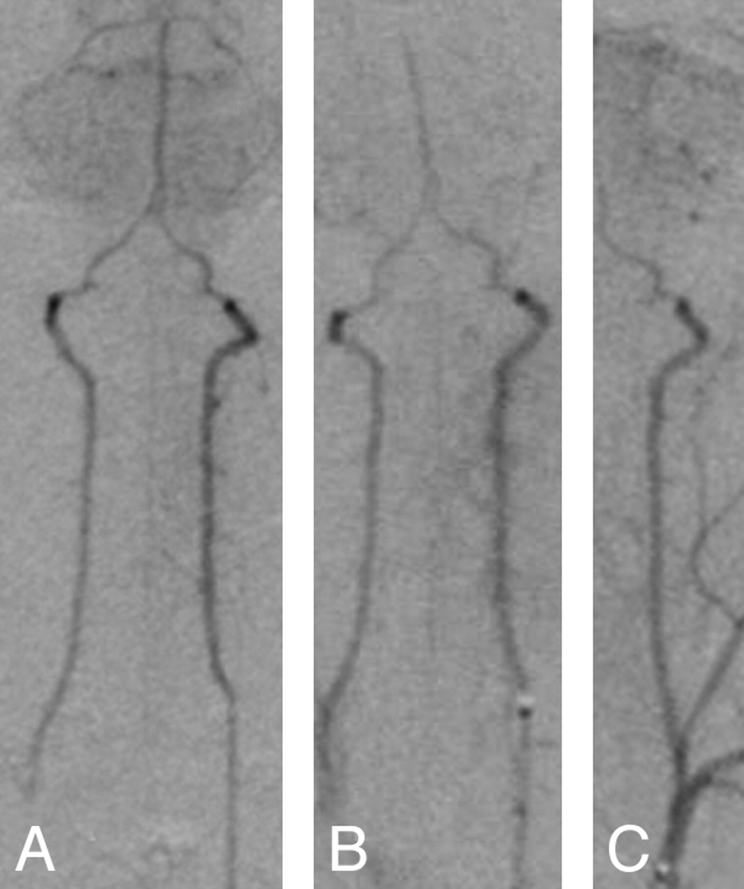
It was possible to selectively catheterize the same 2 arteries in each of the animals for each of the 3 angiographic examinations. These arteries are shown in the Table. Except for a decrease in caliber and an increase in the collateral branches of the femoral arteries that were ligated, there was no evidence of any change in the caliber or distribution of these arteries from 1 examination to the next. The time required for fluoroscopy for catheter manipulation and arterial catheterization never exceeded 10 minutes, and most often it was <5 minutes in all examinations. Figures 1⇓–3 are representative examples of the angiograms obtained as a part of this study.

Mid-arterial phase AP projections following injection of contrast into the abdominal aorta just above the aortic bifurcation. A, Initial study with access by way of the right common femoral artery. B, Second study with access by way of the left femoral artery. Note the increase in collateral vessels on the right where the common femoral artery has been ligated. C, Final study with access again from the right femoral artery, above the site of the original cut-down. Collateral supply of the left has increased following ligation of the femoral artery after the second angiogram.
Mid-arterial phase AP projections following injection of contrast into the origin of the left vertebral artery.

AP projections of abdominal aorta and aortic root injections showing the size of the rat aorta. It approximates the size of the human middle cerebral artery. Images are from the studies of 2 different animals.
Discussion
In this study, we have shown the feasibility of performing sequential selective arterial catheterizations and DSA in rats without causing significant physiologic or behavioral changes. The use of this technique should expand the utility of rat models for a variety of applications. As 1 example, the rat and mouse aorta has been used for the study of vascular responses following stent implantation.9–11 The design and optimization of stents that function as flow dividers for the treatment of human intracranial aneurysms demands a good understanding of their impact on arterial perforators that arise from the segment of artery in which such a device is to be implanted. The lumbar and intercostal arteries arising from the rat aorta approximate the sizes of major perforators arising from the basilar and middle cerebral arteries (Fig 3). In previous studies by using the rat aorta, 1-time access has been gained by insertion of a catheter by way of a carotid cut-down. 10,11 Using the technique that we describe, one could implant these devices in the rat aorta under fluoroscopic guidance, acquire an initial angiogram, and, then at 2 subsequent intervals, perform repeat angiography to assess not only the degree of perforator patency but also the vascular response (eg, intimal hyperplasia to the implant).
As another example, a commonly used rat model of cerebral ischemia involves the insertion, through a neck incision, of a suture into the internal carotid artery and then the blind manipulation of the suture into the middle cerebral artery.12 Using the technique that we describe, one could selectively catheterize the internal carotid artery with a transfemoral approach and then deliver the suture by using fluoroscopy for direct visualization. DSA could be performed before, during, and after middle cerebral artery blockage to document the location and degree of arterial obstruction that was obtained as well as the extent of reperfusion established after removal of the suture. Weidauer et al13 described the use of a microcatheter technique for the study of vasospasm in a rat model; however, they did not discuss or demonstrate a method for use of this technique in serial studies.
While we performed femoral artery access by using a surgical microscope, we believe that it would also be possible to do this simply by using surgical loops. One of the primary operators performing the femoral artery catheterizations (A.B.) had just completed his first year in medical school, and before starting this project, he had no special training in operative techniques. Before the start of the project, he completed a 1-week course in microsurgical techniques and then practiced performing femoral artery catheterizations on 15 rats before start of the study. Thus, with only modest training, researchers should be able to quickly master the techniques that are required to safely and efficiently perform these procedures.
Prior studies have shown that a >10% change in blood volume can alter the normal physiology of rodents. We were careful to avoid blood loss at the time of femoral artery access and to avoid overexpansion of the animal's blood volume, we used the formula described by Lee and Blaufox14 (Total Blood Volume = 0.062 × Body Weight + 0.0012) and then limited the amount of contrast medium and saline injected to values that would cause <5% change in total blood volume.
While the image quality of angiograms obtained by using standard DSA equipment was satisfactory, with such devices, it is not possible either to obtain angiographic images of optimal image quality or to exploit the potential for DSA studies to acquire and display physiologic (eg, blood flow) information. The real and potential advantages of a DSA device designed specifically for use in small animals combined with a microinjector capable of injecting small quantities of contrast medium quickly, accurately, and reproducibly have recently been described.4,6
Conclusions
With the technique described, it is possible to perform sequential serial arterial catheterizations and DSA in rats. This technique will be helpful as an adjunct in the use of rodents for the study of human diseases.
References
- Received December 26, 2009.
- Accepted after revision March 2, 2010.
- Copyright © American Society of Neuroradiology


