Advertisement

Main menu

  • Home
  • Content
    • Current Issue
    • Accepted Manuscripts
    • Article Preview
    • Past Issue Archive
    • Video Articles
    • AJNR Case Collection
    • Case of the Week Archive
    • Case of the Month Archive
    • Classic Case Archive
  • Special Collections
    • AJNR Awards
    • ASNR Foundation Special Collection
    • Most Impactful AJNR Articles
    • Photon-Counting CT
    • Spinal CSF Leak Articles (Jan 2020-June 2024)
  • Multimedia
    • AJNR Podcasts
    • AJNR SCANtastic
    • Trainee Corner
    • MRI Safety Corner
    • Imaging Protocols
  • For Authors
    • Submit a Manuscript
    • Submit a Video Article
    • Submit an eLetter to the Editor/Response
    • Manuscript Submission Guidelines
    • Statistical Tips
    • Fast Publishing of Accepted Manuscripts
    • Graphical Abstract Preparation
    • Imaging Protocol Submission
    • Author Policies
  • About Us
    • About AJNR
    • Editorial Board
    • Editorial Board Alumni
  • More
    • Become a Reviewer/Academy of Reviewers
    • Subscribers
    • Permissions
    • Alerts
    • Feedback
    • Advertisers
    • ASNR Home

User menu

  • Alerts
  • Log in

Search

  • Advanced search
American Journal of Neuroradiology

American Journal of Neuroradiology

ASHNR American Society of Functional Neuroradiology ASHNR American Society of Pediatric Neuroradiology ASSR
  • Alerts
  • Log in

Advanced Search

  • Home
  • Content
    • Current Issue
    • Accepted Manuscripts
    • Article Preview
    • Past Issue Archive
    • Video Articles
    • AJNR Case Collection
    • Case of the Week Archive
    • Case of the Month Archive
    • Classic Case Archive
  • Special Collections
    • AJNR Awards
    • ASNR Foundation Special Collection
    • Most Impactful AJNR Articles
    • Photon-Counting CT
    • Spinal CSF Leak Articles (Jan 2020-June 2024)
  • Multimedia
    • AJNR Podcasts
    • AJNR SCANtastic
    • Trainee Corner
    • MRI Safety Corner
    • Imaging Protocols
  • For Authors
    • Submit a Manuscript
    • Submit a Video Article
    • Submit an eLetter to the Editor/Response
    • Manuscript Submission Guidelines
    • Statistical Tips
    • Fast Publishing of Accepted Manuscripts
    • Graphical Abstract Preparation
    • Imaging Protocol Submission
    • Author Policies
  • About Us
    • About AJNR
    • Editorial Board
    • Editorial Board Alumni
  • More
    • Become a Reviewer/Academy of Reviewers
    • Subscribers
    • Permissions
    • Alerts
    • Feedback
    • Advertisers
    • ASNR Home
  • Follow AJNR on Twitter
  • Visit AJNR on Facebook
  • Follow AJNR on Instagram
  • Join AJNR on LinkedIn
  • RSS Feeds

AJNR is seeking candidates for the AJNR Podcast Editor. Read the position description.

Research ArticlePediatricsE

A Novel Association between RASA1 Mutations and Spinal Arteriovenous Anomalies

R. Thiex, J.B. Mulliken, N. Revencu, L.M. Boon, P.E. Burrows, M. Cordisco, Y. Dwight, E.R. Smith, M. Vikkula and D.B. Orbach
American Journal of Neuroradiology April 2010, 31 (4) 775-779; DOI: https://doi.org/10.3174/ajnr.A1907
R. Thiex
  • Find this author on Google Scholar
  • Find this author on PubMed
  • Search for this author on this site
J.B. Mulliken
  • Find this author on Google Scholar
  • Find this author on PubMed
  • Search for this author on this site
N. Revencu
  • Find this author on Google Scholar
  • Find this author on PubMed
  • Search for this author on this site
L.M. Boon
  • Find this author on Google Scholar
  • Find this author on PubMed
  • Search for this author on this site
P.E. Burrows
  • Find this author on Google Scholar
  • Find this author on PubMed
  • Search for this author on this site
M. Cordisco
  • Find this author on Google Scholar
  • Find this author on PubMed
  • Search for this author on this site
Y. Dwight
  • Find this author on Google Scholar
  • Find this author on PubMed
  • Search for this author on this site
E.R. Smith
  • Find this author on Google Scholar
  • Find this author on PubMed
  • Search for this author on this site
M. Vikkula
  • Find this author on Google Scholar
  • Find this author on PubMed
  • Search for this author on this site
D.B. Orbach
  • Find this author on Google Scholar
  • Find this author on PubMed
  • Search for this author on this site
  • Article
  • Figures & Data
  • Info & Metrics
  • Responses
  • References
  • PDF
Loading

Abstract

BACKGROUND AND PURPOSE: CM-AVM is a recently recognized autosomal dominant disorder associated with mutations in RASA1. Arteriovenous lesions have been reported in the brain, limbs, and the face in 18.5% of patients. We report a novel association between RASA1 mutations and spinal arteriovenous anomalies.

MATERIALS AND METHODS: In a collaborative study, 5 index patients (2 females, 3 males) with spinal AVMs or AVFs and cutaneous multifocal capillary lesions were investigated for the RASA1 gene mutation.

RESULTS: All 5 patients were found to have RASA1 mutation (2 de novo, 3 familial), and all had multifocal capillary malformations at birth. Neurologic deficits developed at ages ranging from infancy to early adulthood. All spinal anomalies (2 AVMs at the conus, 1 AVM at the lumbosacral junction, and 1 cervical and 1 cervicothoracic AVF) were complex, extensive, and fast-flow lesions. All patients required treatment based on the clinical and/or radiologic appearance of the lesions.

CONCLUSIONS: To our knowledge, an association of RASA1 mutation and spinal AVM/AVF has not been described. MR imaging screening of patients with characteristic CMs and neurologic symptoms presenting at a young age may be useful in detecting the presence of fast-flow intracranial or intraspinal arteriovenous anomalies before potentially significant neurologic insult has occurred.

Abbreviations

ASA
anterior spinal artery
AVF
arteriovenous fistula
AVM
arteriovenous malformation
CM
capillary malformation
GTP
guanosine 5′-triphosphate
HHT
hereditary hemorrhagic telangiectasia
n-BCA
n-butyl 2-cyanoacrylate
R
right
SWS
Sturge-Weber syndrome

CM-AVM is a newly recognized autosomal dominant disorder caused by mutations in the RASA1 gene.1,2 The RASA1 gene encodes the protein activator 1 (p120-RasGTPase-activating protein), which is essential for the organization of endothelial cells into highly organized networks,1 switching the active GTP-bound Ras to the inactive GDP-bound form. Murine RASA1 knockout embryos exhibit abnormal vascular development.3,4 More than 18% (18.5%) of patients with a RASA1 mutation have a fast-flow anomaly such as an AVM or AVF in the brain (7.1%), face (7.8%), or limbs (3.6%).2,5 Previously reported intracranial arteriovenous lesions in CM-AVM are typically macrofistulous, usually presenting with neurologic signs at birth or before 1 year of age.3 Small, usually multifocal, and randomly distributed cutaneous capillary malformations are the most prominent feature in patients with RASA1 mutations.3 Atypical features of these cutaneous lesions can lead to further diagnostic evaluation for fast-flow anomalies even before these become clinically manifest. As an interdisciplinary and multi-institutional collaboration, the authors identified 5 patients with RASA1 mutations and fast-flow anomalies in the spine, to our knowledge, a previously undocumented association.

Materials and Methods

Data from patients were collected from 8 centers located in Europe and North and South America. All 5 patients in the series or their legal guardian provided written informed consent according to a protocol approved by the institutional review board committee of Children's Hospital Boston, Harvard Medical School, Boston, and the ethics committee of the medical faculty of Université catholique de Louvain, Brussels, Belgium. Blood was drawn from each participant for molecular genetic screening performed at the Laboratory of Human Molecular Genetics, de Duve Institute, Université catholique de Louvain. A clinical questionnaire was completed by each referring physician that included extent, location, and temporal course of the cutaneous changes, the onset and severity of neurologic symptoms, the radiologic features of the vascular anomalies, and treatment.

Results

Clinical Features

All patients had capillary malformations at birth (Fig 1); these were multifocal in 3 of the 5 patients. Neurologic symptoms occurred at 16 months of age in 1 patient,6 2 years of age in 1 patient, 4 years of age in 1, 6 years of age in 1, and at 23 years of age in 1 patient. In the 2 cases of upper spine AVF, the patients presented with relatively nonspecific symptoms, such as headache. Patients with lower spine lesions presented with severe sensorimotor deficits and flaccid paralysis with neurogenic bladder, as described in the Table.

Fig 1.
  • Download figure
  • Open in new tab
  • Download powerpoint
Fig 1.

Atypical capillary malformation on the patient's thigh and the mother's forearm (A) and the mother's back (B). The patient's uncle had a history of pontine hemorrhage from brain AVM, and genetic screening for a RASA1 mutation was positive in this patient

View this table:
  • View inline
  • View popup

Collaborative collection of patients

Genetic Testing

Two patients were found to have a de novo mutation in the RASA1 gene; the other 3 patients had familial RASA1 mutations (Table). Only 1 patient (case 3 in the Table) was diagnosed with RASA1 mutation on the basis of his atypical capillary malformations before he became neurologically symptomatic with severe headaches and intermittent motor tics involving repetitive jaw thrusting.

Radiographic Appearance, Treatment, and Outcome

One patient presented with headaches and motor tics (case 3 in the Table). Given the known presence of the RASA1 mutation in this patient (Fig 1), there was a low threshold for obtaining cross-sectional imaging, and brain and spine MR imaging revealed marked ectasia of the deep venous system of the posterior fossa and posterior intradural space (Fig 2). Subsequent cerebral and spinal angiography revealed a fast-flow posterior spinal artery pial AVF, and combined endovascular/surgical treatment was undertaken (Figs 3⇓⇓–6).

Fig 2.
  • Download figure
  • Open in new tab
  • Download powerpoint
Fig 2.

Sagittal T2-weighted image of the cervical and upper thoracic spine. Ectatic veins in the posterior intradural space extend from the cervicomedullary junction to T6, causing cervical and thoracic cord compression.

Fig 3.
  • Download figure
  • Open in new tab
  • Download powerpoint
Fig 3.

A, In a frontal view of the early arterial phase, a right vertebral artery injection is seen to opacify a markedly enlarged right posterior spinal artery arising from the right lateral spinal artery and ending in a direct AVF into an ectatic venous pouch at C7-T1. B, Slightly delayed frontal view of the right vertebral artery injection shows rostral reflux of arterialized blood up to the posterior fossa, with marked enlargement of the lateral brain stem venous system. C, Frontal view of a left vertebral artery injection demonstrates a near mirror-image configuration, with opacification of the AVF via the left lateral spinal/posterior spinal artery.

Fig 4.
  • Download figure
  • Open in new tab
  • Download powerpoint
Fig 4.

A, Injection of the right T7 segmental artery reveals brisk opacification of the AVF via a right posterior spinal branch supplying the fistula from below at the right inferolateral aspect of the venous pouch. B and C, Supply from below also comes from the left T7 (B) and right T8 (C) segmental arteries. D, Injection of the left T9 segmental artery opacifies the ASA axis, with the artery of Adamkiewicz originating at this level. The ascending ASA axis is enlarged and provides collateral supply to the posterior spinal AVF, via sulcocommissural branches.

Fig 5.
  • Download figure
  • Open in new tab
  • Download powerpoint
Fig 5.

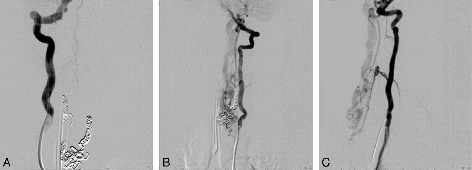
A, Following coil embolization, there is no further opacification of the fistula from the right vertebral arterial injection. A−C, Segmental branches of the left vertebral artery continue to opacify the AVF, albeit less briskly, as seen in frontal (A) and lateral views (C).

Fig 6.
  • Download figure
  • Open in new tab
  • Download powerpoint
Fig 6.

Intraoperative photographs via the operating microscope after opening the dura. A and B, Prominent dilated veins on the dorsal surface of the cord with evidence of coils in a large venous varix are seen preresection (A) and are absent postresection (B). C, Frontal view of a left vertebral artery injection on postoperative angiography demonstrates complete elimination of the AVF. The patient is neurologically intact.

In the other patient (case 2 in the Table) with cervical AVF, the right vertebral artery and thyrocervical trunk provided the main supply to a complex AVM, which was treated by endovascular methods only (Fig 7). The patient had a good recovery.

Fig 7.
  • Download figure
  • Open in new tab
  • Download powerpoint
Fig 7.

Frontal views of a right subclavian (A) and right vertebral artery (B) injection during embolization demonstrate a complex AVF supplied by branches of the right thyrocervical trunk and segmental right vertebral artery branches.

In the 2 patients with flaccid paralysis and bladder dysfunction, a fast-flow AVM was identified at the conus, treated surgically in 1 patient and via n-BCA embolization in the other. The fifth patient, with severe sensorimotor deficits, had a fast-flow AVM at the lumbosacral junction, which was treated with glue embolization. All arteriovenous lesions demonstrated a fast-flow pattern on angiography.

Discussion

CM presents as a characteristic macular stain and has a reported incidence of 0.3% in neonates.7 This slow-flow lesion consists of dermal capillary venular-like channels that are dilated and/or increased in number.7 There are reports of a familial tendency for CM. In a study from the Division of Plastic and Reconstructive Surgery of the Academic Medical Center, Amsterdam, 55 of 280 new patients (19.6%) presenting for treatment had an affected relative.8 When these pedigrees were analyzed, there was no discernable mode of inheritance. While the most common presentation of CM is without association with other anomalies, in some patients, CM can herald the presence of an underlying anomaly of the neurovascular axis.

CM-AVM is a newly described syndrome caused by heterozygous mutations in RASA1, located on 5q13.3.1 The capillary stains in this disorder are phenotypically slightly different from the common-variety CM. They are round or oval and pinkish-tan.9 When solitary, they range in diameter from very small to several centimeters. Multiple lesions also occur and may continue to appear throughout childhood. Often these lesions are surrounded by a pale halo, and even a small stain can exhibit a prominent to-fro murmur by hand-held Doppler examination.9 These atypical cutaneous CMs occur in association with fast-flow anomalies, namely AVM and AVF, in approximately 18.5% of affected families.1,9 They involve the cutis, subcutis, muscles, or bones. Intracranial manifestations include vein of Galen malformations with symptoms at birth or pial AVMs or AVFs, which become symptomatic during the first year of life.9

To the best of our knowledge, an association of CM-AVM and spinal arteriovenous anomalies has not been described. Macro-AVFs are fast-flow direct shunts, fed by 1 or multiple spinal cord arteries, typically ending directly in a giant venous ectasia (characteristic of these lesions), with secondary drainage into the extrinsic and intrinsic venous system of the cord.10 Progressive symptoms are a result either of congestion in the intrinsic and extrinsic venous system of the cord or of direct mass effect inflicted by the ecstatic veins. Bleeding from spinal AVFs is seen more frequently in children than in adults.10 The complexity of these lesions often mandates multidisciplinary treatment, as shown in Figs 3⇑⇑–6.

SWS is the most well-known association of facial CM, often with diffuse capillary staining of the trunk and extremities and ipsilateral vascular anomalies of the leptomeninges and ocular choroid.11,12 In SWS, a metameric distribution of the facial and cerebral venous lesions is seen, which is rarely bilateral. A faint upper facial CM may also indicate a unilateral AVM of the retina and intracranial optic pathway, an entity known by the syndromic terms Bonnet-Dechaume-Blanc in the European literature and Wyburn-Mason in the English literature.13 This, too, is a metameric syndrome affecting the cerebrofacial territory, giving rise to cerebral and facial AVMs. A circular capillary stain of the occipital scalp is often present with heterotopic neural nodules and the “hair collar” sign.14 Enjolras et al15 reported skin anomalies in the cervical area to be indicative of occult spinal dysraphism. CM on the posterior thorax may be a sign of an underlying AVM of the spinal cord.16,17 Cobb syndrome is the spinal expression of the metameric AVM syndrome involving skin, bone, and spinal cord. A capillary stain over the lower lumbar region may overlie spinal dysraphism, lipomeningocele, tethered spinal cord, and diastematomyelia.18

Intracranial AVMs in HHT are usually diagnosed in adulthood, though some pediatric cases have been reported.19 In contrast to CM-AVM, the liver and lungs are typically involved. The mucocutaneous stains of HHT are radiating or arborizing telangiectasias. Macrofistulous spinal cord AVMs have also been reported in patients with HHT.10,20,21 Definite diagnosis of HHT is made clinically if 3 of the 5 Curaçao criteria, established in June 1999 by the Scientific Advisory Board of the HHT Foundation International, are met. The criteria include epistaxis, telangiectasias, visceral AVMs, cerebral or spinal AVMs, and a first-degree relative with HHT.22

Fast-flow fistulas, both intracranial and in the spinal axis, should be considered in patients with the characteristic CMs, who evidence even minor neurologic symptoms. Once fixed neurologic deficits are manifest, functional recovery is much less likely.23 The availability of genetic testing will likely result in an increased reporting incidence of CM-AVM, with 200 individuals thus far identified with RASA1 mutations.9 It is likely that some patients with arteriovenous anomalies of the neural axis, previously labeled as Cobb syndrome or HHT, might, in fact, be more accurately diagnosed with CM-AVM.

Footnotes

  • Indicates Editor's Choices selection

  • Embedded Image

References

  1. 1.↵
    1. Eerola I,
    2. Boon LM,
    3. Mulliken JB,
    4. et al
    . Capillary malformation-arteriovenous malformation: a new clinical and genetic disorder caused by RASA1 mutations. Am J Hum Genet 2003; 73: 1240–49
    CrossRefPubMed
  2. 2.↵
    1. Boon LM,
    2. Mulliken JB,
    3. Vikkula M
    . RASA1: variable phenotype with capillary and arteriovenous malformations. Curr Opin Genet Dev 2005; 15: 265–69
    CrossRefPubMed
  3. 3.↵
    1. Henkemeyer M,
    2. Rossi DJ,
    3. Holmyard DP,
    4. et al
    . Vascular system defects and neuronal apoptosis in mice lacking Ras GTPase-activating protein. Nature 1995; 377: 695–701
    CrossRefPubMed
  4. 4.↵
    1. Lapinski PE,
    2. Bauler TJ,
    3. Brown EJ,
    4. et al
    . Generation of mice with a conditional allele of the p120 Ras GTPase-activating protein. Genesis 2007; 45: 762–67
    CrossRefPubMed
  5. 5.↵
    1. Limaye N,
    2. Boon LM,
    3. Vikkula M
    . From germline towards somatic mutations in the pathophysiology of vascular anomalies. Hum Mol Genet 2009; 18: R65–74
    Abstract/FREE Full Text
  6. 6.↵
    1. Kaplan P,
    2. Hollenberg RD,
    3. Fraser FC
    . A spinal arteriovenous malformation with hereditary cutaneous hemangiomas. Am J Dis Child 1976; 130: 1329–31
    CrossRefPubMed
  7. 7.↵
    1. Jacobs AH,
    2. Walton RG
    . The incidence of birthmarks in the neonate. Pediatrics 1976; 58: 218–22
    Abstract/FREE Full Text
  8. 8.↵
    1. van der Horst CM,
    2. van Eijk TGJ,
    3. de Borgie CA
    . Hereditary port-wine stains: do they exist? Lasers Med Sci 1999; 14: 238–43
    CrossRef
  9. 9.↵
    1. Revencu N,
    2. Boon LM,
    3. Mulliken JB,
    4. et al
    . Parkes Weber syndrome, vein of Galen aneurysmal malformation, and other fast-flow vascular anomalies are caused by RASA1 mutations. Hum Mutat 2008; 29: 959–65
    CrossRefPubMed
  10. 10.↵
    1. Mandzia JL,
    2. terBrugge KG,
    3. Faughnan ME,
    4. et al
    . Spinal cord arteriovenous malformations in two patients with hereditary hemorrhagic telangiectasia. Childs Nerv Syst 1999; 15: 80–83
    CrossRefPubMed
  11. 11.↵
    1. Terdjman P,
    2. Aicardi J,
    3. Sainte-Rose C,
    4. et al
    . Neuroradiological findings in Sturge-Weber syndrome (SWS) and isolated pial angiomatosis. Neuropediatrics 1991; 22: 115–20
    PubMed
  12. 12.↵
    1. Uram M,
    2. Zubillaga C
    . The cutaneous manifestations of Sturge-Weber syndrome. J Clin Neuroophthalmol 1982; 2: 245–48
    PubMed
  13. 13.↵
    1. Schmidt D,
    2. Pache M,
    3. Schumacher M
    . The congenital unilateral retinocephalic vascular malformation syndrome (Bonnet-Dechaume-Blanc syndrome or Wyburn-Mason syndrome): review of the literature. Surv Ophthalmol 2008; 53: 227–49
    CrossRefPubMed
  14. 14.↵
    1. Rogers GF,
    2. Mulliken JB,
    3. Kozakewich HP
    . Heterotopic neural nodules of the scalp. Plast Reconstr Surg 2005; 115: 376–82
    CrossRefPubMed
  15. 15.↵
    1. Enjolras O,
    2. Boukobza M,
    3. Jdid R
    . Cervical occult spinal dysraphism: MRI findings and the value of a vascular birthmark. Pediatr Dermatol 1995; 12: 256–59
    PubMed
  16. 16.↵
    1. Doppman JL,
    2. Wirth FP Jr.,
    3. Di Chiro G,
    4. et al
    . Value of cutaneous angiomas in the arteriographic localization of spinal-cord arteriovenous malformations. N Engl J Med 1969; 281: 1440–44
    PubMed
  17. 17.↵
    1. Jessen RT,
    2. Thompson S,
    3. Smith EB
    . Cobb syndrome. Arch Dermatol 1977; 113: 1587–90
    CrossRefPubMed
  18. 18.↵
    1. Guggisberg D,
    2. Hadj-Rabia S,
    3. Viney C,
    4. et al
    . Skin markers of occult spinal dysraphism in children: a review of 54 cases. Arch Dermatol 2004; 140: 1109–15
    CrossRefPubMed
  19. 19.↵
    1. Kadoya C,
    2. Momota Y,
    3. Ikegami Y,
    4. et al
    . Central nervous system arteriovenous malformations with hereditary hemorrhagic telangiectasia: report of a family with three cases. Surg Neurol 1994; 42: 234–39
    CrossRefPubMed
  20. 20.↵
    1. Morgan T,
    2. McDonald J,
    3. Anderson C,
    4. et al
    . Intracranial hemorrhage in infants and children with hereditary hemorrhagic telangiectasia (Osler-Weber-Rendu syndrome). Pediatrics 2002; 109: E12
    CrossRefPubMed
  21. 21.↵
    1. Rodesch G,
    2. Hurth M,
    3. Alvarez H,
    4. et al
    . Spinal cord intradural arteriovenous fistulae: anatomic, clinical, and therapeutic considerations in a series of 32 consecutive patients seen between 1981 and 2000 with emphasis on endovascular therapy. Neurosurgery 2005; 57: 973–83
    CrossRefPubMed
  22. 22.↵
    1. Shovlin CL,
    2. Guttmacher AE,
    3. Buscarini E,
    4. et al
    . Diagnostic criteria for hereditary hemorrhagic telangiectasia (Rendu-Osler-Weber syndrome). Am J Med Genet 2000; 91: 66–67
    CrossRefPubMed
  23. 23.↵
    1. Cullen S,
    2. Alvarez H,
    3. Rodesch G,
    4. et al
    . Spinal arteriovenous shunts presenting before 2 years of age: analysis of 13 cases. Childs Nerv Syst 2006; 22: 1103–10
    CrossRefPubMed
  • Received July 20, 2009.
  • Accepted after revision September 9, 2009.
  • Copyright © American Society of Neuroradiology
View Abstract
PreviousNext
Back to top

In this issue

American Journal of Neuroradiology: 31 (4)
American Journal of Neuroradiology
Vol. 31, Issue 4
1 Apr 2010
  • Table of Contents
  • Index by author
Advertisement
Print
Download PDF
Email Article

Thank you for your interest in spreading the word on American Journal of Neuroradiology.

NOTE: We only request your email address so that the person you are recommending the page to knows that you wanted them to see it, and that it is not junk mail. We do not capture any email address.

Enter multiple addresses on separate lines or separate them with commas.
A Novel Association between RASA1 Mutations and Spinal Arteriovenous Anomalies
(Your Name) has sent you a message from American Journal of Neuroradiology
(Your Name) thought you would like to see the American Journal of Neuroradiology web site.
CAPTCHA
This question is for testing whether or not you are a human visitor and to prevent automated spam submissions.
Cite this article
R. Thiex, J.B. Mulliken, N. Revencu, L.M. Boon, P.E. Burrows, M. Cordisco, Y. Dwight, E.R. Smith, M. Vikkula, D.B. Orbach
A Novel Association between RASA1 Mutations and Spinal Arteriovenous Anomalies
American Journal of Neuroradiology Apr 2010, 31 (4) 775-779; DOI: 10.3174/ajnr.A1907

Citation Manager Formats

  • BibTeX
  • Bookends
  • EasyBib
  • EndNote (tagged)
  • EndNote 8 (xml)
  • Medlars
  • Mendeley
  • Papers
  • RefWorks Tagged
  • Ref Manager
  • RIS
  • Zotero
0 Responses
Respond to this article
Share
Bookmark this article
A Novel Association between RASA1 Mutations and Spinal Arteriovenous Anomalies
R. Thiex, J.B. Mulliken, N. Revencu, L.M. Boon, P.E. Burrows, M. Cordisco, Y. Dwight, E.R. Smith, M. Vikkula, D.B. Orbach
American Journal of Neuroradiology Apr 2010, 31 (4) 775-779; DOI: 10.3174/ajnr.A1907
del.icio.us logo Twitter logo Facebook logo Mendeley logo
  • Tweet Widget
  • Facebook Like
  • Google Plus One
Purchase

Jump to section

  • Article
    • Abstract
    • Abbreviations
    • Materials and Methods
    • Results
    • Discussion
    • Footnotes
    • References
  • Figures & Data
  • Info & Metrics
  • Responses
  • References
  • PDF

Related Articles

  • No related articles found.
  • PubMed
  • Google Scholar

Cited By...

  • Clarifying the clinical landscape of pediatric spinal arteriovenous shunts: an institutional experience and individual patient-data meta-analysis
  • Parkes Weber Syndrome and Spinal Arteriovenous Malformations
  • Pial arteriovenous fistulae in pediatric patients: associated syndromes and treatment outcome
  • Crossref (83)
  • Google Scholar

This article has been cited by the following articles in journals that are participating in Crossref Cited-by Linking.

  • Management of Stroke in Neonates and Children: A Scientific Statement From the American Heart Association/American Stroke Association
    Donna M. Ferriero, Heather J. Fullerton, Timothy J. Bernard, Lori Billinghurst, Stephen R. Daniels, Michael R. DeBaun, Gabrielle deVeber, Rebecca N. Ichord, Lori C. Jordan, Patricia Massicotte, Jennifer Meldau, E. Steve Roach, Edward R. Smith
    Stroke 2019 50 3
  • RASA1Mutations and Associated Phenotypes in 68 Families with Capillary Malformation-Arteriovenous Malformation
    Nicole Revencu, Laurence M. Boon, Antonella Mendola, Maria Rosa Cordisco, Josée Dubois, Philippe Clapuyt, Frank Hammer, David J. Amor, Alan D. Irvine, Eulalia Baselga, Anne Dompmartin, Samira Syed, Ana Martin-Santiago, Lesley Ades, Felicity Collins, Janine Smith, Sarah Sandaradura, Victoria R. Barrio, Patricia E. Burrows, Francine Blei, Mariarosaria Cozzolino, Nicola Brunetti-Pierri, Asuncion Vicente, Marc Abramowicz, Julie Désir, Catheline Vilain, Wendy K. Chung, Ashley Wilson, Carol A. Gardiner, Yim Dwight, David J.E. Lord, Leona Fishman, Cheryl Cytrynbaum, Sarah Chamlin, Fred Ghali, Yolanda Gilaberte, Shelagh Joss, Maria del C. Boente, Christine Léauté-Labrèze, Marie-Ange Delrue, Susan Bayliss, Loreto Martorell, Maria-Antonia González-Enseñat, Juliette Mazereeuw-Hautier, Brid O'Donnell, Didier Bessis, Reed E. Pyeritz, Aicha Salhi, Oon T. Tan, Orli Wargon, John B. Mulliken, Miikka Vikkula
    Human Mutation 2013 34 12
  • Germline Loss-of-Function Mutations in EPHB4 Cause a Second Form of Capillary Malformation-Arteriovenous Malformation (CM-AVM2) Deregulating RAS-MAPK Signaling
    Mustapha Amyere, Nicole Revencu, Raphaël Helaers, Eleonore Pairet, Eulalia Baselga, Maria Cordisco, Wendy Chung, Josée Dubois, Jean-Philippe Lacour, Loreto Martorell, Juliette Mazereeuw-Hautier, Reed E. Pyeritz, David J. Amor, Annouk Bisdorff, Francine Blei, Hannah Bombei, Anne Dompmartin, David Brooks, Juliette Dupont, Maria Antonia González-Enseñat, Ilona Frieden, Marion Gérard, Malin Kvarnung, Andrea Kwan Hanson-Kahn, Louanne Hudgins, Christine Léauté-Labrèze, Catherine McCuaig, Denise Metry, Philippe Parent, Carle Paul, Florence Petit, Alice Phan, Isabelle Quere, Aicha Salhi, Anne Turner, Pierre Vabres, Asuncion Vicente, Orli Wargon, Shoji Watanabe, Lisa Weibel, Ashley Wilson, Marcia Willing, John B. Mulliken, Laurence M. Boon, Miikka Vikkula
    Circulation 2017 136 11
  • Management of Arteriovenous Malformations
    Arin K. Greene, Darren B. Orbach
    Clinics in Plastic Surgery 2011 38 1
  • RASA1 somatic mutation and variable expressivity in capillary malformation/arteriovenous malformation (CM/AVM) syndrome
    Colleen F. Macmurdo, Whitney Wooderchak‐Donahue, Pinar Bayrak‐Toydemir, Jenny Le, Matthew B. Wallenstein, Carlos Milla, Joyce M. C. Teng, Jonathan A. Bernstein, David A. Stevenson
    American Journal of Medical Genetics Part A 2016 170 6
  • Pathogenesis of Vascular Anomalies
    Laurence M. Boon, Fanny Ballieux, Miikka Vikkula
    Clinics in Plastic Surgery 2011 38 1
  • Somatic mutations in intracranial arteriovenous malformations
    Jeremy A. Goss, August Y. Huang, Edward Smith, Dennis J. Konczyk, Patrick J. Smits, Christopher L. Sudduth, Christopher Stapleton, Aman Patel, Sanda Alexandrescu, Matthew L. Warman, Arin K. Greene, Ajith J. Thomas
    PLOS ONE 2019 14 12
  • Pial arteriovenous fistulae in pediatric patients: associated syndromes and treatment outcome
    Brian P Walcott, Edward R Smith, R Michael Scott, Darren B Orbach
    Journal of NeuroInterventional Surgery 2013 5 1
  • Capillary Malformation—Arteriovenous Malformation Syndrome: Review of the Literature, Proposed Diagnostic Criteria, and Recommendations for Management
    Charisse M. Orme, Lynn M. Boyden, Keith A. Choate, Richard J. Antaya, Brett A. King
    Pediatric Dermatology 2013 30 4
  • Expanding the clinical and molecular findings in RASA1 capillary malformation-arteriovenous malformation
    Whitney L. Wooderchak-Donahue, Peter Johnson, Jamie McDonald, Francine Blei, Alejandro Berenstein, Michelle Sorscher, Jennifer Mayer, Angela E. Scheuerle, Tracey Lewis, J. Fredrik Grimmer, Gresham T. Richter, Marcie A. Steeves, Angela E. Lin, David A. Stevenson, Pinar Bayrak-Toydemir
    European Journal of Human Genetics 2018 26 10

More in this TOC Section

  • SyMRI & MR Fingerprinting in Brainstem Myelination
  • Comparison of Image Quality and Radiation Dose in Pediatric Temporal Bone CT Using Photon-Counting Detector CT and Energy-Integrating Detector CT
  • Venous Sinus Stenosis in Mucopolysaccharidosis I
Show more Pediatrics

Similar Articles

Advertisement

Indexed Content

  • Current Issue
  • Accepted Manuscripts
  • Article Preview
  • Past Issues
  • Editorials
  • Editor's Choice
  • Fellows' Journal Club
  • Letters to the Editor
  • Video Articles

Cases

  • Case Collection
  • Archive - Case of the Week
  • Archive - Case of the Month
  • Archive - Classic Case

Special Collections

  • AJNR Awards
  • ASNR Foundation Special Collection
  • Most Impactful AJNR Articles
  • Photon-Counting CT
  • Spinal CSF Leak Articles (Jan 2020-June 2024)

More from AJNR

  • Trainee Corner
  • Imaging Protocols
  • MRI Safety Corner

Multimedia

  • AJNR Podcasts
  • AJNR Scantastics

Resources

  • Turnaround Time
  • Submit a Manuscript
  • Submit a Video Article
  • Submit an eLetter to the Editor/Response
  • Manuscript Submission Guidelines
  • Statistical Tips
  • Fast Publishing of Accepted Manuscripts
  • Graphical Abstract Preparation
  • Imaging Protocol Submission
  • Evidence-Based Medicine Level Guide
  • Publishing Checklists
  • Author Policies
  • Become a Reviewer/Academy of Reviewers
  • News and Updates

About Us

  • About AJNR
  • Editorial Board
  • Editorial Board Alumni
  • Alerts
  • Permissions
  • Not an AJNR Subscriber? Join Now
  • Advertise with Us
  • Librarian Resources
  • Feedback
  • Terms and Conditions
  • AJNR Editorial Board Alumni

American Society of Neuroradiology

  • Not an ASNR Member? Join Now

© 2025 by the American Society of Neuroradiology All rights, including for text and data mining, AI training, and similar technologies, are reserved.
Print ISSN: 0195-6108 Online ISSN: 1936-959X

Powered by HighWire