
Abstract
BACKGROUND AND PURPOSE: Gamma knife radiosurgery has become an important treatment option for vestibular schwannoma. The effect of treatment can be assessed only by neuroimaging. We analyzed the evolution of follow-up MR imaging findings after gamma knife radiosurgery to provide information for the clinical management of these tumors.
METHODS: Changes in tumor volume and enhancement were assessed visually on 341 follow-up MR studies obtained in 78 of 86 consecutive patients with unilateral vestibular schwannoma who underwent gamma knife radiosurgery.
RESULTS: Follow-up MR studies were obtained between 10 and 63 months (mean, 34 months) after treatment. Tumor control rate was 81%. Changes in tumor volume were classified as temporary enlargement (41%), no change or sustained regression (34%), alternating enlargement and regression (13%), or continuous enlargement (12%). Temporary enlargement occurred within 2 years after radiosurgery. Changes in tumor enhancement were classified as transient loss of enhancement (84%), continuous increase in enhancement (5%), or no change in enhancement (11%). There was no significant correlation between changes in tumor volume and tumor enhancement. Areas of T2 hyperintensity in adjacent brain tissue appeared in 31% of patients.
CONCLUSION: Dynamic changes in vestibular schwannoma are seen on serial follow-up MR studies obtained after gamma knife radiosurgery. An increase in tumor size up to 2 years after radiosurgery is likely to be followed by regression. Changes in contrast enhancement are not predictive of clinical outcome. Neuroimaging follow-up is recommended.
Gamma knife radiosurgery was first used to treat vestibular schwannomas in 1969 (1). The relatively high peripheral radiation dose of about 20 Gy achieved a good rate of tumor control with a low rate of cranial nerve dysfunction as compared with microsurgical resection at that time (2). However, the high radiation doses were associated with unacceptable complication rates relative to the improved results obtained by modern surgical techniques. Subsequently, radiation doses were significantly reduced, and 12 to 16 Gy is now used in many centers for the greatest benefit and least risk of complications (3–12). The good results have encouraged the adoption of gamma knife radiosurgery as an important treatment option for vestibular schwannoma.
The treatment goal of gamma knife radiosurgery for vestibular schwannomas is to arrest tumor growth throughout the patient's life. Follow-up imaging is needed to fully assess the effectiveness of tumor control. Unfortunately, the long-term follow-up results in several series of patients treated with high peripheral doses (7, 8, 13) are difficult to compare with more recent series of patients treated with low peripheral doses and less than 10 years' follow-up (10–12, 14–19). Currently, MR imaging is regarded as the most sensitive neuroimaging technique by which to evaluate tumor response. Optimum clinical management of tumors requires understanding of the changes in MR imaging findings over time and of the implications of these findings for clinical outcome. Previous analyses of serial MR studies obtained after gamma knife radiosurgery for vestibular schwannomas involved only a small series of patients or a small number of follow-up studies per patient (14, 20–22).
We describe the serial MR imaging findings in 78 patients with unilateral vestibular schwannoma who underwent gamma knife radiosurgery and analyze the changes in these studies over time to provide data for the follow-up management of vestibular schwannomas after gamma knife radiosurgery.
Methods
Patients
Eighty-six patients with unilateral vestibular schwannoma underwent gamma knife radiosurgery with a Leksell Gamma Unit (Elekta Instruments, Atlanta, GA) between November 1991 and April 1996. Selection for gamma knife radiosurgery required that the maximum tumor diameter be less than 40 mm. Gamma knife radiosurgery was recommended for patients who were elderly or in poor medical condition, who had a tumor in the only hearing ear, or who had regrowth after partial removal of a tumor. Patients with a tumor that could be treated by either surgical resection or gamma knife radiosurgery were allowed to decide which alternative they preferred after receiving an adequate explanation of the treatment options.
Patients ranged in age from 23 to 79 years (mean, 53 years) and included 31 men and 55 women. Twenty-three patients (27%) had undergone previous surgical resection. Tumor volume varied from 0.2 to 20.1 mm3 (mean, 6.0 mm3) (Fig 1). Sixty-four tumors were solid and 22 had cystic components (mixed tumor).

Scattergram shows the relationship between tumor volume and peripheral dose. # indicates cystic tumor; only solid component was irradiated
Treatment
Stereotactic radiosurgery was performed with the standard gamma knife technique. Axial and coronal contrast-enhanced T1-weighted MR images were used for dose planning in all cases. The imaging technique included 2D Fourier transform sequences with a 3-mm slice thickness and an interslice gap of 1 mm for tumors with a diameter of more than 1 cm and 3D Fourier transform sequences with a 2-mm slice thickness for tumors with a diameter of less than 1 cm. Contrast-enhanced T1-weighted MR images were acquired 5 minutes after bolus injection of contrast medium (0.1 mmol/kg). Dose planning was done in consideration of tumor size, tumor consistency, hearing acuity, and regrowth after surgery (Fig 1). Doses to the periphery of the tumor (peripheral dose) varied from 10.0 to 17.0 Gy (mean, 13.3 Gy). Maximum tumor doses varied from 11.1 to 40.0 Gy (mean, 26.4 Gy). Treatment isodose (ie, that which enveloped the tumor volume with a maximum dose normalized 100%) varied from 30% to 90%. The peripheral dose coincided with the 50% isodose in 44 cases. The number of isocenters varied from one to 12 per tumor (median, four).
Follow-up MR Imaging
Follow-up neuroimaging included contrast-enhanced T1- and T2-weighted sequences at 3, 6, and 12 months during the first year; every 6 months during the second year; and yearly thereafter. If significant changes in tumor size or clinical presentation developed, follow-up studies were increased in frequency to every 3 months until stabilization occurred. Follow-up MR studies were acquired with the same protocol as that used before treatment.
Seventy-eight (91%) of the 86 patients underwent one or more MR studies at least 10 months after gamma knife radiosurgery. The last follow-up MR studies were obtained at less than 12 months in five patients, between 12 and 24 months in 19 patients, between 24 and 36 months in 23 patients, and at more than 36 months in 31 patients (range, 10–63 months; mean, 34 months). Tumor control rate was calculated in these patients. Six patients rejected neuroimaging follow-up. None of these patients showed remarkable symptoms after gamma knife radiosurgery. Two patients, ages 79 and 74 years, respectively, died of unrelated causes after gamma knife radiosurgery for vestibular schwannoma. Sixty-one patients (71%) had serial follow-up MR studies. In all, 341 follow-up studies were available for detailed analysis, with a mean of 5.6 studies per patient.
Analysis of Images
Tumor volume was calculated on each MR study by the following method. Maximum tumor diameter was measured in three dimensions (transverse and longitudinal on the axial images, and vertical on the coronal images) using fine calipers. Measurements were defined as x, y, and z, respectively. Tumor volume was xyz/2 (23).
Tumor control rate was estimated by comparing tumor volume at the time of treatment with that at the last follow-up. A tumor was considered to be controlled if it showed either no change or regression. The rate of measurement error tends to be in inverse proportion to tumor volume; therefore, tumors were categorized according to calculated tumor volume as small (<2 mm3), medium (2–8 mm3), or large (>8 mm3). Significant change was categorized as more than 20%, more than 15%, or more than 10% of tumor volume. These figures correspond to ± 1 mm change in each given volume (ie, a volume of more than 2 mm3 = 20%).
The pattern of changes in tumor volume was examined by plotting the ratio of tumor volume at each follow-up study to that at treatment. Univariate analysis was used to evaluate the correlation between tumor volume and tumor control rate. For the purpose of evaluating the correlation between tumor consistency and tumor control rate, univariate analysis was also used to compare tumor control rates between the solid and mixed tumors.
Changes in contrast enhancement of the tumor over time were analyzed on serial MR studies for each patient; the degree of contrast enhancement of the tumor was assessed visually on each MR study.
T2-weighted images were inspected for new areas of signal hyperintensity in adjacent brain tissue. The correlation between occurrence of hyperintensity and initial tumor volume, peripheral dose, maximum dose, peripheral isodose, and history of prior surgery was evaluated by multivariate analysis.
Statistical analyses were performed using Stat View 4.5J (Abacus Concepts, Berkeley, CA). The χ2-test was used to evaluate the correlation between changes in tumor volume and contrast enhancement. Univariate or multivariate analysis was performed with the Cox proportional hazards model. P values less than .05 were considered significant.
Results
Changes in Tumor Volume
Among the 78 patients in whom follow-up studies were available, 47 showed tumor regression, 16 showed no change, and 15 showed tumor enlargement. In two patients, tumor enlargement occurred after gamma knife radiosurgery, so microsurgical resection was performed at the referral hospital. The tumor control rate was 81%. The correlation between tumor control rate and tumor volume was not statistically significant. The rate of tumor regression was significantly greater for mixed tumors than for solid tumors.
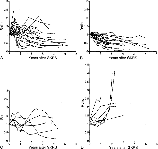
Changes in tumor volume were categorized into the following four patterns: 1) initial enlargement followed by sustained regression (although not necessarily reverting to initial size), termed “temporary enlargement” (n = 25) (Figs 2A and 3); 2) no change, or sustained regression (n = 21) (Figs 2B and 4); 3) repeated alternate enlargement and regression (n = 8) (Figs 2C and 5); and 4) continuous enlargement (n = 7) (Figs 2D and 6). Most tumors that showed temporary enlargement reached their peak within 1 year and regressed within 2 years. Maximum temporary enlargement was double the initial tumor volume in some cases. In six of seven patients with a pattern of continuous tumor enlargement, the enlargement persisted for more than 2 years. Follow-up MR studies in all patients with controlled tumor over 3 years showed no significant change in tumor volume. Five of the eight patients with a pattern of alternating changes had tumors that repeatedly showed enlargement and/or collapse of the cystic component. Follow-up MR studies revealed fluid-fluid levels in five of 22 patients with cystic components in the tumor.

Graphs showing the change in the ratio of tumor volume on follow-up MR examinations to tumor volume at treatment over time.
A, Tumor shows temporary enlargement.
B, Tumor shows no change or sustained regression without temporary enlargement.
C, Tumor shows repeated alternate enlargement and regression.
D, Tumor shows continuous enlargement.
Changes in Contrast Enhancement
Changes in tumor contrast enhancement could be divided into the following three patterns: 1) initial decrease of contrast enhancement followed by recovery of enhancement in the central area, termed “transient loss of contrast enhancement” (n = 51) (Figs 3, 4, and 6); 2) continuous increase in contrast enhancement (n = 3) (Fig 7); and 3) no change in contrast enhancement (n = 7) (Fig 8). The onset of a transient loss of contrast enhancement ranged from 2 to 12 months (median, 4 months), and recovery of contrast enhancement appeared from 7 to 35 months (median, 12 months). The onset of increased contrast enhancement ranged from 5 to 28 months (median, 14 months). We found no significant correlation between contrast enhancement and pattern of change in tumor volume (Fig 9).

Serial contrast-enhanced axial T1-weighted images (450/17/5) in a 51-year-old man. Note that the tumor shows temporary enlargement with transient loss of contrast enhancement 3 months after treatment. GKRS indicates gamma knife radiosurgery; mos., months after gamma knife radiosurgery
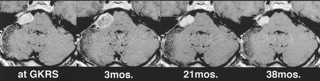
Serial contrast-enhanced axial T1-weighted images (450/17/5) in a 25-year-old woman. Note that the tumor shows no change in size with transient loss of contrast enhancement 6 months after treatment and thereafter the tumor shows continuous regression with recovery of contrast enhancement. GKRS indicates gamma knife radiosurgery; mos., months after gamma knife radiosurgery

Serial contrast-enhanced axial T1-weighted images (450/17/5) in a 64-year-old woman show enlargement of the cystic component and transient loss of contrast enhancement in the solid component at 3 months; regression of the cystic component, slight enlargement and recovery of contrast enhancement of the solid component, and slight regression of the overall tumor at 18 months; further enlargement of the solid component, no change in the cystic component, and regression of the overall tumor at 24 months; and remarkable regression of the tumor at 50 months. GKRS indicates gamma knife radiosurgery; mos., months after gamma knife radiosurgery

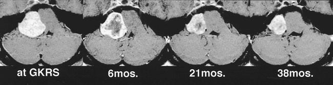
Serial contrast-enhanced axial T1-weighted images (250/25/1 at treatment, 400/15/2 at 6 months, and 400/15/4 at 12 months and 18 months after treatment) in a 55-year-old woman. Note the continuous enlargement of the tumor, although with transient loss of contrast enhancement. GKRS indicates gamma knife radiosurgery; mos., months after gamma knife radiosurgery
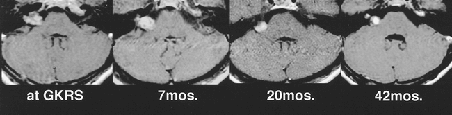
Serial contrast-enhanced axial T1-weighted images (250/30/1 at treatment, 400/17/3 at 7 months, 340/16/3 at 20 months, and 250/30/1 at 42 months after treatment) in a 42-year-old woman showing continuous increase in contrast enhancement without transient loss of contrast enhancement. GKRS indicates gamma knife radiosurgery; mos., months after gamma knife radiosurgery

Serial contrast-enhanced axial T1-weighted images (450/17/5 at treatment and 500/15/2 at each follow up) in a 52-year-old woman show tumor regression but almost no change in contrast enhancement. GKRS indicates gamma knife radiosurgery; mos., months after gamma knife radiosurgery
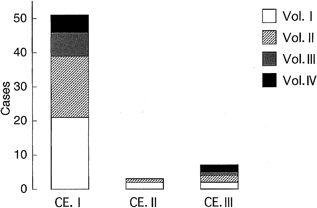
Bar graph shows the correlation between changes in tumor volume and contrast enhancement. Vol. I indicates temporary enlargement; Vol. II, no change or sustained regression; Vol. III, alternate enlargement and regression repeatedly; Vol. IV, continuous enlargement; CE. I, transient loss of contrast enhancement followed by recovery of enhancement; CE. II, continuous increase in enhancement; CE. III, no change in enhancement
Changes in Adjacent Brain
In 19 patients, T2-weighted MR images showed new areas of hyperintensity in adjacent brain tissue, which appeared from 3 to 37 months (mean, 12 months) after gamma knife radiosurgery (Fig 10). These findings disappeared or improved partially in 14 patients and persisted for over 2 years in three patients. These areas of hyperintensity did not correlate significantly with initial tumor volume, peripheral dose, maximum dose, peripheral isodose, or history of previous surgery.

Serial axial T2-weighted images (2000/110/2 at treatment, 3400/100/4 at 6 months and 4000/100/3 at 10 months and 36 months after treatment) in a 52-year-old woman. Note the hyperintensity in the adjacent brain tissue at 6 months and remarkable improvement at 10 months. GKRS indicates gamma knife radiosurgery; mos., months after gamma knife radiosurgery
Discussion
In our series, the tumor control rate was 81%. However, eight of the 13 tumors judged as enlarged at the last follow-up had regressed after temporary enlargement. If these tumors are also considered to be controlled, then the control rate becomes 91%. These results are comparable to those of other series. Flickinger et al (5) treated 134 patients with 136 vestibular schwannomas with 12 to 20 Gy (median, 17 Gy) to the periphery of the tumors and found that tumor regressed in 42% and enlarged in 3% at a mean of 24 months' follow-up (range, 6–56 months). The actuarial tumor control rate was 89.2 ± 6.0%. In another study (13), evaluation of 174 patients, including those treated with high doses (25 to 35 Gy), followed up for a mean of 54 months (range, 12–206 months), found tumor regression in 56%, no change in 32%, and enlargement in 12%, with a tumor control rate of 88%. The outcomes for low- and high-dose treatment were not given separately; however, the early results of low-dose treatment showed no decrease in tumor control rate as compared with the results of high-dose treatment. Technological advancements in computerized dose planning and MR imaging techniques, which were not available in the early period, may have compensated for the reduction of delivered doses.
An understanding of the patterns of tumor behavior observed during follow-up MR imaging after gamma knife radiosurgery is important to offset the limited follow-up periods available for patients undergoing low-dose treatment and to predict the long-term effects of treatment. Initial enlargement followed by arrest of growth or regression (ie, temporary enlargement, as defined in our series) has been reported (7, 17, 20, 22, 24). Temporary enlargement was reported in 6% of 193 unilateral vestibular schwannomas (7) and in two of 10 tumors in another series (24). In our study, as many as 41% of tumors treated by gamma knife radiosurgery showed temporary enlargement. This high rate of temporary enlargement detected by close neuroimaging follow-up of the patients in our series suggests that temporary enlargement is more common after gamma knife radiosurgery for vestibular schwannomas than previously reported.
Microsurgical resection of tumors that have enlarged after gamma knife radiosurgery has shown hyalinized thrombosis, thickening of the vascular wall, vascular obstruction, and granulomatous change (16, 17). We believe that these “failures” in fact represent a stage of temporary enlargement that follows treatment. Such temporary enlargement is a biological response to radiation and complicates the interpretation of MR images in the early period after gamma knife radiosurgery. Therefore, we recommend that tumor enlargement identified at follow-up neuroimaging not associated with aggravation of signs and symptoms be followed closely by serial neuroimaging for at least 2 years after radiosurgery. In most patients, tumor volume tends to stabilize after 3 years (Fig 2), indicating the importance of monitoring tumor size with neuroimaging within the first 3 years after treatment.
Large tumors were treated with lower doses than those applied to small tumors (Fig 1), but large tumors responded to gamma knife radiosurgery as well as small tumors in our series. The effectiveness of gamma knife radiosurgery for cystic tumors compared favorably with that for solid tumors. However, cystic tumors tended to show alternating increase and decrease of tumor volume with time. Two of seven patients with continuous enlargement of the tumor showed enlargement of only the cystic component, with continuous regression of the solid component. Enlargement of the cystic component of the tumor may occur regardless of the effectiveness of gamma knife radiosurgery. Therefore, changes in the cystic component cause the alternating changes in the tumor volume and the unpredictable behavior of the tumor. Minor hemorrhage from the cyst wall may be responsible for enlargement of the cystic component, because some tumors show fluid-fluid levels on follow-up MR studies.
Transient loss of contrast enhancement occurred in 84% of the tumors in our series. Others have reported transient loss of contrast enhancement in 70% (8) and 79% (21) of treated schwannomas, and this was found to be a good prognostic indicator of tumor regression (25). In our series, continuous enlargement was found in five tumors that showed transient loss of contrast enhancement, a condition that may result from radiation-induced vascular injury and occlusion. Obliteration of blood supply to the tumor may be one of the major mechanisms controlling tumor growth after radiosurgery (25). However, five of seven tumors with no change in contrast enhancement showed regression, so other mechanisms must be important for controlling tumor growth. We believe that a transient loss of contrast enhancement does not necessarily predict subsequent tumor regression.
Conclusion
Serial MR studies of unilateral vestibular schwannomas treated with gamma knife radiosurgery showed that temporary enlargement of tumor occurred in 41% of cases. Temporary enlargement occurred mostly within the first 2 years after radiosurgery. Subsequent regression of tumor volume occurred during and after the second year following radiosurgery. Because such enlargement within 2 years after gamma knife radiosurgery is usually followed by regression, close follow-up with neuroimaging is desirable in these cases. Transient loss of contrast enhancement after gamma knife radiosurgery was recognized in most cases, but was not necessarily a useful prognostic sign. Gamma knife radiosurgery for vestibular schwannomas was effective for both small and large tumors up to 4 cm in largest diameter and for solid tumors and tumors with cystic components. However, the behavior of the cystic component can make the tumor volume unpredictable. Serial MR studies may show that the tumor is stable after the third year following radiosurgery.
Footnotes
↵1 Address reprint requests to Hiroyuki Nakamura, MD, Department of Neurosurgery, Tohoku University School of Medicine, 1–1 Seiryo-machi, Aoba-ku, Sendai 980–8574, Japan.
References
- Received July 6, 1999.
- Accepted after revision February 20, 2000.
- Copyright © American Society of Neuroradiology
In this issue


Jump to section
Related Articles
Cited By...
- Significant Temporal Evolution of Diffusion Anisotropy for Evaluating Early Response to Radiosurgery in Patients with Vestibular Schwannoma: Findings from Functional Diffusion Maps
- Diagnostic Accuracy of the Constructive Interference in Steady State Sequence Alone for Follow-Up Imaging of Vestibular Schwannomas