
Abstract
Summary: We report the case of a 9-year-old male patient with idiopathic intracranial hypertension without papilledema for which MR imaging of the optic nerves and pituitary gland provided important clues for the diagnosis of idiopathic intracranial hypertension and showed a return to normal appearance after normalization of CSF pressure.
Idiopathic intracranial hypertension (IIH) is a headache syndrome characterized by raised CSF pressure in the absence of an intracranial mass lesion or ventricular dilation; normal CSF composition; usually normal results of neurologic examination, except for papilledema and occasional CN VI palsy; and a normal level of consciousness (1). The hallmark of IIH is papilledema, which may be bilateral, asymmetrical, or even unilateral; however, IIH can occur in the absence of papilledema. The diagnosis of IIH is, therefore, not always simply achieved. In this report, we describe the case of a 9-year-old male patient with IIH without papilledema for which MR imaging of the optic nerves and pituitary gland provided important clues to the diagnosis of IIH and showed a return to normal appearance after normalization of CSF pressure.
Case Report
A previously healthy 9-year-old slender male patient was referred because of severe headache and weight loss. He had loss of appetite and intermittent frontal headache, which lasted nearly all day and occurred at intervals of several days. These symptoms progressively worsened over 4 months, and significant weight loss (−20%) was recognized. The patient complained of transient obscuration of vision occurring once. At admission, the results of neurologic examinations were normal and neck stiffness was not observed. Blood pressure was normal (88/52 mm Hg). An ophthalmologic test revealed normal visual acuity and a normal visual field. Fundoscopic examination performed by an ophthalmologist revealed tortuous retinal vessels but no papilledema or disk atrophy. The results of routine blood and urine laboratory studies were all within normal limits. Electroencephalographic results were also normal.
Cranial MR imaging performed at the time of admission with a 1.5-T superconducting magnet showed small cortical veins and superior sagittal, straight, transverse, and sigmoid sinuses but no intracranial mass lesion, ventricular dilation, or sinus thrombosis (Fig 1A). Fat-saturated T2-weighted images revealed flattening of the posterior sclera, vertical tortuosity and elongation of the orbital optic nerves, and distension of the perioptic subarachnoid space (Fig 1B). Sagittal T1-weighted images also revealed a partially empty sella (Fig 1C). Two-dimensional phase-contrast MR velocity measurement of the superior sagittal sinus showed a peak systolic volume of 240 mL/min and an end diastolic volume of 210 mL/min. Three-dimensional phase-contrast MR venography (encoding velocity, 40 cm/s) revealed no sinus thrombosis. A lumbar puncture taken when the patient was in the decubitus position and at rest revealed an elevated CSF pressure of 106 cm H2O and a normal composition (ie, cell count, one leukocyte; protein, 35 mg/dL; and glucose, 68 mg/dL). A clinical diagnosis of IIH without papilledema was made. After initial lumbar puncture, the clinical symptoms gradually improved, and the patient's parents rejected medication such as acetazolamide or prednisolone.

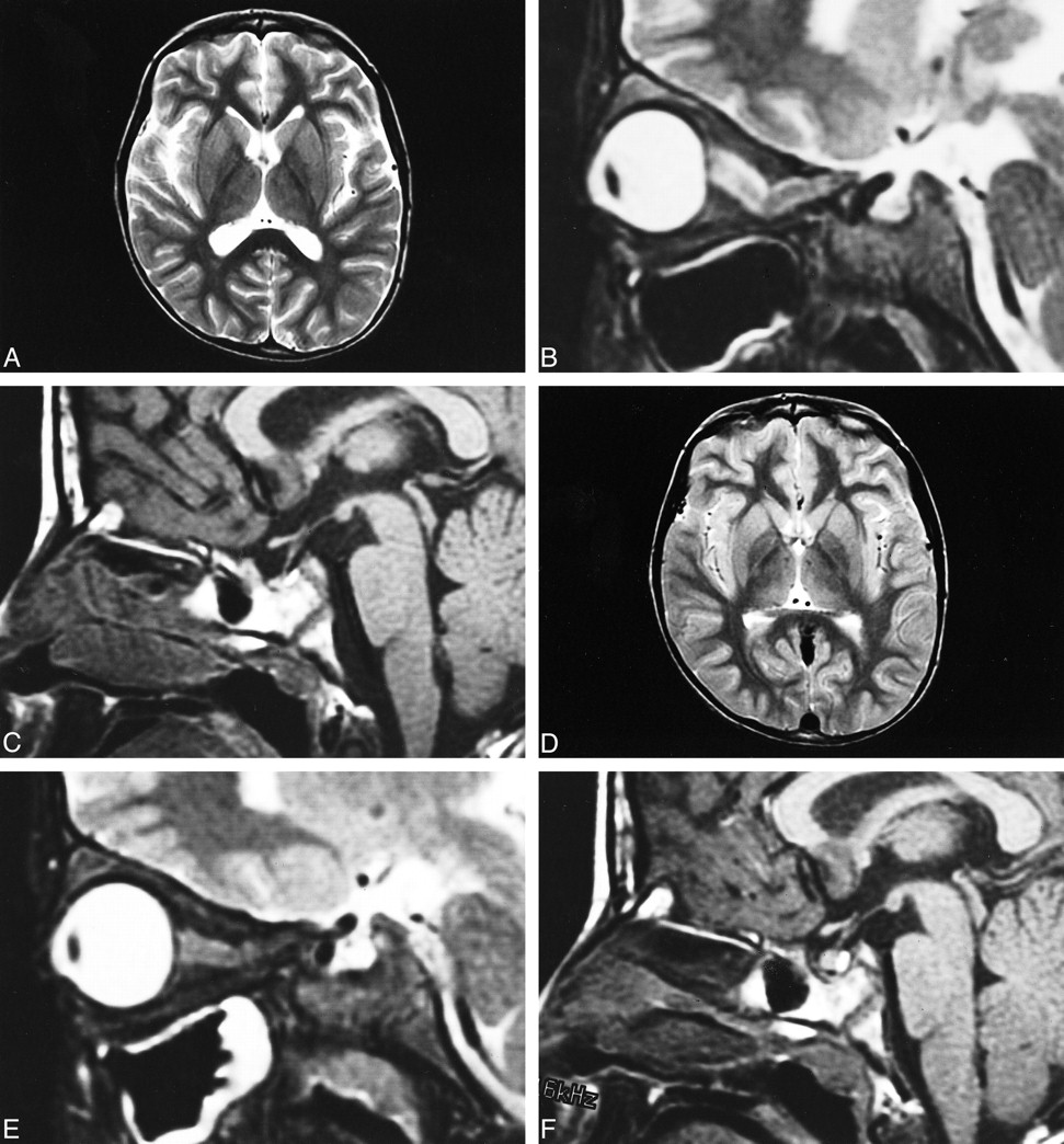
MR images from the case of a 9-year-old male patient with IIH without papilledema.
A, Transverse T2-weighted image (4000/96/2 [TR/TE/excitations]), obtained at admission, shows small cortical veins and superior sagittal, straight, transverse and sigmoid sinuses but no intracranial mass lesion, ventricular dilation, or sinus thrombosis.
B, Sagittal fat-saturated T2-weighted image (4000/100/2) of the right optic nerve, obtained at admission, shows flattening of the posterior sclera, vertical tortuosity and elongation of the nerve, and distension of the perioptic subarachnoid space.
C, Sagittal T1-weighted image (500/9/2), obtained at admission, shows a partially empty sella.
D, Transverse T2-weighted image, obtained 4 months after admission and after three lumbar punctures and improvement of the clinical symptomatology, reveals a decrease in the subarachnoid space and normalization of the sizes of the cortical veins and superior sagittal, straight, transverse, and sigmoid sinuses.
E, Sagittal fat-saturated T2-weighted image of the right optic nerve, obtained 4 months after admission and after three lumbar punctures and improvement of the clinical symptomatology, shows a normally round orbit and a normally straight nerve.
F, Sagittal T1-weighted image, obtained 4 months after admission and after three lumbar punctures and improvement of the clinical symptomatology, shows that the previously compressed pituitary gland had reexpanded to fill the sella turcica.
Because CSF pressure at the time of initial lumbar puncture was markedly elevated, even considered as IIH, lumbar puncture was performed three times in addition to therapy to monitor CSF pressure. CSF pressure had decreased to 35 and 15 cm H2O, respectively, at the times of the second and third taps (ie, 1 and 4 months after initial lumbar puncture). Also, the headache and appetite loss disappeared completely. The volume of CSF removed during each puncture was approximately 20 mL. MR imaging performed at 4 months after admission showed a decrease in the subarachnoid space and normalization of the sizes of the cortical veins and superior sagittal, straight, transverse, and sigmoid sinuses (Fig 1D), straightening of the optic nerves (Fig 1E), and a reexpanded pituitary gland (Fig 1F). Two-dimensional phase-contrast MR velocity measurement showed almost normal blood flow volume in the superior sagittal sinus (ie, peak systolic volume of 390 mL/min and end diastolic volume of 250 mL/min).
Discussion
IIH, also known as pseudotumor cerebri and benign intracranial hypertension, is a syndrome characterized by increased CSF pressure and papilledema in patients without focal neurologic findings, except for occasional CN VI palsy. It is a diagnosis of exclusion, and radiologic examinations are traditionally performed to help exclude lesions that produce intracranial hypertension, such as obstructive hydrocephalus, tumor, chronic meningitis, arteriovenous fistula, internal jugular vein stenosis, and dural sinus thrombosis.
CSF pressure of 106 cm H2O (78 mm Hg) during initial lumbar puncture was almost as high as the systolic blood pressure, which should result in a mortal metabolic disturbance and in a lack of energy in the CNS. Johnston et al (2) continuously monitored CSF pressure in 20 IIH patients by inserting an intraventricular catheter. According to their report, the mean pressure levels were less than 35 mm Hg (48 cm H2O); however, plateau waves, varying in amplitude from 50 to 80 mm Hg (68–109 cm H2O) and in duration from 5 to 20 minutes, were seen in eight of the 20 patients. The plateau waves were not accompanied by headache or any other change in the patient's clinical state. It is therefore speculated that the markedly elevated CSF pressure at the time of initial lumbar puncture reflected plateau waves.
Direct transmission of the elevated CSF pressure results in distension of the perioptic subarachnoid space and ballooning of the optic papilla, causing it to protrude physically into the posterior aspect of the globe (1, 3–5). The long-standing effect of pulsatile CSF under high pressure also leads to downward herniation of an arachnocele through a defect in the diaphragma sella (6, 7). The use of high-resolution, thin-slice MR imaging improves the visualization of the optic nerves and pituitary gland. In one study, MR imaging disclosed flattening of the posterior sclera in 80%, an empty sella in 70%, distension of the perioptic subarachnoid space in 45%, enhancement of the prelaminar optic nerve in 50%, vertical tortuosity of the orbital optic nerve in 40%, and intraocular protrusion of the prelaminar optic nerve in 30% of 20 patients with IIH (3). The present patient actually exhibited flattening of the posterior sclera, distension of the perioptic subarachnoid space, vertical tortuosity of the orbital optic nerve, and a partially empty sella on initial MR images, from which a diagnosis of IIH was strongly suspected. CSF pressure measurement via the lumbar route was therefore performed. For patients with severe headache associated with papilledema, lumbar puncture is always performed after CT or MR imaging to exclude intracranial mass lesions. However, the procedure is not always performed for those without papilledema when the results of initial neuroimaging are grossly normal. Soler et al (1) reported that four of 22 pediatric patients with IIH showed increased CSF pressure in the absence of papilledema. Therefore, attention to the optic nerves and pituitary gland should be given to MR images of patients with severe headache so as not to miss IIH without papilledema.
Zagardo et al (6) repeated MR imaging in two patients with IIH and found that the previously compressed pituitary gland had reexpanded to fill the sella turcica after normalization of CSF pressure. This suggests that acute or subacute elevation of CSF pressure may be sufficient to compress the pituitary gland. Repeat MR imaging of the present patient also showed reversibility of a partially empty sella and normalization of the volume of the optic nerve sheaths, which had not been previously reported. The return to a normal appearance of the pituitary gland and optic nerves on MR images may indicate a positive response to therapy and possibly denote a corresponding decrease in CSF pressure. Technical difficulties, distressing to the patient, and the theoretical risk of the development of intraspinal epidermoid tumors and low back pain have discouraged us from using lumbar puncture repeatedly. Therefore, recovery from IIH is often assumed with the resolution of papilledema, and is thought to be synonymous with the return of CSF pressure to normal. MR imaging that is focused on optic nerves and the pituitary gland might also be used as a secondary outcome measure for monitoring patients with IIH, especially those without papilledema.
To the best of our knowledge, there have been no reports illustrating normalization of the sizes of cerebral veins and dural sinuses after decreasing CSF pressure in patients with IIH. Gideon et al (8) and Mattle et al (9), by means of MR flow measurement, showed mean blood flows in the superior sagittal sinus of 457 mL/min and 420 mL/min, respectively, in healthy control volunteers and also showed a tendency toward a lower mean flow volume in the superior sagittal sinus (mean, 345 mL/min) in patients with IIH than in control volunteers (8). The flow volume in the superior sagittal sinus in the present patient was low at the time of admission and increased to within normal range, probably because of resolution of the intracranial hypertension. On the other hand, MR imaging in cases of spontaneous intracranial hypotension may reflect an increase in the venous volume throughout the brain, which shows diffuse dural enhancement, prominent dural sinuses, and an engorged epidural venous plexus, with a return to normal appearance with elevation of CSF pressure (10, 11). Fishman and Dillon (10) speculated that the changes were a consequence of the Monro-Kellie rule (10, 12), which states that CSF volume fluctuates reciprocally with changes in intracranial blood volume. We also speculate that normalization in the sizes of cerebral sinuses and veins and in the flow volume of the superior sagittal sinus after normalization of CSF pressure are a consequence of reductions in CSF volume and pressure.
In conclusion, MR imaging of the optic nerves and pituitary gland may provide important clues for the diagnosis of IIH, with a return to normal appearance after normalization of CSF pressure. In the present case, follow-up MR imaging revealed normalization in the sizes of cerebral sinuses and veins and in the flow volume of the superior sagittal sinus, which could be a consequence of reduced CSF volume and pressure. Further evaluation of CNS circulation, including intracranial sinuses, is necessary for identifying the pathogenesis of IIH.
Acknowledgments
We thank K. Okumura, Y. Nakano, T. Isobe, and F. Morita for excellent technical support.
Footnotes
↵1 Address reprint requests to Jun-ichi Takanashi, Department of Pediatrics, Faculty of Medicine, Chiba University, 1-8-1 Inohana, Chuo-ku, Chiba-shi, Chiba 260-8677, Japan.
References
- Received January 3, 2000.
- Copyright © American Society of Neuroradiology
In this issue


Jump to section
Related Articles
Cited By...
- Hyperintense Optic Nerve Heads on Diffusion-Weighted Imaging: A Potential Imaging Sign of Papilledema
- Incidence of Cerebellar Tonsillar Ectopia in Idiopathic Intracranial Hypertension: A Mimic of the Chiari I Malformation
- Pseudotumor Cerebri: Brief Review of Clinical Syndrome and Imaging Findings
- Cerebrospinal fluid dynamics between the basal cisterns and the subarachnoid space of the optic nerve in patients with papilloedema