Advertisement

Main menu

  • Home
  • Content
    • Current Issue
    • Accepted Manuscripts
    • Article Preview
    • Past Issue Archive
    • Video Articles
    • AJNR Case Collection
    • Case of the Week Archive
    • Case of the Month Archive
    • Classic Case Archive
  • Special Collections
    • AJNR Awards
    • ASNR Foundation Special Collection
    • Most Impactful AJNR Articles
    • Photon-Counting CT
    • Spinal CSF Leak Articles (Jan 2020-June 2024)
  • Multimedia
    • AJNR Podcasts
    • AJNR SCANtastic
    • Trainee Corner
    • MRI Safety Corner
    • Imaging Protocols
  • For Authors
    • Submit a Manuscript
    • Submit a Video Article
    • Submit an eLetter to the Editor/Response
    • Manuscript Submission Guidelines
    • Statistical Tips
    • Fast Publishing of Accepted Manuscripts
    • Graphical Abstract Preparation
    • Imaging Protocol Submission
    • Author Policies
  • About Us
    • About AJNR
    • Editorial Board
    • Editorial Board Alumni
  • More
    • Become a Reviewer/Academy of Reviewers
    • Subscribers
    • Permissions
    • Alerts
    • Feedback
    • Advertisers
    • ASNR Home

User menu

  • Alerts
  • Log in

Search

  • Advanced search
American Journal of Neuroradiology

American Journal of Neuroradiology

ASHNR American Society of Functional Neuroradiology ASHNR American Society of Pediatric Neuroradiology ASSR
  • Alerts
  • Log in

Advanced Search

  • Home
  • Content
    • Current Issue
    • Accepted Manuscripts
    • Article Preview
    • Past Issue Archive
    • Video Articles
    • AJNR Case Collection
    • Case of the Week Archive
    • Case of the Month Archive
    • Classic Case Archive
  • Special Collections
    • AJNR Awards
    • ASNR Foundation Special Collection
    • Most Impactful AJNR Articles
    • Photon-Counting CT
    • Spinal CSF Leak Articles (Jan 2020-June 2024)
  • Multimedia
    • AJNR Podcasts
    • AJNR SCANtastic
    • Trainee Corner
    • MRI Safety Corner
    • Imaging Protocols
  • For Authors
    • Submit a Manuscript
    • Submit a Video Article
    • Submit an eLetter to the Editor/Response
    • Manuscript Submission Guidelines
    • Statistical Tips
    • Fast Publishing of Accepted Manuscripts
    • Graphical Abstract Preparation
    • Imaging Protocol Submission
    • Author Policies
  • About Us
    • About AJNR
    • Editorial Board
    • Editorial Board Alumni
  • More
    • Become a Reviewer/Academy of Reviewers
    • Subscribers
    • Permissions
    • Alerts
    • Feedback
    • Advertisers
    • ASNR Home
  • Follow AJNR on Twitter
  • Visit AJNR on Facebook
  • Follow AJNR on Instagram
  • Join AJNR on LinkedIn
  • RSS Feeds

AJNR is seeking candidates for the AJNR Podcast Editor. Read the position description.

Research ArticleBRAIN

Diffuse Lesions in the CNS Revealed by MR Imaging in a Case of Whipple Disease

Stéphane Kremer, Gérard Besson, Bruno Bonaz, Basile Pasquier, Jean-F. Le Bas and Sylvie Grand
American Journal of Neuroradiology March 2001, 22 (3) 493-495;
Stéphane Kremer
  • Find this author on Google Scholar
  • Find this author on PubMed
  • Search for this author on this site
Gérard Besson
  • Find this author on Google Scholar
  • Find this author on PubMed
  • Search for this author on this site
Bruno Bonaz
  • Find this author on Google Scholar
  • Find this author on PubMed
  • Search for this author on this site
Basile Pasquier
  • Find this author on Google Scholar
  • Find this author on PubMed
  • Search for this author on this site
Jean-F. Le Bas
  • Find this author on Google Scholar
  • Find this author on PubMed
  • Search for this author on this site
Sylvie Grand
  • Find this author on Google Scholar
  • Find this author on PubMed
  • Search for this author on this site
  • Article
  • Figures & Data
  • Info & Metrics
  • Responses
  • References
  • PDF
Loading

Abstract

Summary: We report a case of Whipple disease involving the brain, optic chiasm, posterior fossa, and spinal cord. We evaluate the role of MR imaging for initial evaluation and for long-term follow-up of Whipple disease involving the CNS. We also discuss the semiologic characteristics of the lesions.

Whipple disease is a systemic disorder affecting particularly the small bowel and inducing a malabsorption syndrome (1). Osteoarticular, cardiac, and CNS involvements are also common (1, 2). The responsibility of a gram-positive bacillus, Tropheryma whippelii, has been recently shown (3). The pathogenesis and epidemiology, however, remain obscure. The most common neurologic symptoms include cognitive changes, supranuclear gaze palsy, altered level of consciousness, psychiatric signs, and upper motor neurons signs (4). Postmortem studies (5) have shown that these neurologic symptoms are associated with a granulomatous infiltration of the different levels of the CNS, particularly the brain, chiasm, pituitary gland, pons, and spinal cord. The brain lesions are essentially confined to the gray matter, preferentially the basal part of the telencephalon, the hypothalamus, and the thalamus (6).

However, diagnosis of Whipple disease involving the CNS remains difficult. It is often delayed, particularly if it occurs without digestive signs, because 20% of the patients present with neurologic features alone and no other manifestations of the disease (4, 7–9). In these cases, neuroimaging, and particularly MR imaging, has been able to reveal, in a noninvasive manner, involvement of the CNS (7, 10–12). We report the first case of Whipple disease in which MR imaging revealed lesions affecting the different levels of the CNS simultaneously; namely the chiasm, brain, posterior fossa, and spinal cord.

Case Report

A 68-year-old man was admitted for walking difficulties, memory loss, and incontinence that had been progressing for 5 months. For 12 years, he had complained of arthralgias of the hands and wrists. The diagnosis of rheumatoid polyarthritis had been made.

A neurologic examination revealed a tetrapyramidal syndrome associated with apathy and memory loss. Blood tests indicated an inflammatory syndrome with an elevation of the erythrocyte sedimentation rate (80 mm at the first hour) and an increase of proteins. The results of antinuclear antibody, HIV, B and C hepatitis, latex, and Waler Rose tests were negative. The CSF test showed an increase in the protein rate (1.95 g/L) with an absolute increase of immunoglobulin G (205 mg/L), but the cell count and glucose level were normal. Polymerase chain reaction for Tropheryma whippelii was negative.

MR images of the brain (Fig 1A–D and F) had areas of T2 hyperintensity in the right and left mediobasal temporal lobe, anterior commissure, right and left mamillary bodies, left cerebral peduncle, right middle cerebellar peduncle, bulb, right lobe of the cerebellum, and optic chiasm. These lesions showed no mass effect. After IV infusion of a contrast agent, mild enhancement of the right temporal lesion could be seen. MR imaging of the spinal cord was also performed, showing T2 panmedullary central hyperintensity.

fig 1.
  • Download figure
  • Open in new tab
  • Download powerpoint
fig 1.

Images from the case of a 68-year-old man who was admitted for walking difficulties, memory loss, and incontinence that had been progressing for 5 months.

A, Axial T2-weighted MR image (4347/100/4) of the brain shows hyperintensity in the mediobasal part of the right and left temporal lobes (arrows), in the mamillary bodies (star), and in the optic chiasm (arrowhead).

B, Axial T2-weighted fluid-attenuated inversion recovery MR image (11000/140/2 [TR/TE/excitaitons]) of the brain shows diffuse hyperintensity involving the right mediobasal and left mediotemporal lobes (arrows), as well as involvement of the left cerebral peduncle (arrowhead). Note the lack of mass effect despite the size of the lesion.

C, Coronal fluid-attenuated inversion recovery MR image (11000/140/2) of the brain shows hyperintensity in the mediobasal part of right and left temporal lobes (arrows), brain stem (star), and right middle cerebellar peduncle (arrowhead).

D, Coronal T1-weighted MR image (535/14/2) of the brain shows mild enhancement of the right temporal lesions after the IV administration of a contrast agent (arrow).

E, Sagittal T2-weighted MR image (2745/120/6) of the spine shows hyperintensity in the central part of the spinal cord (arrow).

F, Coronal T2- weighted fluid-attenuated inversion recovery MR image (11000/140/2) of the brain, obtained after 1 year of treatment, shows nearly complete resolution of the right temporal lesion (arrow).

The patient underwent a duodenal biopsy, which showed macrophages that stained positively with periodic acid-Schiff stain (Fig 2). Polymerase chain reaction for Tropheryma whippelii was positive on the duodenal biopsy.

fig 2.
  • Download figure
  • Open in new tab
  • Download powerpoint
fig 2.

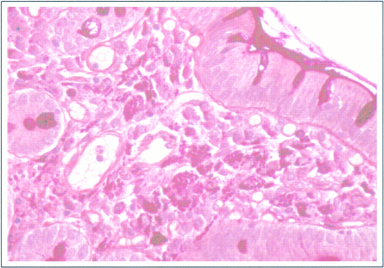
Duodenal biopsy was performed. Note the characteristic foamy macrophages containing periodic acid-Schiff stain-positive rod-shaped structures in the submucosal region

The patient was treated for 2 weeks with the IV administration of amoxicillin and amikacin sulfate and then with continuous orally administered sulfamethoxazole-trimethoprim. One year later, the neurologic abnormalities had improved, with nearly complete resolution of the tetrapyramidal syndrome and complete resolution of the cognitive disorders. This clinical improvement correlated with MR investigations that showed nearly complete regression of the right temporal lesion (Fig 1F). Complete resolution of the posterior fossa, optic chiasm, and spinal cord lesions was shown. Moderate cerebral and optic chiasm atrophy remained.

Discussion

A review of 84 cases described in the literature (4) revealed that clinically detectable involvement of the CNS ranges from 6% to 43% of patients suffering from Whipple disease. In 43 of the 84 reported cases, CT or MR imaging of the head was performed, and in 23, focal abnormalities were found (4).

However, Dobbins (1) postulates that all patients suffering from Whipple disease have anatomic-pathologic CNS involvement (13). It consists of multiple small circular or oval lesions, measuring an average of 2 mm in diameter, disseminated throughout the gray matter and characterized by the accumulation of macrophages, staining very intensely with periodic acid-Schiff stain, as shown in postmortem studies (6). Electron microscopy shows that the macrophages contain a bacilliform organism, named Tropheryma whippelii by Relman et al (3), revealing the bacterial cause of the disease (2, 6). The same lesions are found in other organs when they are affected by the disease (2, 6). Recently, molecular biology allowed the detection of the presence of Tropheryma whippelii in the affected organs by using polymerase chain reaction (3). Previous studies have shown a good correlation between preferential sites of anatomic-pathologic lesions and the lesions revealed by MR imaging and CT (5, 8, 9, 14).

The first neuroradiologic descriptions of CNS involvement were reported as seen on CT scans (8, 14), and they presented no specific characteristics (2). The CT scan can be normal, regardless of the clinical features, or can reveal focalized lesions, which can be hypo- or hyperdense, contrast enhanced or not, and with or without mass effect (2). However, MR imaging has proved its superiority for the detection of small lesions (3). In a review of the literature presented by Louis et al (4), 43 patients had undergone CT or MR imaging and three patients with normal CT findings had focal abnormalities on their MR images (4).

These lesions, which occur in 53% of the cases (4), often consist of T1 hypointensity and T2 hyperintensity, show no mass effect, and are located in the medial part of the temporal lobes, in the hypothalamic region, or in the pons (9, 10). These lesions are sometimes enhanced after infusion of contrast media (4, 8, 9, 14). Moreover, associated moderate atrophy occurs in 42% of the cases (4).

Multiple mass lesions have rarely been described. They usually appear hypointense on T1-weighted images and hyperintense on T2-weighted images and enhance after infusion of contrast media (12). However, a small number of patients have normal-appearing MR images (4).

The involvement of the spinal cord has rarely been reported. We found only one report of myelopathy secondary to Whipple disease, diagnosed by using MR imaging (11). It was located in the cervical region, and it appeared hyperintense on T2-weighted images after IV injection of gadolinium.

Involvement of the optic chiasm has also rarely been revealed by neuroradiologic examination, although it is more often discovered during postmortem studies (9). We found only one report with an increased T2 signal intensity located in the optic tracts (9). However, clinical optical manifestations are common in cases of Whipple disease (4, 5).

Early treatment of Whipple disease leads to improvement of the lesions revealed by neuroradiologic examination (4, 9, 12), as shown in our case, but diagnosis is often difficult. Diagnosis is often difficult to determine on the basis of polymerase chain reaction or histologic findings (98%); more precisely, 63% of patients underwent biopsy (88% of which were obtained from the small bowel (4). More rarely, the diagnosis is made based on lymph node biopsy or cerebral biopsy. However, polymerase chain reaction has shown sensitivity and specificity to confirm a diagnosis of Whipple disease (15), and recent studies have revealed genetic material of Tropheryma whippelii in peripheral blood mononuclear cells and in cells of pleural effusion (16), suggesting that the diagnosis can sometimes be made on the basis of polymerase chain reaction of peripheral blood (17). The diagnosis can also be obtained by conducting an analysis of the CSF for periodic acid-Schiff stain-positive cells (2), but the value of polymerase chain reaction in CSF has not yet been evaluated because of the rarity of the disease.

Treatment is often delayed, however, resulting in death or irreversible cerebral lesions, such as atrophy (9, 13). Moreover, relapse of the disease is frequent, leading to the reappearance of the CNS lesions on MR images (4).

In conclusion, this case report shows the importance of neuroradiologic examinations, particularly MR imaging, for the detection of CNS lesions in the initial evaluation of patients suspected of having Whipple disease and for the long-term follow-up of these patients. It is necessary to perform MR imaging of the brain and spinal cord to ascertain the different locations of these lesions.

Footnotes

  • ↵1 Address reprint requests to Dr. Sylvie Grand, Magnetic Resonance Imaging Unit, Centre Hospitalier et Universitaire de Grenoble, Hôpital Michallon, 38043 Grenoble Cedex 09, France.

References

  1. ↵
    Dobbins WO. Whipple's disease. Mayo Clin Proc 1988;63:623-624
    PubMed
  2. ↵
    Durand DV, Lecomte C, Cathébras P, Rousset H, Godeau P. Whipple disease: clinical review of 52 cases. Medicine (Baltimore) 1997;76:170-184
    CrossRefPubMed
  3. ↵
    Relman DA, Schmidt TM, Mac Dermott RP, Falcow S. Identification of the uncultured bacillus of Whipple's disease. N Engl J Med 1992;311:293-301
  4. ↵
    Louis ED, Lynch T, Kaufman P, Fahn S, Odel J. Diagnostic guidelines in central nervous system Whipple's disease. Ann Neurol 1996;40:561-568
    CrossRefPubMed
  5. ↵
    Knox DL, Green WR, Troncoso JC, Yardley JH, Hsu J, Zee DS. Cerebral ocular Whipple's disease. Neurology 1995;45:617-625
    Abstract/FREE Full Text
  6. ↵
    Romanul FCA, Radvany J, Rosales RK. Whipple's disease confined to the brain: a case studied clinically and pathologically. J Neurol Neurosurg Psychiatry 1977;40:901-909
    Abstract/FREE Full Text
  7. ↵
    Davion T, Rosat P, Sevestre H, et al. MR imaging of CNS relapse of Whipple disease. J Comput Assist Tomogr 1990;14:815-817
    PubMed
  8. ↵
    Ludwig B, Bohl J, Haferkamp G. Central nervous system involvement in Whipple's disease. Neuroradiology 1981;21:289-293
    CrossRefPubMed
  9. ↵
    Schnider P, Trattnig S, Kolleger H, Auff E. MR of cerebral Whipple disease. AJNR Am J Neuroradiol 1995;16:1328-1329
    Abstract
  10. Brown AP, Lane JC, Murayama S, Vollmer DG. Whipple's disease presenting with isolated neurological symptoms. J Neurosurg 1990;73:623-627
    PubMed
  11. ↵
    Clarke CE, Falope ZF, Abdelhadi HA, Franks AJ. Cervical myelopathy caused by Whipple's disease. Neurology 1998;50:1505-1506
  12. ↵
    Wroe SJ, Pires M, Harding B, Youl BD, Shorvon S. Whipple's disease confined to the CNS presenting with multiple intracerebral mass lesions. J Neurol Neurosurg Psychiatry 1991;54:989-992
    Abstract/FREE Full Text
  13. ↵
    Schnider PJ, Reisinger EC, Gerschlager W, et al. Long-term follow-up in cerebral Whipple's disease. Eur J Gastroenterol Hepatol 1996;8:899-903
    PubMed
  14. Halperin JJ, Landis DMD, Kleinman GMK. Whipple disease of the nervous system. Neurology 1982;32:612-617
    Abstract/FREE Full Text
  15. ↵
    Ramzan NN, Loftus E, Burgart LJ, et al. Diagnosis and monitoring of Whipple disease by polymerase chain reaction. Ann Intern Med 1997;126:520-527
    CrossRefPubMed
  16. ↵
    Müller C, Stain C, Burghuber O. Tropheryma whippelii in peripheral blood mononuclear cells and cells of pleural effusion. Lancet 1993;341:701
    PubMed
  17. ↵
    Lowsky R, Archer GL, Fyles G, et al. Brief report: diagnosis of Whipple's disease by molecular analysis of peripheral blood. N Engl J Med 1994;331:1343-1346
    CrossRefPubMed
  • Received March 7, 2000.
  • Copyright © American Society of Neuroradiology
View Abstract
PreviousNext
Back to top

In this issue

American Journal of Neuroradiology
Vol. 22, Issue 3
1 Mar 2001
  • Table of Contents
  • Index by author
Advertisement
Print
Download PDF
Email Article

Thank you for your interest in spreading the word on American Journal of Neuroradiology.

NOTE: We only request your email address so that the person you are recommending the page to knows that you wanted them to see it, and that it is not junk mail. We do not capture any email address.

Enter multiple addresses on separate lines or separate them with commas.
Diffuse Lesions in the CNS Revealed by MR Imaging in a Case of Whipple Disease
(Your Name) has sent you a message from American Journal of Neuroradiology
(Your Name) thought you would like to see the American Journal of Neuroradiology web site.
CAPTCHA
This question is for testing whether or not you are a human visitor and to prevent automated spam submissions.
Cite this article
Stéphane Kremer, Gérard Besson, Bruno Bonaz, Basile Pasquier, Jean-F. Le Bas, Sylvie Grand
Diffuse Lesions in the CNS Revealed by MR Imaging in a Case of Whipple Disease
American Journal of Neuroradiology Mar 2001, 22 (3) 493-495;

Citation Manager Formats

  • BibTeX
  • Bookends
  • EasyBib
  • EndNote (tagged)
  • EndNote 8 (xml)
  • Medlars
  • Mendeley
  • Papers
  • RefWorks Tagged
  • Ref Manager
  • RIS
  • Zotero
0 Responses
Respond to this article
Share
Bookmark this article
Diffuse Lesions in the CNS Revealed by MR Imaging in a Case of Whipple Disease
Stéphane Kremer, Gérard Besson, Bruno Bonaz, Basile Pasquier, Jean-F. Le Bas, Sylvie Grand
American Journal of Neuroradiology Mar 2001, 22 (3) 493-495;
del.icio.us logo Twitter logo Facebook logo Mendeley logo
  • Tweet Widget
  • Facebook Like
  • Google Plus One

Jump to section

  • Article
    • Abstract
    • Case Report
    • Discussion
    • Footnotes
    • References
  • Figures & Data
  • Info & Metrics
  • Responses
  • References
  • PDF

Related Articles

  • No related articles found.
  • PubMed
  • Google Scholar

Cited By...

  • Invasion of the Central Nervous System by Intracellular Bacteria
  • Spinal Cord Involvement in CNS Whipple Disease: An Ongoing Experience in the Magnetic Resonance Imaging Era
  • Crossref
  • Google Scholar

This article has not yet been cited by articles in journals that are participating in Crossref Cited-by Linking.

More in this TOC Section

  • Usefulness of Quantitative Susceptibility Mapping for the Diagnosis of Parkinson Disease
  • White Matter Alterations in the Brains of Patients with Active, Remitted, and Cured Cushing Syndrome: A DTI Study
  • Qualitative and Quantitative Analysis of MR Imaging Findings in Patients with Middle Cerebral Artery Stroke Implanted with Mesenchymal Stem Cells
Show more BRAIN

Similar Articles

Advertisement

Indexed Content

  • Current Issue
  • Accepted Manuscripts
  • Article Preview
  • Past Issues
  • Editorials
  • Editor's Choice
  • Fellows' Journal Club
  • Letters to the Editor
  • Video Articles

Cases

  • Case Collection
  • Archive - Case of the Week
  • Archive - Case of the Month
  • Archive - Classic Case

Special Collections

  • AJNR Awards
  • ASNR Foundation Special Collection
  • Most Impactful AJNR Articles
  • Photon-Counting CT
  • Spinal CSF Leak Articles (Jan 2020-June 2024)

More from AJNR

  • Trainee Corner
  • Imaging Protocols
  • MRI Safety Corner

Multimedia

  • AJNR Podcasts
  • AJNR Scantastics

Resources

  • Turnaround Time
  • Submit a Manuscript
  • Submit a Video Article
  • Submit an eLetter to the Editor/Response
  • Manuscript Submission Guidelines
  • Statistical Tips
  • Fast Publishing of Accepted Manuscripts
  • Graphical Abstract Preparation
  • Imaging Protocol Submission
  • Evidence-Based Medicine Level Guide
  • Publishing Checklists
  • Author Policies
  • Become a Reviewer/Academy of Reviewers
  • News and Updates

About Us

  • About AJNR
  • Editorial Board
  • Editorial Board Alumni
  • Alerts
  • Permissions
  • Not an AJNR Subscriber? Join Now
  • Advertise with Us
  • Librarian Resources
  • Feedback
  • Terms and Conditions
  • AJNR Editorial Board Alumni

American Society of Neuroradiology

  • Not an ASNR Member? Join Now

© 2025 by the American Society of Neuroradiology All rights, including for text and data mining, AI training, and similar technologies, are reserved.
Print ISSN: 0195-6108 Online ISSN: 1936-959X

Powered by HighWire