Advertisement

Main menu

  • Home
  • Content
    • Current Issue
    • Accepted Manuscripts
    • Article Preview
    • Past Issue Archive
    • Video Articles
    • AJNR Case Collection
    • Case of the Week Archive
    • Case of the Month Archive
    • Classic Case Archive
  • Special Collections
    • AJNR Awards
    • ASNR Foundation Special Collection
    • Most Impactful AJNR Articles
    • Photon-Counting CT
    • Spinal CSF Leak Articles (Jan 2020-June 2024)
  • Multimedia
    • AJNR Podcasts
    • AJNR SCANtastic
    • Trainee Corner
    • MRI Safety Corner
    • Imaging Protocols
  • For Authors
    • Submit a Manuscript
    • Submit a Video Article
    • Submit an eLetter to the Editor/Response
    • Manuscript Submission Guidelines
    • Statistical Tips
    • Fast Publishing of Accepted Manuscripts
    • Graphical Abstract Preparation
    • Imaging Protocol Submission
    • Author Policies
  • About Us
    • About AJNR
    • Editorial Board
    • Editorial Board Alumni
  • More
    • Become a Reviewer/Academy of Reviewers
    • Subscribers
    • Permissions
    • Alerts
    • Feedback
    • Advertisers
    • ASNR Home

User menu

  • Alerts
  • Log in

Search

  • Advanced search
American Journal of Neuroradiology

American Journal of Neuroradiology

ASHNR American Society of Functional Neuroradiology ASHNR American Society of Pediatric Neuroradiology ASSR
  • Alerts
  • Log in

Advanced Search

  • Home
  • Content
    • Current Issue
    • Accepted Manuscripts
    • Article Preview
    • Past Issue Archive
    • Video Articles
    • AJNR Case Collection
    • Case of the Week Archive
    • Case of the Month Archive
    • Classic Case Archive
  • Special Collections
    • AJNR Awards
    • ASNR Foundation Special Collection
    • Most Impactful AJNR Articles
    • Photon-Counting CT
    • Spinal CSF Leak Articles (Jan 2020-June 2024)
  • Multimedia
    • AJNR Podcasts
    • AJNR SCANtastic
    • Trainee Corner
    • MRI Safety Corner
    • Imaging Protocols
  • For Authors
    • Submit a Manuscript
    • Submit a Video Article
    • Submit an eLetter to the Editor/Response
    • Manuscript Submission Guidelines
    • Statistical Tips
    • Fast Publishing of Accepted Manuscripts
    • Graphical Abstract Preparation
    • Imaging Protocol Submission
    • Author Policies
  • About Us
    • About AJNR
    • Editorial Board
    • Editorial Board Alumni
  • More
    • Become a Reviewer/Academy of Reviewers
    • Subscribers
    • Permissions
    • Alerts
    • Feedback
    • Advertisers
    • ASNR Home
  • Follow AJNR on Twitter
  • Visit AJNR on Facebook
  • Follow AJNR on Instagram
  • Join AJNR on LinkedIn
  • RSS Feeds

AJNR is seeking candidates for the AJNR Podcast Editor. Read the position description.

Research ArticleHEAD AND NECK

Intraosseous Hemangioma of the Zygoma: CT and MR Findings

Sandra L. Moore, Jin K. Chun, Sandra A. Mitre and Peter M. Som
American Journal of Neuroradiology August 2001, 22 (7) 1383-1385;
Sandra L. Moore
  • Find this author on Google Scholar
  • Find this author on PubMed
  • Search for this author on this site
Jin K. Chun
  • Find this author on Google Scholar
  • Find this author on PubMed
  • Search for this author on this site
Sandra A. Mitre
  • Find this author on Google Scholar
  • Find this author on PubMed
  • Search for this author on this site
Peter M. Som
  • Find this author on Google Scholar
  • Find this author on PubMed
  • Search for this author on this site
  • Article
  • Figures & Data
  • Info & Metrics
  • Responses
  • References
  • PDF
Loading

Abstract

Summary: Intraosseous hemangiomas are uncommon, constituting less than 1% of all osseous tumors. The most frequent sites are the calvaria and the vertebral column. Involvement of the facial bones is rare, and occurs most commonly in the maxilla, mandible, and nasal bones. Only 20 cases of zygomatic involvement have been reported in the English-language literature. We report a case of an intraosseous hemangioma of the zygoma documented by CT and MR studies.

Intraosseous hemangiomas of the zygoma are rare. The diagnosis in our case was suggested by the CT and MR imaging characteristics. Preoperative awareness of the vascular nature of this lesion is crucial, as routine bone biopsy and curettage may lead to significant hemorrhage. For this reason, we also obtained an angiogram.

Case Report

A 31-year-old woman presented with a 3-month history of progressive dystopia and a 1-year history of painless swelling of the left cheek. She denied any prior trauma. Physical examination was significant for a hard, immobile mass of the malar eminence. A contrast-enhanced CT study of the paranasal sinuses showed a 2.5-cm expansile, rounded, well-defined bony lesion in the left zygoma (Fig 1A–C). Posteriorly, the lesion tapered into the anterior zygomatic arch. There was no associated soft tissue mass, either in the overlying soft tissues of the cheek or in the infratemporal fossa. The lesion had an overall spokewheel appearance formed by radiating fine trabeculae, and the inner and outer cortices were intact but thinned. A residual “ghost” of the cortex was seen, which argued for a benign lesion, as a destructive process would not respect the original cortex. There was no periosteal reaction. The mass impressed on the inferolateral aspect of the orbit, resulting in slight upward and medial displacement of the globe. The preliminary diagnosis, based on the CT appearance, was intraosseous hemangioma of the zygoma.

fig 1.
  • Download figure
  • Open in new tab
  • Download powerpoint
fig 1.

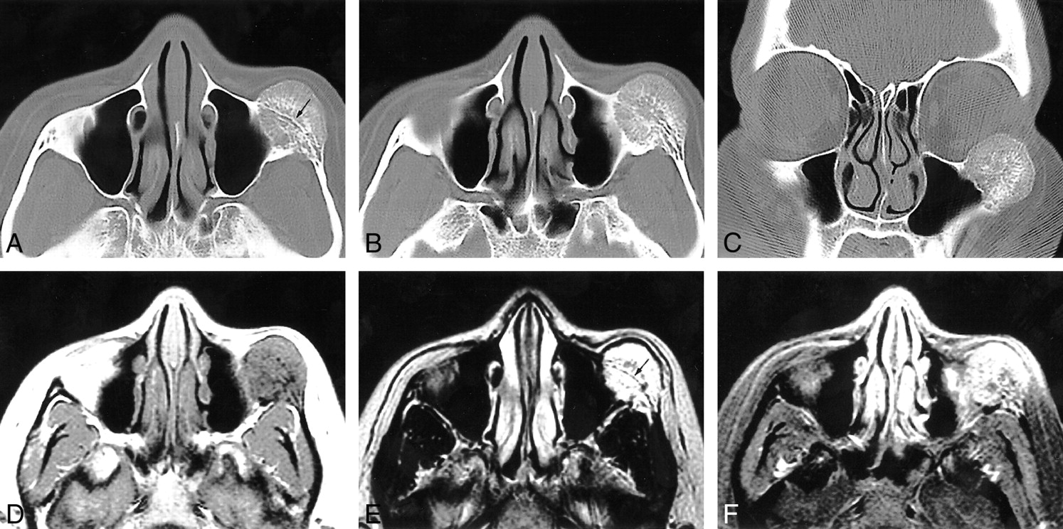
31-year-old woman with a 3-month history of progressive dystopia and a 1-year history of painless swelling of the left cheek.

A–C, Axial caudal (A), cranial (B), and coronal (C) CT scans, viewed at wide window settings.

A, A 2.5-cm round lesion arises from the zygoma. The inner and outer cortices, although thinned, are preserved. The trabeculae radiate in a spokewheel-type pattern, and a residual trace of the original cortex is seen (arrow).

B, The trabeculae of the lesion taper into the zygomatic arch. There is no associated soft tissue mass.

C, The rounded lesion encroaches on the inferolateral orbit.

D–F, Axial T1-weighted (500/9/2) (D), T2-weighted (3600/95/2) (E), and contrast-enhanced, fat-suppressed T1-weighted (450/9/2) (F) MR images.

D, The lesion is isointense with muscle and shows fine radiating lines of high signal intensity that are interspersed with the trabecular signal voids.

E, Encroachment on the inferolateral aspect of the orbit is seen. No extraosseous mass is evident. A line of signal representing the original cortex is visible (arrow).

F, The mass enhances; no large regional vessels are evident.

MR imaging of the paranasal sinuses confirmed the CT localization of the lesion (Fig 1D–F). The mass had an overall intermediate T1 signal intensity and a high T2 signal intensity. Within the mass were areas of signal void that corresponded to the trabeculae seen on the CT study. There was marked enhancement after intravenous administration of 15 mL of gadopentetate dimeglumine. No extraosseous soft tissue component was apparent.

Based on the imaging diagnosis, a decortication incisional biopsy of the lesion was performed, which precipitated moderate bleeding that was controlled with bone wax. The diagnosis of this tissue was consistent with hemangioma.

A carotid arteriogram showed a hypervascular lesion in the left zygoma, and a delayed persistent blush with a “cluster of grapes” appearance. The lesion was supplied predominantly by the left external carotid artery via the infraorbital and posterosuperior alveolar arteries, the anterior deep temporal artery, and the left facial artery. Supraselective embolization of these vessels was performed with polyvinyl alcohol particles, resulting in nearly complete devascularization.

The surgical approach was via a subciliary incision, with exposure of the zygoma and orbital floor. An en bloc resection of the lesion was performed with reconstruction of the malar bone using Medpor, and of the orbital floor using titanium mesh. The open maxillary sinus was repaired with an Alloderm patch. Intraoperative bleeding was minimal; the surgeons felt that the preoperative embolization limited the bleeding and optimized a clean field for resection.

Discussion

Intraosseous hemangiomas of the facial bones are rare and most commonly arise in the maxilla, mandible, and nasal bones. Only 20 cases of zygomatic involvement have been reported in the English-language literature (1–10). Patients usually present in about the fourth decade of life with a tender or painless swelling of the malar eminence. There is a 3:1 female predominance. The lesion is more common on the left side, and there may be associated prior trauma. Clinical diagnosis may be difficult; in our case, the initial clinical impression favored fibrous dysplasia. Establishing the vascular nature of the lesion is critical in presurgical planning, as routine bone biopsy and curettage may result in severe hemorrhage.

Hemangiomas, which were originally classified as vascular neoplasms, are now thought to be hamartomas with an anomalous proliferation of endothelial-lined vascular channels. Malignant transformation has only been reported after radiation treatment. Osseous hemangiomas are slow growing, and treatment is not always necessary. Indications for surgery include correction of a mass effect (in our case, elevation of the eye), control of hemorrhage, and cosmesis. Because curettage may have a high morbidity due to uncontrolled hemorrhage, en bloc surgical excision, including a rim of normal bone, is usually the definitive treatment. Recurrence is rare. Radiotherapy is not recommended because of scar formation, impairment of regional bone growth in children, and, rarely, development of a malignant transformation.

CT is considered the most useful imaging technique because of its excellent characterization of trabecular and cortical detail. The CT appearance is variable, and in the calvaria most commonly shows a characteristic sharply marginated expansile lesion with intact inner and outer tables and a sunburst pattern of radiating trabeculae. “Soap bubble” and “honeycomb” configurations may also occur. MR imaging provides information about any associated soft tissue elements. The MR signal characteristics of a hemangioma are dependent on the quantity of slow-moving venous blood and on the ratio of red marrow to converted fatty marrow present within the lesion. Low or high signal may be seen on T1-weighted images, and high signal on water-sensitive sequences (11). Small lesions may appear bright on T1-weighted images; however, in larger lesions, the trabeculae typically show low signal on T1-weighted images. In cases in which a round, potentially benign bone lesion is initially detected on MR images, the lesion may be confused with other disease processes, including malignancy. The addition of a CT scan is recommended to evaluate the cortex and periosteum.

In our case, the diagnosis of an intraosseous hemangioma of the zygoma was suggested on the basis of the morphologic similarity of the lesion to a calvarial hemangioma. An intraosseous meningioma was thought unlikely, as the trabeculae were not perpendicular and the zygoma is an unlikely site for that lesion. Osteogenic sarcoma was also thought unlikely, as the cortex, although thinned, was preserved and there was no periostitis or associated soft tissue mass.

The usefulness of preoperative angiography and selective embolization has been debated in the literature. Although many authors recommend either embolization or external carotid ligation (1, 2, 4, 8), others state that proximal vascular control is unnecessary if an adequate margin of normal bone is resected (9, 10).

Conclusion

Intraosseous hemangiomas of the zygoma often show a characteristic cross-sectional configuration, with radiating trabeculae and intact, smooth cortices, as reported in six cases studied with CT and in two studied with MR imaging (1–3, 5, 6, 8). This appearance is pathognomonic, and allows for confident preoperative diagnosis.

Footnotes

  • 1 Address reprint requests to Sandra Moore, MD, Department of Radiology, Mount Sinai Hospital, One Gustave Levy Place, Box 1234, New York, NY 10029.

References

  1. ↵
    Clauser L, Meneghini A, Riga M, et al. Haemangioma of the zygoma: report of two cases with a review of the literature. J Craniomaxillofac Surg 1991;19:353-358
    PubMed
  2. Cuesta-Gil M, Navarro-Villa V. Intraosseous hemangioma of the zygomatic bone: a case report. Int J Oral Maxillofac Surg 1992;21:287-291
    PubMed
  3. Har-El G, Hadar T, Zirkin HY, et al. Hemangioma of the zygoma. Ann Plast Surg 1987;18:533-540
    CrossRefPubMed
  4. Jeter TS, Hackney FL, Aufdemorte TB. Cavernous hemangioma of the zygoma: report of cases. J Oral Maxillofac Surg 1990;48:508-512
    CrossRefPubMed
  5. Pinna V, Clauser L, Marchi M, Castellan L. Haemangioma of the zygoma: a case report. Neuroradiology 1997;39:216-218
    PubMed
  6. Savastano G, Russo A, Dell Aquila A. Osseous hemangioma of the zygoma: a case report. J Oral Maxillofac Surg 1997;55:1352-1356
    PubMed
  7. Schmidt GH. Haemangioma in the zygoma. Ann Plast Surg 1982;9:330-332
    PubMed
  8. Warman S, Myssiorek D. Hemangioma of the zygomatic bone. Ann Otol Rhinol Laryngol 1989;98:655-658
    PubMed
  9. ↵
    Konior R, Kelly T, Hemmer D. Intraosseous zygomatic hemangioma. Otolaryngol Head Neck Surg 1999;121:122-125
    Abstract/FREE Full Text
  10. Marshak G. Hemangioma of the zygomatic bone. Arch Otolaryngol 1980;106:581-582
    CrossRefPubMed
  11. ↵
    Firooznia H, Golimbu C, Rafii M, Rauchning W, Weinreb J. MRI and CT of the Musculoskeletal System. St Louis: Mosby; 1992:630
  • Received September 29, 2000.
  • Accepted after revision January 3, 2001.
  • Copyright © American Society of Neuroradiology
View Abstract
PreviousNext
Back to top

In this issue

American Journal of Neuroradiology: 22 (7)
American Journal of Neuroradiology
Vol. 22, Issue 7
1 Aug 2001
  • Table of Contents
  • Index by author
Advertisement
Print
Download PDF
Email Article

Thank you for your interest in spreading the word on American Journal of Neuroradiology.

NOTE: We only request your email address so that the person you are recommending the page to knows that you wanted them to see it, and that it is not junk mail. We do not capture any email address.

Enter multiple addresses on separate lines or separate them with commas.
Intraosseous Hemangioma of the Zygoma: CT and MR Findings
(Your Name) has sent you a message from American Journal of Neuroradiology
(Your Name) thought you would like to see the American Journal of Neuroradiology web site.
CAPTCHA
This question is for testing whether or not you are a human visitor and to prevent automated spam submissions.
Cite this article
Sandra L. Moore, Jin K. Chun, Sandra A. Mitre, Peter M. Som
Intraosseous Hemangioma of the Zygoma: CT and MR Findings
American Journal of Neuroradiology Aug 2001, 22 (7) 1383-1385;

Citation Manager Formats

  • BibTeX
  • Bookends
  • EasyBib
  • EndNote (tagged)
  • EndNote 8 (xml)
  • Medlars
  • Mendeley
  • Papers
  • RefWorks Tagged
  • Ref Manager
  • RIS
  • Zotero
0 Responses
Respond to this article
Share
Bookmark this article
Intraosseous Hemangioma of the Zygoma: CT and MR Findings
Sandra L. Moore, Jin K. Chun, Sandra A. Mitre, Peter M. Som
American Journal of Neuroradiology Aug 2001, 22 (7) 1383-1385;
del.icio.us logo Twitter logo Facebook logo Mendeley logo
  • Tweet Widget
  • Facebook Like
  • Google Plus One

Jump to section

  • Article
    • Abstract
    • Case Report
    • Discussion
    • Conclusion
    • Footnotes
    • References
  • Figures & Data
  • Info & Metrics
  • Responses
  • References
  • PDF

Related Articles

  • No related articles found.
  • PubMed
  • Google Scholar

Cited By...

  • Intraosseous Venous Malformations of the Head and Neck
  • Symptomatic Intraosseous Vascular Malformation of Infraorbital Rim: A Case Report With Literature Survey
  • Intraosseous cavernous haemangioma of the inferior turbinate
  • Crossref
  • Google Scholar

This article has not yet been cited by articles in journals that are participating in Crossref Cited-by Linking.

More in this TOC Section

  • Correlation of Apparent Diffusion Coefficient at 3T with Prognostic Parameters of Retinoblastoma
  • Parathyroid Lesions: Characterization with Dual-Phase Arterial and Venous Enhanced CT of the Neck
  • Efficacy of Diffusion-Weighted Imaging for the Differentiation between Lymphomas and Carcinomas of the Nasopharynx and Oropharynx: Correlations of Apparent Diffusion Coefficients and Histologic Features
Show more HEAD AND NECK

Similar Articles

Advertisement

Indexed Content

  • Current Issue
  • Accepted Manuscripts
  • Article Preview
  • Past Issues
  • Editorials
  • Editor's Choice
  • Fellows' Journal Club
  • Letters to the Editor
  • Video Articles

Cases

  • Case Collection
  • Archive - Case of the Week
  • Archive - Case of the Month
  • Archive - Classic Case

Special Collections

  • AJNR Awards
  • ASNR Foundation Special Collection
  • Most Impactful AJNR Articles
  • Photon-Counting CT
  • Spinal CSF Leak Articles (Jan 2020-June 2024)

More from AJNR

  • Trainee Corner
  • Imaging Protocols
  • MRI Safety Corner

Multimedia

  • AJNR Podcasts
  • AJNR Scantastics

Resources

  • Turnaround Time
  • Submit a Manuscript
  • Submit a Video Article
  • Submit an eLetter to the Editor/Response
  • Manuscript Submission Guidelines
  • Statistical Tips
  • Fast Publishing of Accepted Manuscripts
  • Graphical Abstract Preparation
  • Imaging Protocol Submission
  • Evidence-Based Medicine Level Guide
  • Publishing Checklists
  • Author Policies
  • Become a Reviewer/Academy of Reviewers
  • News and Updates

About Us

  • About AJNR
  • Editorial Board
  • Editorial Board Alumni
  • Alerts
  • Permissions
  • Not an AJNR Subscriber? Join Now
  • Advertise with Us
  • Librarian Resources
  • Feedback
  • Terms and Conditions
  • AJNR Editorial Board Alumni

American Society of Neuroradiology

  • Not an ASNR Member? Join Now

© 2025 by the American Society of Neuroradiology All rights, including for text and data mining, AI training, and similar technologies, are reserved.
Print ISSN: 0195-6108 Online ISSN: 1936-959X

Powered by HighWire