Advertisement

Main menu

  • Home
  • Content
    • Current Issue
    • Accepted Manuscripts
    • Article Preview
    • Past Issue Archive
    • Video Articles
    • AJNR Case Collection
    • Case of the Week Archive
    • Case of the Month Archive
    • Classic Case Archive
  • Special Collections
    • AJNR Awards
    • ASNR Foundation Special Collection
    • Most Impactful AJNR Articles
    • Photon-Counting CT
    • Spinal CSF Leak Articles (Jan 2020-June 2024)
  • Multimedia
    • AJNR Podcasts
    • AJNR SCANtastic
    • Trainee Corner
    • MRI Safety Corner
    • Imaging Protocols
  • For Authors
    • Submit a Manuscript
    • Submit a Video Article
    • Submit an eLetter to the Editor/Response
    • Manuscript Submission Guidelines
    • Statistical Tips
    • Fast Publishing of Accepted Manuscripts
    • Graphical Abstract Preparation
    • Imaging Protocol Submission
    • Author Policies
  • About Us
    • About AJNR
    • Editorial Board
    • Editorial Board Alumni
  • More
    • Become a Reviewer/Academy of Reviewers
    • Subscribers
    • Permissions
    • Alerts
    • Feedback
    • Advertisers
    • ASNR Home

User menu

  • Alerts
  • Log in

Search

  • Advanced search
American Journal of Neuroradiology

American Journal of Neuroradiology

ASHNR American Society of Functional Neuroradiology ASHNR American Society of Pediatric Neuroradiology ASSR
  • Alerts
  • Log in

Advanced Search

  • Home
  • Content
    • Current Issue
    • Accepted Manuscripts
    • Article Preview
    • Past Issue Archive
    • Video Articles
    • AJNR Case Collection
    • Case of the Week Archive
    • Case of the Month Archive
    • Classic Case Archive
  • Special Collections
    • AJNR Awards
    • ASNR Foundation Special Collection
    • Most Impactful AJNR Articles
    • Photon-Counting CT
    • Spinal CSF Leak Articles (Jan 2020-June 2024)
  • Multimedia
    • AJNR Podcasts
    • AJNR SCANtastic
    • Trainee Corner
    • MRI Safety Corner
    • Imaging Protocols
  • For Authors
    • Submit a Manuscript
    • Submit a Video Article
    • Submit an eLetter to the Editor/Response
    • Manuscript Submission Guidelines
    • Statistical Tips
    • Fast Publishing of Accepted Manuscripts
    • Graphical Abstract Preparation
    • Imaging Protocol Submission
    • Author Policies
  • About Us
    • About AJNR
    • Editorial Board
    • Editorial Board Alumni
  • More
    • Become a Reviewer/Academy of Reviewers
    • Subscribers
    • Permissions
    • Alerts
    • Feedback
    • Advertisers
    • ASNR Home
  • Follow AJNR on Twitter
  • Visit AJNR on Facebook
  • Follow AJNR on Instagram
  • Join AJNR on LinkedIn
  • RSS Feeds

AJNR is seeking candidates for the AJNR Podcast Editor. Read the position description.

OtherPEDIATRICS

Rapid Development of Optic Glioma in a Patient with Hybrid Phakomatosis: Neurofibromatosis Type 1 and Tuberous Sclerosis

Sami H. Erbay, Stephen A. Oljeski and Rafeeque Bhadelia
American Journal of Neuroradiology January 2004, 25 (1) 36-38;
Sami H. Erbay
  • Find this author on Google Scholar
  • Find this author on PubMed
  • Search for this author on this site
Stephen A. Oljeski
  • Find this author on Google Scholar
  • Find this author on PubMed
  • Search for this author on this site
Rafeeque Bhadelia
  • Find this author on Google Scholar
  • Find this author on PubMed
  • Search for this author on this site
  • Article
  • Figures & Data
  • Info & Metrics
  • Responses
  • References
  • PDF
Loading

Abstract

Summary: Increased propensity for tumor formation in neurofibromatosis and tuberous sclerosis exists because of defective tumor-suppressor genes. Although different tumor-suppressor genes may be involved in neurofibromatosis and tuberous sclerosis, at the cellular level these genes share rather common enzymatic pathways. We believe these genetic malfunctions have resulted in a cumulative or additive effect for rapid growth of optic glioma in the following unusual case that has hybrid phakomatosis.

Optic glioma is commonly seen in the setting of neurofibromatosis type 1 (NF-1) and has a favorable course in childhood due to relatively benign disease (1, 2); however, coexistence of more than one phakomatosis such as neurofibromatosis and tuberous sclerosis (TS) can augment the phenotypic expression, which in our case is optic glioma (OG), because of additive or synergistic effect at the genetic level. Recent evidence indicates that formation of neoplasm in both conditions can be due to deficiency of tumor-suppressor genes. In this article, we present a case of an infant with combined NF-1 and TS who had a worsening course due to rapidly growing OG.

Case Report

The infant has history of maternal NF-1 and paternal TS. Her sister has NF-1, and the father’s mother and sister have TS. The patient was delivered by cesarean section at 39 weeks because of breech presentation. Otherwise, her pre- and perinatal course was unremarkable. At birth, her physical examination was significant for café-au-lait spots, hypopigmented macules, and a 3–4-cm Shagreen patch on the back of her right thigh. She was normocephalic with soft and flat fontanel. Ear pits were prominent, but otherwise the auriculocular structures were symmetric and normal in position. She had normal genitalia. The kidneys were normal at sonography. Her back was straight. Soon after birth, she developed right-sided proptosis. The cornea and pupil were also larger on the right, although the pupillary reflexes were preserved. Both optic nerves were normal on fundoscopic examination. The ocular pressures were also normal. MR imaging of brain at age 3 months revealed multiple subependymal nodules, T1 hyperintensities in the white matter, and a left foramina Monro lesion. The myelination pattern was normal. Orbits were assessed with a dedicated MR imaging study. There was right-sided proptosis, but the optic nerves were symmetric without any discrete enhancing mass. At 4 months of age, she was able to hold her head up, make eye contact, follow, smile, and blow bubbles. She was rolling over and starting to crawl at 5 months old.

The patient returned to the radiology department at age 8 months for a follow-up MR imaging study. The multiple subependymal nodules and left foramina Monro nodule were found to be stable. The white matter T1 hyperintensities appeared more conspicuous, with two new foci of similar signal intensity abnormalities being seen in the left superior frontal region.

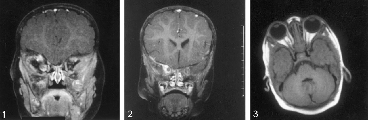
Normal progression of myelination was present. The right optic nerve was now enlarged with intense enhancement (Fig 1). The appearance was consistent with developing OG. The neuroophthalmology team chose to follow the patient closely as per the family request. Nonetheless, the OG continued to show rapid growth and enhancement with a worsening proptosis (Figs 2 and 3). This led to the institution of chemotherapy at age 2 years with gradual response of the tumor (not shown).

Fig 1.
  • Download figure
  • Open in new tab
  • Download powerpoint
Fig 1.

Coronal T1-weighted images (TR/TE: 523/14) with contrast enhancement and fat suppression and obtained through the orbits reveal an enhancing right-sided OG at age 8 months.

Fig 2.
  • Download figure
  • Open in new tab
  • Download powerpoint
Fig 2.

Coronal T1-weighted images (TR/TE: 523/14) with contrast enhancement and fat suppression and obtained through the orbits demonstrate interval worsening of right-sided OG at age 18 months.

Fig 3.
  • Download figure
  • Open in new tab
  • Download powerpoint
Fig 3.

Axial postcontrast T1-weighted images (TR/TE: 670/14) with fat saturation demonstrate marked worsening of right-sided proptosis due to rapid growth of the right OG at age 23 months).

Discussion

NF-1 and TS are neuroectodermal dysplasias, namely, phakomatosis with characteristic lesions and multiple clinical stigmata. To the best of our knowledge, there have been only eight cases reported in the literature (3–5) in which both conditions have occurred simultaneously in the same patient. Ours is the ninth such case described. For the purpose of this article, we named the coexistence of these dysplasias in the same patient “hybrid phakomatosis.” Although our patient did not undergo genetic testing, separate diagnosis for NF-1 and TS was established by family background, as well as specific clinical and imaging criteria as specified elsewhere (6, 7).

Some light has recently been shed on the genetic basis of phakomatosis. The determination of a full genetic profile has been hindered by several factors. The NF-1 gene has increased susceptibility to spontaneous mutation (8) that is, in turn, responsible for most of these cases. The patients can be homo- or heterozygotic. In addition, NF-1 has a variable expression, possibly because of genetic mosaics, making it very hard to determine which patients will develop the disease or its severe complications (9). Within the past several years, however, the NF-1 gene has been identified on chromosome segment 17q11.2. The NF-1 gene encodes a protein called “neurofibromin.” The recent evidence from animal research focused on this gene indicates that, among its many roles, the most important function can be the tumor suppression through the cellular GTPase systems. The homozygotic deletion can accelerate the tumor formation in patients with NF-1, especially in plexiform neurofibroma and malignant tumor (10). Excellent review of the subject has been provided in the literature (11).

There is also growing evidence in the literature indicating a similar genetic basis for TS-induced neoplasms. The recently mapped TSC1 and TSC2 genes are believed to function as tumor suppressors through the same cellular systems. Complete inactivation of TSC2 gene has been implicated in the formation of hamartomas and possibly renal cell carcinomas (12, 13).

Approximately 15–20% of patients with NF-1 will develop OG, sometimes as the first sign of subtle NF-1. In Lund and Skovby’s series (14), the mean age of diagnosis was 6.4 years, with the average of duration of visual symptoms before diagnosis being 2 years. MR imaging has been shown to be far more superior to visual field testing, fundoscopy, and visually evoked potentials to detect these lesions (15). Therefore, routine cranial imaging is now recommended to exclude OG in patients with NF-1 in the first few years of life (16). OG has a favorable histologic course and slow growth in NF-1 compared with OG of the non-NF-1 variety (1), with most of these being pilocytic astrocytomas. It has been reported in a long-term follow-up of more than 20 years that tumor growth and the difference between treated and untreated lesions can be hardly noticeable (2). Aggressive malignant behavior is seen less than 20% of patients (17), especially for tumors detected in infancy (18). It has been hypothesized that, in patients with NF-1, there are fundamental pathophysiologic differences between patients with and those without OG (19). A striking association has been observed between the OG and the other CNS neoplasms, independent of age.

Nonetheless, it has been reported that patients with NF-1 who have had prior negative findings for OG are still at risk for forming OG later in life. In a series of neurofibromatosis-related OGs, Massry et al (20) described two cases among seven for which prior neuroimaging findings were negative. They suggested that prior normal CT or MR findings of orbits do not exclude the future development of OG, because slightly older cases took much longer (ie, 2–3 years) to form OG. Conversely, a 15-mm OG in our case had developed only 8 months after initial imaging (Fig 1).

Although the TS and NF1 genes are not located closely at the genetic locus, their functions at the cellular level are intimately related. They both suppress the oncogenes through the GTPase systems (21). We believe this might have resulted in additive or synergistic effect of dysfunctional tumor-suppressor genes in rapid formation of OG in this unusual patient.

Conclusion

We report a rare occurrence of neurofibromatosis and tuberosclerosis in the same patient. These are genetically transmitted disorders. Their phenotypic expression reflects underlying dysfunction of specific genes, also known as tumor suppressor genes. At the cellular level, tumor suppressor genes may share the same enzymatic pathways. We believe their additive or synergistic effects in our patient resulted in an aggressive course of a rapidly developing infantile optic glioma.

References

  1. ↵
    Deliganis AV, Geyer JR, Berger MS. Prognostic significance of type 1 neurofibromatosis (von Recklinghausen disease) in childhood optic glioma. Neurosurgery 1996;38:1114–1118
    CrossRefPubMed
  2. ↵
    Pascual-Castroviejo I, Martinez Bermojo A, Lopez Marin V, et al. Optic gliomas in neurofibromatosis type 1. Neurologia 1994;9:173–177
    PubMed
  3. ↵
    Lee TC, Sung ML, Chen JS. Tuberous sclerosis associated with neurofibromatosis: report of a case. J Formos Med Assoc 1994;93:797–801
    PubMed
  4. Piccoli F, Demma MP, Pastore I, et al. A case of tuberous sclerosis associated with Recklinghausen’s disease: Attempted review of the problem of phacomatoses. Acta Neurol (Napoli) 1997;32:389–398
  5. ↵
    Sanchez SL, Oliva AH, Castro TA, et al. Combination of tuberous sclerosis, neurofibromatosis and polycystic kidney. Rev Clin Exp 1968;108:311–318
  6. ↵
    National Institute of Health Consensus Development Conference. Neurofibromatosis: conference statement. Arch Neurol 1988;45:575–578
    CrossRefPubMed
  7. ↵
    Gomez MR. 00000000. In: Gomez ED, ed. Tuberous sclerosis. 2nd ed. New York: Raven Press;1985 :9–20
  8. ↵
    Hulsebos TJ. Molecular genetic aspects of neurofibromatosis. Ned Tijdschr Geneeskd 1997;141:620–623
    PubMed
  9. ↵
    Riva P, Corrado L, Natacci F, et al. NF1 microdeletion syndrome: refined FISH characterization of sporadic and familial deletions with locus specific probes. Am J Hum Genet 2000;66:100–109
    CrossRefPubMed
  10. ↵
    Rasmussen SA, Overman J, Thomson SA et al. Chromosome 17 loss-of-heterozygosity studies in benign and malignant tumors in neurofibromatosis type 1. Genes Chromosomes Cancer 2000;28:425–431
    CrossRefPubMed
  11. ↵
    von Deimling A, Krone W, Menon AG. Neurofibromatosis type 1: pathology, clinical features and molecular genetics. Brain Pathol 1995;52:153–162
  12. ↵
    Au KS, Hebert AA, Roach ES, Northrup H. Complete inactivation of the TSC2 gene leads to formation of hamartomas. Am J Hum Genet 1999;65:1790–1795
    CrossRefPubMed
  13. ↵
    Ohno K. The results of recent research in neurocutaneous syndromes. Brain Dev 1998;30:109–114
  14. ↵
    Lund AM, Skovby F. Optic gliomas in children with neurofibromatosis type 1. Eur J Pediatr 1991;150:853–858
  15. ↵
    Mulvihill JJ. Neurofibromatosis 1 (Recklinghausen disease) and neurofibromatosis 2 (bilateral acoustic neurofibromatosis): an update. Ann Intern Med 1990;113:39–52
  16. ↵
    Elster AD. Radiologic screening in the neurocutaneous syndromes: strategies and controversies. AJNR Am J Neuroradiol 1992;13:1078–1082
    FREE Full Text
  17. ↵
    Imes RK, Hoyt WF. Childhood chiasmal gliomas: update on the fate patients in the 1969 San Francisco study. Br J Opthalmol 1986;70:179–182
    Abstract/FREE Full Text
  18. ↵
    Kanamori M, Shibuya M, Yoshida J, et al. Long-term follow-up of patients with optic glioma. Childs Nerv Syst 1985;1:272–278
    CrossRefPubMed
  19. ↵
    Friedman JM, Birch P. An association between optic glioma and other tumors of the central nervous system in neurofibromatosis type 1. Neuropediatrics 1997;28:131–132
    PubMed
  20. ↵
    Massry GG, Morgan CF, Chung SM. Evidence of optic pathway gliomas after previously negative neuroimaging. Ophthalmology 1997;104:930–935
    PubMed
  21. ↵
    Gutmann D. H. Parallels between tuberous sclerosis complex and neurofibromatosis 1:common threads in the same tapestry. Semin Pediatr Neurol 1998;5:276–286
    CrossRefPubMed
  • Received December 18, 2002.
  • Accepted after revision April 14, 2003.
  • Copyright © American Society of Neuroradiology
PreviousNext
Back to top

In this issue

American Journal of Neuroradiology: 25 (1)
American Journal of Neuroradiology
Vol. 25, Issue 1
1 Jan 2004
  • Table of Contents
  • Index by author
Advertisement
Print
Download PDF
Email Article

Thank you for your interest in spreading the word on American Journal of Neuroradiology.

NOTE: We only request your email address so that the person you are recommending the page to knows that you wanted them to see it, and that it is not junk mail. We do not capture any email address.

Enter multiple addresses on separate lines or separate them with commas.
Rapid Development of Optic Glioma in a Patient with Hybrid Phakomatosis: Neurofibromatosis Type 1 and Tuberous Sclerosis
(Your Name) has sent you a message from American Journal of Neuroradiology
(Your Name) thought you would like to see the American Journal of Neuroradiology web site.
CAPTCHA
This question is for testing whether or not you are a human visitor and to prevent automated spam submissions.
Cite this article
Sami H. Erbay, Stephen A. Oljeski, Rafeeque Bhadelia
Rapid Development of Optic Glioma in a Patient with Hybrid Phakomatosis: Neurofibromatosis Type 1 and Tuberous Sclerosis
American Journal of Neuroradiology Jan 2004, 25 (1) 36-38;

Citation Manager Formats

  • BibTeX
  • Bookends
  • EasyBib
  • EndNote (tagged)
  • EndNote 8 (xml)
  • Medlars
  • Mendeley
  • Papers
  • RefWorks Tagged
  • Ref Manager
  • RIS
  • Zotero
0 Responses
Respond to this article
Share
Bookmark this article
Rapid Development of Optic Glioma in a Patient with Hybrid Phakomatosis: Neurofibromatosis Type 1 and Tuberous Sclerosis
Sami H. Erbay, Stephen A. Oljeski, Rafeeque Bhadelia
American Journal of Neuroradiology Jan 2004, 25 (1) 36-38;
del.icio.us logo Twitter logo Facebook logo Mendeley logo
  • Tweet Widget
  • Facebook Like
  • Google Plus One

Jump to section

  • Article
    • Abstract
    • Case Report
    • Discussion
    • Conclusion
    • References
  • Figures & Data
  • Info & Metrics
  • Responses
  • References
  • PDF

Related Articles

  • No related articles found.
  • PubMed
  • Google Scholar

Cited By...

  • No citing articles found.
  • Crossref
  • Google Scholar

This article has not yet been cited by articles in journals that are participating in Crossref Cited-by Linking.

More in this TOC Section

  • SyMRI & MR Fingerprinting in Brainstem Myelination
  • Comparison of Image Quality and Radiation Dose in Pediatric Temporal Bone CT Using Photon-Counting Detector CT and Energy-Integrating Detector CT
  • Neonatal Hypocalcemia: Cerebral MRI Abnormalities
Show more Pediatrics

Similar Articles

Advertisement

Indexed Content

  • Current Issue
  • Accepted Manuscripts
  • Article Preview
  • Past Issues
  • Editorials
  • Editor's Choice
  • Fellows' Journal Club
  • Letters to the Editor
  • Video Articles

Cases

  • Case Collection
  • Archive - Case of the Week
  • Archive - Case of the Month
  • Archive - Classic Case

Special Collections

  • AJNR Awards
  • ASNR Foundation Special Collection
  • Most Impactful AJNR Articles
  • Photon-Counting CT
  • Spinal CSF Leak Articles (Jan 2020-June 2024)

More from AJNR

  • Trainee Corner
  • Imaging Protocols
  • MRI Safety Corner

Multimedia

  • AJNR Podcasts
  • AJNR Scantastics

Resources

  • Turnaround Time
  • Submit a Manuscript
  • Submit a Video Article
  • Submit an eLetter to the Editor/Response
  • Manuscript Submission Guidelines
  • Statistical Tips
  • Fast Publishing of Accepted Manuscripts
  • Graphical Abstract Preparation
  • Imaging Protocol Submission
  • Evidence-Based Medicine Level Guide
  • Publishing Checklists
  • Author Policies
  • Become a Reviewer/Academy of Reviewers
  • News and Updates

About Us

  • About AJNR
  • Editorial Board
  • Editorial Board Alumni
  • Alerts
  • Permissions
  • Not an AJNR Subscriber? Join Now
  • Advertise with Us
  • Librarian Resources
  • Feedback
  • Terms and Conditions
  • AJNR Editorial Board Alumni

American Society of Neuroradiology

  • Not an ASNR Member? Join Now

© 2025 by the American Society of Neuroradiology All rights, including for text and data mining, AI training, and similar technologies, are reserved.
Print ISSN: 0195-6108 Online ISSN: 1936-959X

Powered by HighWire