Advertisement

Main menu

  • Home
  • Content
    • Current Issue
    • Accepted Manuscripts
    • Article Preview
    • Past Issue Archive
    • Video Articles
    • AJNR Case Collection
    • Case of the Week Archive
    • Case of the Month Archive
    • Classic Case Archive
  • Special Collections
    • AJNR Awards
    • ASNR Foundation Special Collection
    • Most Impactful AJNR Articles
    • Photon-Counting CT
    • Spinal CSF Leak Articles (Jan 2020-June 2024)
  • Multimedia
    • AJNR Podcasts
    • AJNR SCANtastic
    • Trainee Corner
    • MRI Safety Corner
    • Imaging Protocols
  • For Authors
    • Submit a Manuscript
    • Submit a Video Article
    • Submit an eLetter to the Editor/Response
    • Manuscript Submission Guidelines
    • Statistical Tips
    • Fast Publishing of Accepted Manuscripts
    • Graphical Abstract Preparation
    • Imaging Protocol Submission
    • Author Policies
  • About Us
    • About AJNR
    • Editorial Board
    • Editorial Board Alumni
  • More
    • Become a Reviewer/Academy of Reviewers
    • Subscribers
    • Permissions
    • Alerts
    • Feedback
    • Advertisers
    • ASNR Home

User menu

  • Alerts
  • Log in

Search

  • Advanced search
American Journal of Neuroradiology

American Journal of Neuroradiology

ASHNR American Society of Functional Neuroradiology ASHNR American Society of Pediatric Neuroradiology ASSR
  • Alerts
  • Log in

Advanced Search

  • Home
  • Content
    • Current Issue
    • Accepted Manuscripts
    • Article Preview
    • Past Issue Archive
    • Video Articles
    • AJNR Case Collection
    • Case of the Week Archive
    • Case of the Month Archive
    • Classic Case Archive
  • Special Collections
    • AJNR Awards
    • ASNR Foundation Special Collection
    • Most Impactful AJNR Articles
    • Photon-Counting CT
    • Spinal CSF Leak Articles (Jan 2020-June 2024)
  • Multimedia
    • AJNR Podcasts
    • AJNR SCANtastic
    • Trainee Corner
    • MRI Safety Corner
    • Imaging Protocols
  • For Authors
    • Submit a Manuscript
    • Submit a Video Article
    • Submit an eLetter to the Editor/Response
    • Manuscript Submission Guidelines
    • Statistical Tips
    • Fast Publishing of Accepted Manuscripts
    • Graphical Abstract Preparation
    • Imaging Protocol Submission
    • Author Policies
  • About Us
    • About AJNR
    • Editorial Board
    • Editorial Board Alumni
  • More
    • Become a Reviewer/Academy of Reviewers
    • Subscribers
    • Permissions
    • Alerts
    • Feedback
    • Advertisers
    • ASNR Home
  • Follow AJNR on Twitter
  • Visit AJNR on Facebook
  • Follow AJNR on Instagram
  • Join AJNR on LinkedIn
  • RSS Feeds

AJNR is seeking candidates for the AJNR Podcast Editor. Read the position description.

OtherSPINE

Spinal Schwannoma with Acute Subarachnoid Hemorrhage: A Diagnostic Challenge

Hemant Parmar, Boon Chuang Pang, C. C. Tchoyoson Lim, Soke Miang Chng and Kheng Kooi Tan
American Journal of Neuroradiology May 2004, 25 (5) 846-850;
Hemant Parmar
  • Find this author on Google Scholar
  • Find this author on PubMed
  • Search for this author on this site
Boon Chuang Pang
  • Find this author on Google Scholar
  • Find this author on PubMed
  • Search for this author on this site
C. C. Tchoyoson Lim
  • Find this author on Google Scholar
  • Find this author on PubMed
  • Search for this author on this site
Soke Miang Chng
  • Find this author on Google Scholar
  • Find this author on PubMed
  • Search for this author on this site
Kheng Kooi Tan
  • Find this author on Google Scholar
  • Find this author on PubMed
  • Search for this author on this site
  • Article
  • Figures & Data
  • Info & Metrics
  • Responses
  • References
  • PDF
Loading

Abstract

Summary: We report a case of spinal intradural schwannoma presenting with intracranial subarachnoid hemorrhage. Cerebral angiography studies were negative, but MR imaging of the spine revealed a large hemorrhagic tumor in the thoraco-lumbar junction. The tumor was misdiagnosed as ependymoma of the conus medullaris. This case illustrates the importance of a high index of suspicion for spinal disease in angiographically-negative subarachnoid hemorrhage and pitfalls in MR diagnosis of thoraco-lumbar tumors.

Patients with spinal abnormalities infrequently present with intracranial subarachnoid hemorrhage (SAH), accounting for less than 1% of all patients with SAH. The more common causes include spinal trauma, arteriovenous malformations, and saccular aneurysms of spinal arteries (1, 2). On occasion, spinal cord tumors, either primary or metastatic, may cause cranial SAH, with ependymoma of the conus medullaris accounting for most of these cases (1, 3). Spinal nerve sheath tumors such as schwannomas only rarely cause SAH, especially in the absence of spinal cord or nerve root symptoms. We report a case of thoraco-lumbar schwannoma presenting clinically with intracranial SAH and illustrate the diagnostic difficulties for the neuroradiologist and neurosurgeon.

Case Report

A 56-year-old man presented with fever, neck pain, and altered mental status. Except for nuchal rigidity, physical examination was normal. CT of the head revealed hyperattenuation in the cortical sulci bilaterally consistent with acute SAH (Fig 1). There was no other intracranial abnormality. A lumbar puncture yielded bloody and xanthochromic CSF. Cerebral angiography was performed on two occasions, 1 and 12 days after admission, but was negative for aneurysm, vascular malformations, or tumor on both occasions. No vasospasm was detected. On direct systemic review of spinal symptoms, the patient gave a history of vague generalized backache for many years, without precipitating or aggravating events. He also had mild symptoms of hesitancy and poor stream of urine. Spinal neurologic examination, however, was again normal, and digital rectal examination revealed a moderately enlarged prostate gland. An MR examination of the whole spine was performed. A 5 × 2 × 2-cm mass at the conus medullaris was found extending from T11 to L1 vertebral levels (Fig 2). It was centrally located and was indistinguishable from the conus medullaris (Fig 2D). The lesion showed heterogeneous signal intensity and susceptibility effect on gradient-recalled echo images, consistent with blood degradation products. There was thick, irregular rim enhancement after intravenous contrast, medium administration (Fig 2C). Immediately caudal to the mass, there were two separate abnormal focal collections of increased signal intensity on T1- and T2-weighted images compared with the spinal cord and nerve roots. These were located intradurally, both anterior and posterior to the cauda equina, and clearly separated form the hyperintense CSF and the epidural fat (Fig 3), consistent with subdural hematoma (SDH) (4). A diagnosis of conus ependymoma causing SAH and SDH was made and the patient proceeded to surgery.

Fig 1.
  • Download figure
  • Open in new tab
  • Download powerpoint
Fig 1.

SAH. Unenhanced axial CT section of the brain (A) showing hyperattenuated cortical sulci (arrow) and (B) fourth ventricle (arrow) consistent with acute subarachnoid hemorrhage.

Fig 2.
  • Download figure
  • Open in new tab
  • Download powerpoint
Fig 2.

Thoraco-lumbar spinal tumor. Sagittal T2- (A) and T1-weighted images (B) show a large mass at the conus medullaris with heterogeneous signal intensity (arrows). The two hyperintense SHDs may be seen located caudal to the mass (arrowheads).

After intravenous contrast material injection (C and D), there is heterogeneous enhancement of the mass.

Fig 3.
  • Download figure
  • Open in new tab
  • Download powerpoint
Fig 3.

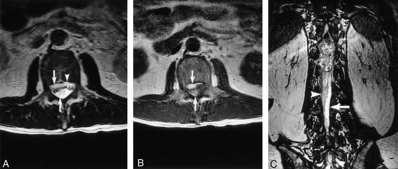
Spinal SDH. Axial T2- (A) and T1-weighted (B) images show hyperintense subdural blood collections located both anterior and posterior (arrows) to the cauda equina. Anteriorly, the subdural blood is separate from the epidural fat signal intensity (A, arrowhead). Coronal fast imaging employing steady-state acquisition (C) shows the posterior blood collection (arrowhead) is clearly separate from the hyperintense CSF (arrow).

A grayish white, intradural-extramedullary tumor was found at surgery, with intrathecal blood clot surrounding its lower end. Gross pathology showed a poorly circumscribed mass with whitish capsular tissue. Cut section showed tanned tissue with focal firm whitish areas. On histopathology, the tumor was found to be composed of spindle cells arranged in vaguely alternating cellular and loosely textured areas (Antoni A and B, respectively) with the former predominating. The cellular areas revealed fascicles and whorls of spindle cells with nuclear palisades and tight aggregates of Verocay bodies. Hyalinized vessels, perivascular siderophage deposition, and ectatic blood vessels were also seen. There was mild to moderate cellular pleomorphism, but no features of malignant transformation. The final diagnosis was schwannoma with degenerative changes.

Discussion

Spinal causes of SAH are rare, representing 1.5% of all cases of SAH (5). Halpern et al (6) reported 0.6% of their cases of SAH to have spinal origin, whereas Sahs et al (7) reported an even lower incidence, 0.05%. SAH from spinal tumors tends to occur in younger patients between the 2nd and 4th decades of life (8, 9, 10). The clinical presentation depends on the amount of bleeding, with massive hemorrhage resulting in sudden, rapidly evolving classic symptoms of back pain with or without neurologic deficit (3). Michon (11) in 1928 gave the first description of spinal SAH, which he likened to being stabbed in the spine (le coup de poignard rachidien). In the absence of pain, predominant motor and spinchteric deficits have been reported (12).

The more common causes of spinal SAH include trauma and vascular malformations (1, 2, 9, 13). Among the spinal tumors, conus ependymoma accounts for most of the cases of SAH (1, 3). Nerve sheath tumors may also cause SAH, but it is exceedingly rare for these intradural lesions to come to clinical attention with only intracranial SAH without spinal symptoms. In our literature review, we identified 20 reports of spinal nerve sheath tumors that caused SAH, of which only seven cases (28%) presented exclusively with intracranial symptoms like our patient (6, 8–10, 13, 14–27) (Table).

View this table:
  • View inline
  • View popup

Spinal nerve sheath tumors presenting with intracranial subarachnoid hemorrhage

Schwannomas are well-circumscribed intradural or extradural or combined intraextradural tumors located on peripheral nerves or spinal nerve roots (28). Hemorrhage is unusual in this tumor, and various theories, including tumor location and histologic features, have been proposed to explain this phenomenon (8, 22, 25, 29). According to the vascular theory, ectatic and hyalinized vessels of the tumor may undergo spontaneous thrombosis, followed by distal tumor necrosis and hemorrhage (25). The mechanical theory (8, 13, 22, 25), on the other hand, suggests that hemorrhage into subarachnoid space occurs when there is trauma at the interface between the tumor and the normal neural tissue, particularly at the conus medullaris and the cauda equina (13, 25). This may be due to the differences in inertia between the tumor of the spinal cord and the cauda equina, resulting in abnormal movement at their interface. Traction on vascular attachments to nerve roots may also occur, leading to disruption of the blood vessels on the surface of the tumor (30). Such tractional forces occur most commonly in areas of high mobility, and this probably explains why the onset of symptoms in 50% of cases is related to exertion (25, 27). Other causes of hemorrhage due to central ischemic necrosis associated with tumor growth (28, 29) or malignant transformation and neovascularization can further increase the susceptibility of such tumors to hemorrhage by accelerating the process mentioned above, especially within large schwannomas (20).

In our case, there was no clear history of exertion or pathologic evidence of malignant transformation. We believe that the degenerative changes, along with the mechanical stress at the conus, caused rupture of the fragile, ectatic tumor vessels and subsequent dissection of the blood into the subarachnoid space. This probably led to the loss of the typical plane of demarcation that separates the tumor from the normal conus medullaris at MR imaging, giving rise to misdiagnosis of a conus ependymoma. Even without tumor hemorrhage, ependymomas of the conus medullaris and cauda equina and schwannomas may be indistinguishable by imaging features alone (31). Although ependymomas tend to have more cystic and hemorrhagic changes and are more commonly associated with SAH (30, 31), these findings were not helpful in our case. In retrospect, the presence of spinal SDH immediately caudal to the tumor is interesting and may have been helpful in differential diagnosis (4). Nontraumatic spinal SDH is rare and may rarely be related to spinal tumors. In our literature review, schwannomas (32, 33, 34), as well as an ependymoma (35), have been described. In hindsight, it should have been possible to suggest nerve sheath tumor as a strong differential diagnosis.

Our patient presented a diagnostic challenge in clinical presentation of SAH without spinal symptoms. Conventional cerebral angiography is the method of choice to diagnose arterial aneurysm, the most common cause of SAH. Intracranial and systemic causes of SAH with negative cerebral angiographic findings include microaneurysms of perforating vessels, thrombosed or obliterated aneurysms, cryptic arteriovenous malformations, cranial tumors, trauma, blood dyscrasias, and infection (14, 31, 36). Although the number of aneurysms detected at repeat angiography is low (37, 38), at our institution all patients with an initial negative angiogram undergo a second study 1–2 weeks later. If the second angiographic study is also negative, and other abnormalities are excluded, the patient is followed up clinically without further investigation because patients with negative angiograms are known to have a better prognosis compared with those with ruptured aneurysms (39). Although a few authors have recommended MR imaging of the spine in patients with negative angiograms (8, 14), this is not the routine practice at our institution in patients without spinal signs or symptoms. In our patient, the vague symptom of backache without localizing neurologic deficits was only forthcoming on direct questioning and was not deemed significant by the patient himself. We believe this case demonstrates the importance of a thorough systemic review and a high index of clinical suspicion for spinal disease.

References

  1. ↵
    Cummings TM, Johnson MH. Neurofibroma manifested by spinal subarachnoid hemorrhage. AJR Am J Roentgenol 1994;162:959–960
    PubMed
  2. ↵
    Saunders FW, Birchard D, Willimer J. Spinal artery aneurysm. Surg Neurol 1987;27:269–272
    CrossRefPubMed
  3. ↵
    Scotti G, Filizzolo F, Scialfa G, Tampieri D. Repeated subarachnoid hemorrhages from a cervical meningiomas. J Neurosurg 1987;66:779–781
    PubMed
  4. ↵
    Post MJ, Becerra JL, Parley WM, et al. Acute spinal subdural hematoma: MR and CT findings with pathologic correlates. AJNR Am J Neuroradiol 1994;15:1895–1905
    Abstract/FREE Full Text
  5. ↵
    Walton JN. Subarachnoid hemorrhage of unusual aetiology. Neurology 1953;3:517–543
  6. ↵
    Halpren J, Feldman S, Peyser E. Subarachnoid hemorrhage with papilledema due to spinal neurofibroma. Arch Neurol Psychiatry 1958;79:138–141
    CrossRef
  7. ↵
    Sahs AL, Perret GE, Locksley HB, Nishioka H, eds. Intracranial aneurysm and subarachnoid hemorrhage. Philadelphia: JB Lippincott;1969
  8. ↵
    De Divitiis E, Maiuri F, Corriero G, Donzelli R. Subarachnoid hemorrhage due to spinal neurinoma. Surg Neurol 1985;24:187–190
    PubMed
  9. ↵
    Bruni P, Esposito S, Oddi G, et al. Subarachnoid hemorrhage from multiple neurofibromas of the cauda equina: case report. Neurosurgery 1991;28:910–913
    PubMed
  10. ↵
    Djindjian M, Djindjian R, Huth M, et al. Les hemorrhagies meningees spinales tumorales: a propos de 5 cas arterographies. Rev Neurol (Paris) 1978;134:685–692
    PubMed
  11. ↵
    Michon P. Le coup de poignard rachidien: symptome initial de certaines hemorragies sousarachnoidiennes. Essai sur les hemorragies meningees spinales Presse Med 1928;36:964–966
  12. ↵
    Campbell FG. Painless tumors of the region of the cauda equina: a case report. Neurology 1963;13:341–343
  13. ↵
    Cordon T, Bekar A, Yaman O. Spinal subarachnoid hemorrhage attributable to schwannoma of the cauda equina. Surg Neurol 1999;51:373–375
    PubMed
  14. ↵
    Chalif DJ, Black K, Rosenstein D. Intradural spinal cord tumour presenting as a subarachnoid hemorrhage: magnetic resonance imaging diagnosis. Neurosurgery 1990;27:631–634
    PubMed
  15. Andre-Thomas F, Schaeffer H, De Martel T. Syndrome d’hemorrhagie meningee realise par une tumeur de la queue de cheval. Paris Med 1930;77:292–296
  16. Krayenbuhl H. Spontane spinale Subarachnoidalblutung und akute Ruckenmarkskompression bei intraduralem spinalem Neurinom. Schweiz Med Wochenschr 1947;77:692–694
    PubMed
  17. Fincher EF. Spontaneous subarachnoid hemorrhage in intradural tumours of the lumber sac. J Neurosurg 1951;8:576–584
    PubMed
  18. Prieto A, Cantu RC. Spinal subarachnoid hemorrhage associated with neurofibroma of cauda equina. J Neurosurg 1967;27:63–69
    PubMed
  19. Fortuna A, La Torre E. Neurinoma della cauda con emorragia subarcnoidea circoscritta. Il Lavaro Neuropsichiatrico 1968;43:1157–64
  20. ↵
    Bernell WR, Kepes JJ, Clough CA. Subarachnoid hemorrhage from malignant schwannoma of the cauda equina. Tex Med 1973;69:101–104
    PubMed
  21. Grollmuss J. Spinal subarachnoid hemorrhage with schwannoma. Acta Neurochir (Wein) 1975;31:253–256
  22. ↵
    Luxon LM, Harrison MJ. Subarachnoid hemorrhage and papilledema due to a cervical neurilemmoma: case report. J Neurosurg 1978;48:1015–1018
    PubMed
  23. Muhtaroglu U, Strenge H. Recurrent subarachnoid haemorrhage with spinal neurinoma [author’s translation]. Neurochirurgia (Stuttg) 1980;23:151–155
  24. Motomochi M, Makita Y, Nabeshima S, et al. Spinal subarachnoid hemorrhage due to a thoracic neurinoma during anticoagulation therapy: a case report. Neurol Med Chir (Tokyo) 1981;21:781–784
    PubMed
  25. ↵
    Mills B, Marks PV, Nixon JM. Spinal subarachnoid haemorrhage from an “ancient” schwannoma of the cervical spine. Br J Neurosurg 1993;7:557–559
    PubMed
  26. Bonicki W, Koszewski W, Marchel A, Sherif A. Caudal tumors as a rare cause of subarachnoid hemorrhage. Neurol Neurochir Pol 1993;27:599–603
    PubMed
  27. ↵
    Correiro G, Iacopino DG, Valentini S, Lanza PL. Cervical neuroma presenting as a subarachnoid hemorrhage: case report. Neurosurgery 1996;39:1046–1049
    PubMed
  28. ↵
    Parmar H, Patkar D, Gadani S, Shah J. Cystic lumbar nerve sheath tumours: MRI features. Australas Radiol 2001;45:123–127
    PubMed
  29. ↵
    Sakamato M, Harayama K, Furufu T. A case of cystic spinal schwannoma presented unusual clinical findings. Orthopedics 1985;3:1185–1189
  30. ↵
    Argyropoulou PI, Argyropoulou MI, Tsampoulas C, et al. Myxopapillary ependymoma of the conus medullaris with subarachnoid haemorrhage: MRI in two cases. Neuroradiology 2001;43:489–491
    PubMed
  31. ↵
    Alexander MS, Dias PS, Uttley D. Spontaneous sunarachnoid hemorrhage and negative cerebral panangiography. J Neurosurg 1986;64:537–542
    PubMed
  32. ↵
    Ng PY. Schwannoma of the cervical spine presenting with acute haemorrhage. J Clin Neurosci 2001;8:277–278
    PubMed
  33. ↵
    Vazquez-Barquero A, Pascual J, Quintana F, et al. Cervical schwannoma presenting as a spinal subdural haematoma. Br J Neurosurg 1994;8:739–741
    PubMed
  34. ↵
    Roscoe MW, Barrington TW. Acute spinal subdural hematoma: a case report and review of literature. Spine 1984;9:672–675
    PubMed
  35. ↵
    Smith RA. Spinal subdural hematoma, neurilemmoma and acute transverse myelopathy. Surg Neurol 1985;23:367–370
    PubMed
  36. ↵
    Hayward RD. Subarachnoid hemorrhage of unknown etiology: a clinical and radiological study of 51 cases. J Neurol Neurosurg Psychiatry 1977;40:926–931
    Abstract/FREE Full Text
  37. ↵
    Forster DM, Steiner L, Hakason S, Bergvall U. The value of repeat panangiography in cases of unexplained subarachnoid hemorrhage. J Neurosurg 1978;48:712–716
    PubMed
  38. ↵
    Juul R, Fredriksen TA, Ringkjob R. Prognosis in subarachnoid hemorrhage of unknown etiology. J Neurosurg 1986;64:359–362
    PubMed
  39. ↵
    Eskesen V, Sorensen EB, Rosenorn J, Schmidt K. The prognosis in subarachnoid hemorrhage of unknown etiology. J Neurosurg 1984;61:1029–1031
    PubMed
  • Received September 11, 2003.
  • Accepted after revision September 22, 2003.
  • Copyright © American Society of Neuroradiology
View Abstract
PreviousNext
Back to top

In this issue

American Journal of Neuroradiology: 25 (5)
American Journal of Neuroradiology
Vol. 25, Issue 5
1 May 2004
  • Table of Contents
  • Index by author
Advertisement
Print
Download PDF
Email Article

Thank you for your interest in spreading the word on American Journal of Neuroradiology.

NOTE: We only request your email address so that the person you are recommending the page to knows that you wanted them to see it, and that it is not junk mail. We do not capture any email address.

Enter multiple addresses on separate lines or separate them with commas.
Spinal Schwannoma with Acute Subarachnoid Hemorrhage: A Diagnostic Challenge
(Your Name) has sent you a message from American Journal of Neuroradiology
(Your Name) thought you would like to see the American Journal of Neuroradiology web site.
CAPTCHA
This question is for testing whether or not you are a human visitor and to prevent automated spam submissions.
Cite this article
Hemant Parmar, Boon Chuang Pang, C. C. Tchoyoson Lim, Soke Miang Chng, Kheng Kooi Tan
Spinal Schwannoma with Acute Subarachnoid Hemorrhage: A Diagnostic Challenge
American Journal of Neuroradiology May 2004, 25 (5) 846-850;

Citation Manager Formats

  • BibTeX
  • Bookends
  • EasyBib
  • EndNote (tagged)
  • EndNote 8 (xml)
  • Medlars
  • Mendeley
  • Papers
  • RefWorks Tagged
  • Ref Manager
  • RIS
  • Zotero
0 Responses
Respond to this article
Share
Bookmark this article
Spinal Schwannoma with Acute Subarachnoid Hemorrhage: A Diagnostic Challenge
Hemant Parmar, Boon Chuang Pang, C. C. Tchoyoson Lim, Soke Miang Chng, Kheng Kooi Tan
American Journal of Neuroradiology May 2004, 25 (5) 846-850;
del.icio.us logo Twitter logo Facebook logo Mendeley logo
  • Tweet Widget
  • Facebook Like
  • Google Plus One

Jump to section

  • Article
    • Abstract
    • Case Report
    • Discussion
    • References
  • Figures & Data
  • Info & Metrics
  • Responses
  • References
  • PDF

Related Articles

  • No related articles found.
  • PubMed
  • Google Scholar

Cited By...

  • Intralesional and subarachnoid bleeding of a spinal schwannoma presenting with acute cauda equina syndrome
  • Crossref
  • Google Scholar

This article has not yet been cited by articles in journals that are participating in Crossref Cited-by Linking.

More in this TOC Section

  • MP2RAGE 7T in MS Lesions of the Cervical Spine
  • Bern Score Validity for SIH
  • Resisted Inspiration for CSF-Venous Fistula
Show more Spine

Similar Articles

Advertisement

Indexed Content

  • Current Issue
  • Accepted Manuscripts
  • Article Preview
  • Past Issues
  • Editorials
  • Editor's Choice
  • Fellows' Journal Club
  • Letters to the Editor
  • Video Articles

Cases

  • Case Collection
  • Archive - Case of the Week
  • Archive - Case of the Month
  • Archive - Classic Case

Special Collections

  • AJNR Awards
  • ASNR Foundation Special Collection
  • Most Impactful AJNR Articles
  • Photon-Counting CT
  • Spinal CSF Leak Articles (Jan 2020-June 2024)

More from AJNR

  • Trainee Corner
  • Imaging Protocols
  • MRI Safety Corner

Multimedia

  • AJNR Podcasts
  • AJNR Scantastics

Resources

  • Turnaround Time
  • Submit a Manuscript
  • Submit a Video Article
  • Submit an eLetter to the Editor/Response
  • Manuscript Submission Guidelines
  • Statistical Tips
  • Fast Publishing of Accepted Manuscripts
  • Graphical Abstract Preparation
  • Imaging Protocol Submission
  • Evidence-Based Medicine Level Guide
  • Publishing Checklists
  • Author Policies
  • Become a Reviewer/Academy of Reviewers
  • News and Updates

About Us

  • About AJNR
  • Editorial Board
  • Editorial Board Alumni
  • Alerts
  • Permissions
  • Not an AJNR Subscriber? Join Now
  • Advertise with Us
  • Librarian Resources
  • Feedback
  • Terms and Conditions
  • AJNR Editorial Board Alumni

American Society of Neuroradiology

  • Not an ASNR Member? Join Now

© 2025 by the American Society of Neuroradiology All rights, including for text and data mining, AI training, and similar technologies, are reserved.
Print ISSN: 0195-6108 Online ISSN: 1936-959X

Powered by HighWire