Advertisement

Main menu

  • Home
  • Content
    • Current Issue
    • Accepted Manuscripts
    • Article Preview
    • Past Issue Archive
    • Video Articles
    • AJNR Case Collection
    • Case of the Week Archive
    • Case of the Month Archive
    • Classic Case Archive
  • Special Collections
    • AJNR Awards
    • ASNR Foundation Special Collection
    • Most Impactful AJNR Articles
    • Photon-Counting CT
    • Spinal CSF Leak Articles (Jan 2020-June 2024)
  • Multimedia
    • AJNR Podcasts
    • AJNR SCANtastic
    • Trainee Corner
    • MRI Safety Corner
    • Imaging Protocols
  • For Authors
    • Submit a Manuscript
    • Submit a Video Article
    • Submit an eLetter to the Editor/Response
    • Manuscript Submission Guidelines
    • Statistical Tips
    • Fast Publishing of Accepted Manuscripts
    • Graphical Abstract Preparation
    • Imaging Protocol Submission
    • Author Policies
  • About Us
    • About AJNR
    • Editorial Board
    • Editorial Board Alumni
  • More
    • Become a Reviewer/Academy of Reviewers
    • Subscribers
    • Permissions
    • Alerts
    • Feedback
    • Advertisers
    • ASNR Home

User menu

  • Alerts
  • Log in

Search

  • Advanced search
American Journal of Neuroradiology

American Journal of Neuroradiology

ASHNR American Society of Functional Neuroradiology ASHNR American Society of Pediatric Neuroradiology ASSR
  • Alerts
  • Log in

Advanced Search

  • Home
  • Content
    • Current Issue
    • Accepted Manuscripts
    • Article Preview
    • Past Issue Archive
    • Video Articles
    • AJNR Case Collection
    • Case of the Week Archive
    • Case of the Month Archive
    • Classic Case Archive
  • Special Collections
    • AJNR Awards
    • ASNR Foundation Special Collection
    • Most Impactful AJNR Articles
    • Photon-Counting CT
    • Spinal CSF Leak Articles (Jan 2020-June 2024)
  • Multimedia
    • AJNR Podcasts
    • AJNR SCANtastic
    • Trainee Corner
    • MRI Safety Corner
    • Imaging Protocols
  • For Authors
    • Submit a Manuscript
    • Submit a Video Article
    • Submit an eLetter to the Editor/Response
    • Manuscript Submission Guidelines
    • Statistical Tips
    • Fast Publishing of Accepted Manuscripts
    • Graphical Abstract Preparation
    • Imaging Protocol Submission
    • Author Policies
  • About Us
    • About AJNR
    • Editorial Board
    • Editorial Board Alumni
  • More
    • Become a Reviewer/Academy of Reviewers
    • Subscribers
    • Permissions
    • Alerts
    • Feedback
    • Advertisers
    • ASNR Home
  • Follow AJNR on Twitter
  • Visit AJNR on Facebook
  • Follow AJNR on Instagram
  • Join AJNR on LinkedIn
  • RSS Feeds

AJNR is seeking candidates for the AJNR Podcast Editor. Read the position description.

OtherHEAD AND NECK

Second Brachial Cleft Cyst Mimic: Case Report

Philip D. Lanham and Curtis Wushensky
American Journal of Neuroradiology August 2005, 26 (7) 1862-1864;
Philip D. Lanham
  • Find this author on Google Scholar
  • Find this author on PubMed
  • Search for this author on this site
Curtis Wushensky
  • Find this author on Google Scholar
  • Find this author on PubMed
  • Search for this author on this site
  • Article
  • Figures & Data
  • Info & Metrics
  • Responses
  • References
  • PDF
Loading

Abstract

Summary: We report the CT findings in a patient with a lateral neck mass histologically shown to be a schwannoma but having certain radiographic features commonly considered pathognomonic for a type III second branchial cleft cyst. Our case, therefore, represents an exception to this long-established rule.

Branchial cleft cysts (BCCs) are both the most common cysts to arise in the neck and the most common congenital masses of the lateral neck (1–6). Other common benign cystic lateral neck masses that can mimic BCCs include thyroglossal duct cysts, ectopic thymic cysts, lymphangiomas, dermoid and epidermoid cysts, and cystic nerve sheath tumors (3, 4). Abscesses and necrotic adenopathy also can be difficult to distinguish from a BCC, particularly if it has previously been infected (3–5). Both BCCs and their benign cystic mimics are typically soft, slow growing, and painless, making clinical distinction difficult (3–6). Cross-sectional imaging has become the mainstay of diagnosis for these lesions (3, 4). This is a case report of a cystic schwannoma demonstrating the classic location and CT signs of a type III second BCC.

Case Report

Clinical History and Physical Examination Findings

A 44-year-old man presented to his family physician with a several-year history of a slowly enlarging, painless mass in the lateral right neck. The measured attenuation postcontrast was 35 HU. Evaluation by an otolaryngologist revealed a nontender, 3–4-cm, palpable, slightly mobile, level II–region mass. The remainder of the examination was unremarkable.

Radiographic Findings

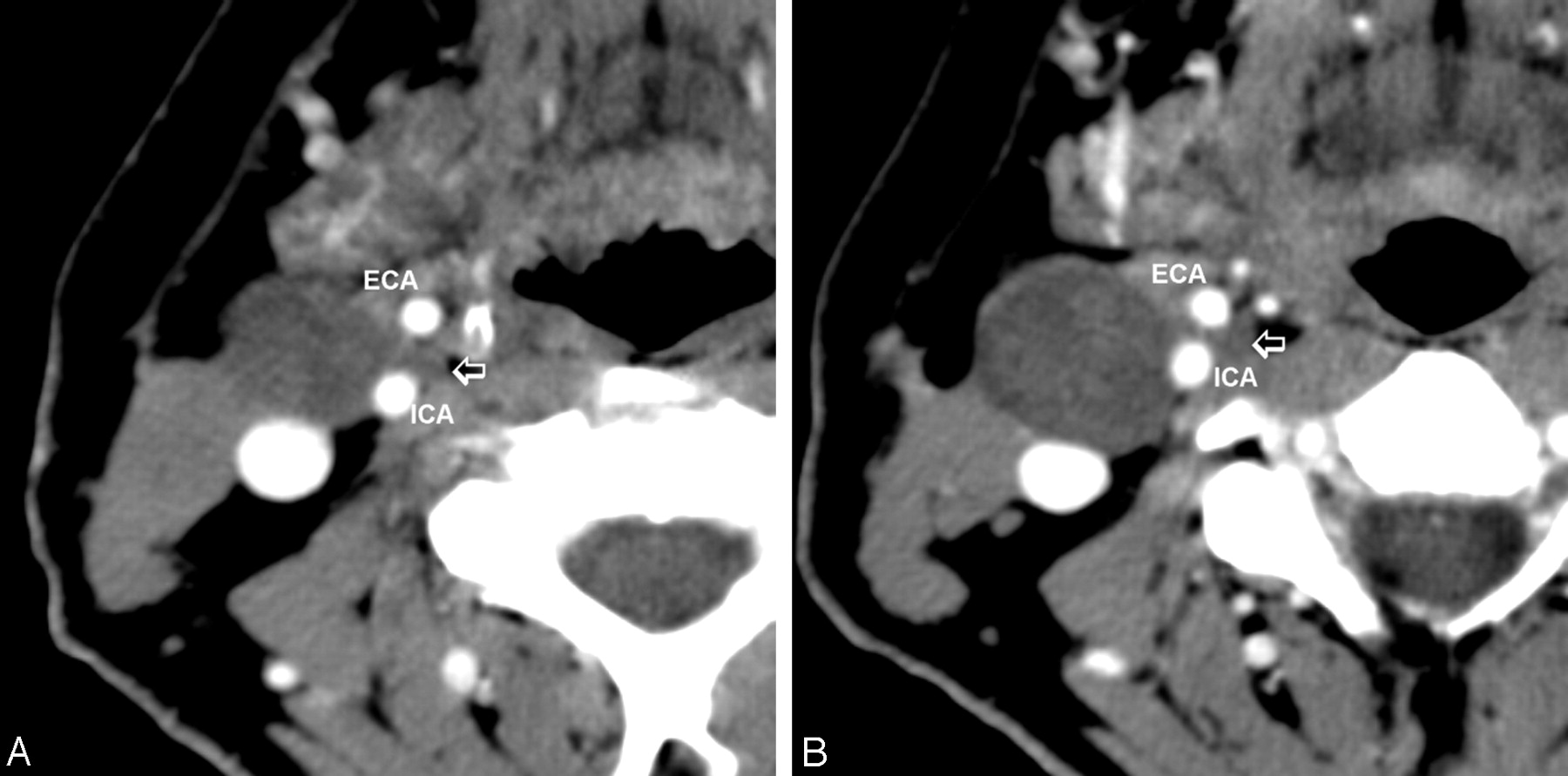
A contrast CT scan of the neck revealed a well-defined low-attenuation lesion anterior to the right sternocleidomastoid muscle without contrast enhancement. The lesion demonstrated a small “tail” extending between the internal carotid artery and the external carotid artery just above the carotid bifurcation (Figs 1 and 2). Mild posterior displacement of the internal jugular vein was also noted. A radiographic diagnosis of type III second BCC was made.

Fig 1.
  • Download figure
  • Open in new tab
  • Download powerpoint
Fig 1.

Contiguous postcontrast CT images of the neck, showing the cystic tumor (small arrow) extending between the internal (ICA) and external (ECA) carotid arteries.

Fig 2.
  • Download figure
  • Open in new tab
  • Download powerpoint
Fig 2.

Four types of second branchial cleft cyst. The cysts progressively move medially along a path between the anterior margin of the sternocleidomastoid muscle and the tonsillar fossa.

Histologic Findings

Histologic examination of the tumor was consistent with a cystic schwannoma. Antoni B areas were noted, which consisted of hypocellular tissue separated by edematous stroma. Verocay bodies and S-100 protein were present. No mitoses or areas of necrosis were identified. The cytology was bland.

Discussion

Second BCCs are by far the most common of the brachial cleft apparatus anomalies, representing about 95% of cases (1–4). Bailey and colleagues originally classified four types of BCC (3, 7). Type I is just deep to platysma muscle and the anterior lateral surface of sternocleidomastoid muscle. The type II BCC, more medial than type I, is seen along the anterior surface of the sternocleidomastoid muscle, just lateral to carotid space and posterior to the submandibular glands, arising on a line between the skin of the lateral neck and the ipsilateral faucial tonsil. Type III BCCs extend farther medially between the internal carotid artery and external carotid artery at the carotid bifurcation. Visualization of the cyst’s extension or “tail” between the internal carotid artery and external carotid artery has been considered pathognomonic for type III BCCs (2, 3, 8). Type IV is the least common and arises in the pharyngeal mucosal space just deep to the palatine tonsil, often extending upward from tonsil toward the skull base (8).

Schwannomas of the neck are, by contrast, uncommon tumors (9–11). They can arise from any peripheral, cranial, or autonomic nerve. As with BCCs, they commonly present clinically as insidious, slow-growing, painless masses, characteristics that often lead to an incorrect diagnosis (11). Most of these tumors are benign, but, if symptomatic, they may require surgical excision to preserve nerve function (10, 11). Diagnosis usually requires a combination of radiographic assessment and surgical pathologic correlation. The classic imaging appearance for a schwannoma of the neck is a large, sharply demarcated, fusiform enhancing mass in the region of the jugular foramen or parapharyngeal space (10, 11). In our review of the literature, this is the first reported case of a schwannoma extending between the internal and external carotid arteries mimicking a type III second BCC. Although rare, schwannoma should therefore be included in the differential diagnosis when a type III cyst is considered.

Several imaging techniques are available that can help differentiate between a schwannoma of the neck and a second BCC. These include MR spectroscopy (12–14) and MR diffusion-weighted imaging (15) or, alternatively, color Doppler sonography (16).

Cho et al evaluated the metabolic pattern of schwannomas and demonstrated (with MR spectroscopy) increased myoinositol (mI) in 11 cases of the 13 schwannomas they evaluated (12). This pattern alone appears to be very sensitive for the diagnosis of schwannoma. Cho et al observed elevated mI in almost every schwannoma they examined (12). In addition, Kinoshita and Yokota have noted enhanced mI from in vitro proton MR spectroscopy (13).

Wang et al were able to differentiate head and neck lesions with diffusion-weighted echo-planar MR imaging (15). They calculated the apparent diffusion coefficients from diffusion-weighted echo-planar MR images and noted that the mean of such coefficients of benign solid tumors was significantly smaller than for those of benign cystic lesions. This protocol could help differentiate a benign or malignant schwannoma from a BCC.

If further diagnostic accuracy is required, color Doppler sonography may be useful in distinguishing between a schwannoma and a BCC. Gritzmann et al reviewed the utility of sonography in evaluating soft-tissue masses of the neck. Their review concluded that BCCs have variable echogenicity, but on color Doppler imaging no vascularity is demonstrated inside the lesion (16). By contrast, schwannomas when evaluated with color Doppler imaging show internal vascularity that can easily be identified (16).

Conclusion

We present the case of a cystic schwannoma of the lateral neck, demonstrating what has been considered the classic CT findings of a type III second BCC. Consequently, although this “tail sign” is still strongly suggestive of a type III second BCC, it can no longer be considered pathognomonic for that disease entity.

References

  1. ↵
    Gold B. Second brachial cleft cyst and fistula. AJR Am J Roentgenol 1980;134:1067–1069
    PubMed
  2. ↵
    Lev S, Lev MH. Imaging of cystic lesions. Radiol Clin North Am 2000;38:1013–1027
    CrossRefPubMed
  3. ↵
    Harnsberger H, Manusco A, Muraki A, et al. Branchial cleft anomalies and their mimics: computed tomographic evaluation. Radiology 1984;152:739–748
    PubMed
  4. ↵
    Coppens F, Peene P, Lemahieu SF. Diagnosis and differential diagnosis of branchial cleft cysts by CT scan. J Belge Radiol 1990;73:189–196
    PubMed
  5. ↵
    Herman TE, McAlister WH, Siegel MJ. Branchial fistula: CT manifestations. Pediatr Radiol 1992;22:152–153
    CrossRefPubMed
  6. ↵
    Albers GD. Brachial anomalies. JAMA 1963;183:399–409
  7. ↵
    Bailey H. Branchial cysts and other essays on surgical subjects in the facio-cervical region. London: HK Lewis;1929
  8. ↵
    Koeller KK, Alamo L, Adair CF, et al. Congential cystic masses of the neck: radiologic-pathologic correlation. Radiographics 1999;19:121
    CrossRefPubMed
  9. ↵
    Weber AL, Montandon C, Robson CD. Neurogenic tumors of the neck. Radiol Clin North Am 2000;38:1077–1090
    CrossRefPubMed
  10. ↵
    Colreavy MO, Lacy PD, Hughes J, et al. Head and neck schwannomas: a 10 years’ review. J Laryngol Otol 2000;114:119–124
    PubMed
  11. ↵
    Leu Y-S, Chang K-C. Extracranial head and neck schwannomas: a review of 8 years’ experience. Acta Otolaryngol 2002;122:435–437
    CrossRefPubMed
  12. ↵
    Cho YD, Choi GH, Lee SP, et al. (1)H-MRS metabolic patterns for distinguishing between meningiomas and other brain tumors. Magn Reson Imaging 2003;21:663–672
    CrossRefPubMed
  13. ↵
    Kinoshita Y, Yokota A. Absolute concentrations of metabolites in the human brain tumors using in vitro proton magnetic resonance spectroscopy. NMR Biomed 1997;10 ;2–12
    CrossRefPubMed
  14. ↵
    Gill S, Thomas G, van Bruggen N, et al. Proton MR spectroscopy of intracranial tumors: in vivo and in vitro studies. J Comput Assist Tomogr 1990;14:497–504
    PubMed
  15. ↵
    Wang J, Takashima S, Takayama F, et al. Head and neck lesions: characterization with diffusion-weighted echo-planar MR imaging. Radiology 2001;220:621–630
    CrossRefPubMed
  16. ↵
    Gritzmann N, Hollerweger A, Macheiner P, et al. Sonography of soft tissue masses of the neck. Clin Ultrasound 2002;30:356–373
  • Received May 6, 2003.
  • Accepted after revision September 24, 2004.
  • Copyright © American Society of Neuroradiology
View Abstract
PreviousNext
Back to top

In this issue

American Journal of Neuroradiology: 26 (7)
American Journal of Neuroradiology
Vol. 26, Issue 7
1 Aug 2005
  • Table of Contents
  • Index by author
Advertisement
Print
Download PDF
Email Article

Thank you for your interest in spreading the word on American Journal of Neuroradiology.

NOTE: We only request your email address so that the person you are recommending the page to knows that you wanted them to see it, and that it is not junk mail. We do not capture any email address.

Enter multiple addresses on separate lines or separate them with commas.
Second Brachial Cleft Cyst Mimic: Case Report
(Your Name) has sent you a message from American Journal of Neuroradiology
(Your Name) thought you would like to see the American Journal of Neuroradiology web site.
CAPTCHA
This question is for testing whether or not you are a human visitor and to prevent automated spam submissions.
Cite this article
Philip D. Lanham, Curtis Wushensky
Second Brachial Cleft Cyst Mimic: Case Report
American Journal of Neuroradiology Aug 2005, 26 (7) 1862-1864;

Citation Manager Formats

  • BibTeX
  • Bookends
  • EasyBib
  • EndNote (tagged)
  • EndNote 8 (xml)
  • Medlars
  • Mendeley
  • Papers
  • RefWorks Tagged
  • Ref Manager
  • RIS
  • Zotero
0 Responses
Respond to this article
Share
Bookmark this article
Second Brachial Cleft Cyst Mimic: Case Report
Philip D. Lanham, Curtis Wushensky
American Journal of Neuroradiology Aug 2005, 26 (7) 1862-1864;
del.icio.us logo Twitter logo Facebook logo Mendeley logo
  • Tweet Widget
  • Facebook Like
  • Google Plus One

Jump to section

  • Article
    • Abstract
    • Case Report
    • Discussion
    • Conclusion
    • References
  • Figures & Data
  • Info & Metrics
  • Responses
  • References
  • PDF

Related Articles

  • No related articles found.
  • PubMed
  • Google Scholar

Cited By...

  • Cystic schwannoma of the recurrent laryngeal nerve: a rare finding posing diagnostic difficulties
  • CT Features of an Unusual Calcified Schwannoma of the Superior Laryngeal Nerve
  • Crossref
  • Google Scholar

This article has not yet been cited by articles in journals that are participating in Crossref Cited-by Linking.

More in this TOC Section

  • Correlation of Apparent Diffusion Coefficient at 3T with Prognostic Parameters of Retinoblastoma
  • Parathyroid Lesions: Characterization with Dual-Phase Arterial and Venous Enhanced CT of the Neck
  • MR Diagnosis of Facial Neuritis: Diagnostic Performance of Contrast-Enhanced 3D-FLAIR Technique Compared with Contrast-Enhanced 3D-T1-Fast-Field Echo with Fat Suppression
Show more HEAD AND NECK

Similar Articles

Advertisement

Indexed Content

  • Current Issue
  • Accepted Manuscripts
  • Article Preview
  • Past Issues
  • Editorials
  • Editor's Choice
  • Fellows' Journal Club
  • Letters to the Editor
  • Video Articles

Cases

  • Case Collection
  • Archive - Case of the Week
  • Archive - Case of the Month
  • Archive - Classic Case

Special Collections

  • AJNR Awards
  • ASNR Foundation Special Collection
  • Most Impactful AJNR Articles
  • Photon-Counting CT
  • Spinal CSF Leak Articles (Jan 2020-June 2024)

More from AJNR

  • Trainee Corner
  • Imaging Protocols
  • MRI Safety Corner

Multimedia

  • AJNR Podcasts
  • AJNR Scantastics

Resources

  • Turnaround Time
  • Submit a Manuscript
  • Submit a Video Article
  • Submit an eLetter to the Editor/Response
  • Manuscript Submission Guidelines
  • Statistical Tips
  • Fast Publishing of Accepted Manuscripts
  • Graphical Abstract Preparation
  • Imaging Protocol Submission
  • Evidence-Based Medicine Level Guide
  • Publishing Checklists
  • Author Policies
  • Become a Reviewer/Academy of Reviewers
  • News and Updates

About Us

  • About AJNR
  • Editorial Board
  • Editorial Board Alumni
  • Alerts
  • Permissions
  • Not an AJNR Subscriber? Join Now
  • Advertise with Us
  • Librarian Resources
  • Feedback
  • Terms and Conditions
  • AJNR Editorial Board Alumni

American Society of Neuroradiology

  • Not an ASNR Member? Join Now

© 2025 by the American Society of Neuroradiology All rights, including for text and data mining, AI training, and similar technologies, are reserved.
Print ISSN: 0195-6108 Online ISSN: 1936-959X

Powered by HighWire