
Abstract
BACKGROUND AND PURPOSE: Blister aneurysms of the supraclinoid ICA represent a rare but well-documented cause of subarachnoid hemorrhage. These aneurysms are difficult to detect, and their surgical treatment is challenging, with high morbidity and mortality rates. The reports currently in the literature that describe the surgical and endovascular treatment of these aneurysms offer no clear consensus on the optimal treatment. We describe a staged endovascular treatment entailing stenting using a stent-in-stent technique, as well as planned but delayed embolization as the aneurysm increases in size to allow the introduction of coils.
MATERIALS AND METHODS: We performed a retrospective review of all cerebral angiograms performed at our institution over an 8-month period for evaluation of subarachnoid hemorrhage, identifying 6 ICA blister aneurysms.
RESULTS: All 6 blister aneurysms were located in the supraclinoid ICA. The stent-in-stent technique was used for the initial treatment of all patients. Three patients had no residual or recurrent aneurysm following initial treatment. Three patients required retreatment with coils after continued growth of the aneurysm, identified on follow-up angiography. Five patients had good recovery (average mRS score of 1), and 1 patient had poor neurologic recovery (mRS score of 3) due to a large hemorrhagic infarction.
CONCLUSIONS: Our case series suggests that staged endovascular treatment entailing the use of a stent-in-stent technique, augmented with subsequent coil embolization as necessary for progressive disease, is a viable endovascular option for treating ruptured supraclinoid blister aneurysms, allowing for parent artery preservation.
Abbreviations
- CTA
- CT angiography
- DSA
- digital subtraction angiography
- ICA
- internal carotid artery
- mRS
- modified Rankin Scale
- SAH
- subarachnoid hemorrhage
First described by Sundt and Murphey,1 the term “blister aneurysm” (also known as blister-type or dorsal wall aneurysm) has been used to describe aneurysms arising from nonbranching sites of the wall of the supraclinoid ICA. As a result of their anatomy and histopathology, which appear to differ from saccular aneurysms, these rare lesions offer challenges in both diagnosis and treatment.
As the literature supports, routine microsurgical and endovascular treatment approaches carry a relatively high morbidity and mortality,2 leading to attempts at numerous novel treatment techniques. Endovascular techniques usually employ some combination of coil embolization and/or stent placement, with definitive endovascular treatment limited to parent artery occlusion, with or without surgical bypass.3–7
We report our experience with a series of 6 patients presenting with blister aneurysms of the supraclinoid ICA treated with a staged endovascular treatment entailing stent placement by using a stent-in-stent technique, and planned but delayed embolization as the aneurysm increased in size to allow the introduction of coils.
Materials and Methods
Patients and Techniques
Following internal review board approval, we performed a retrospective review of all catheter cerebral angiograms performed at our institution from July 2007 through March 2008 for evaluation of subarachnoid hemorrhage, identifying 6 cases diagnosed as ruptured ICA blister aneurysms. We reviewed all relevant imaging studies and records pertaining to the diagnosis and treatment of these aneurysms, including admission and discharge summaries, progress notes, initial diagnostic imaging studies, procedural images and reports, and follow-up imaging studies. The review was performed by 2 interventional neuroradiologists (M.E.J., A.J.E.) and 2 neurosurgeons (D.H., A.S.D.).
In each of these patients, the blister aneurysm was initially too small and shallow to allow for coils to be introduced on the first day of treatment. Consequently, the treatment for this cohort included a staged endovascular protocol entailing stent placement by using a stent-in-stent technique, with a planned coiling procedure to follow in the case of expected aneurysm enlargement. In 3 patients the aneurysm enlarged as expected. Two of these aneurysms were treated successfully with coiling, one with coiling and then subsequent parent vessel occlusion.
Endovascular stent placement has been reported to be a potentially effective treatment of uncoilable intradural pseudoaneurysms.8 The aneurysms were deemed at the time of initial treatment to be too small to safely coil, even after the placement of the endovascular stents. Therefore, the patients were initially treated with endovascular stent placement by using a stent-in-stent technique, and all patients subsequently underwent short-term (7–13 days) follow-up conventional angiography. Given our knowledge of the malignant natural history of blister aneurysms, the expectation at follow-up angiography was to perform adjunct coil embolization or parent vessel occlusion, though 3 patients showed improvement in the aneurysms at follow-up and were therefore observed.
The stent-in-stent technique entails the deployment of 2 endovascular stents, a longer stent and a shorter stent. The longer stent is deployed first, spanning a large portion of the supraclinoid ICA segment and covering the aneurysm neck. A second, shorter stent is then deployed within the lumen of the longer stent, also spanning the aneurysm neck. Enterprise stents (Cordis Endovascular, Miami Lakes, Florida) were used in 5 cases, and Neuroform stents (Boston Scientific, Natick, Massachusetts) were used in 1 case. The stent selection was based solely on operator preference. All patients were initiated on dual antiplatelet therapy following the procedure, consisting of aspirin (325 mg) and clopidogrel (75 mg) daily.
Results
Demographics are summarized in Table 1.
Demographics
All 6 patients presented with subarachnoid hemorrhage, diagnosed by unenhanced head CT. Each patient underwent CTA followed by DSA in the diagnostic evaluation of subarachnoid hemorrhage. Of the 6 blister aneurysms, 4 (67%) were identified prospectively on the CTA, and all were identified with DSA. The average interval between the diagnostic and initial treatment procedures was 1.5 days (range, 0–4 days). Importantly, 4 of the aneurysms showed interval growth and change in the size and configuration of the dome between the diagnostic DSA and initial treatment procedure.
All 6 patients were treated with endovascular stent placement, by using the above-described stent-in-stent technique. Short-term (7–13 days) follow-up DSA was performed during the inpatient hospital stay in all 6 cases to evaluate for residual disease and/or vasospasm. Additional short-term follow-up was performed as deemed necessary to monitor the disease status. This subsequent follow-up ranged from 7 days to 3 months. Subsequent intermediate follow-up (routinely performed at 6 months) DSA imaging has been performed on all 6 patients, with an average follow-up interval of 6.3 months (range, 20 days to 12 months).
Results are summarized in Table 2. Contrary to our expectations, 3 of the aneurysms (50%) did not enlarge to the point that coils could be placed. In fact in these 3 patients, no residual or recurrent aneurysms were detected at early or intermediate follow-up (Fig 1). On the other hand, 3 of the aneurysms did enlarge and subsequent interventions were performed as detailed below. Five of the 6 patients (83%) currently have no aneurysm recurrence at intermediate follow-up, with the sixth patient having undergone definitive treatment with aneurysm trapping and parent vessel occlusion. Five patients had good recovery (average mRS score of 1), and the patient who re-bled had poor neurologic recovery (mRS score of 3), due to a large hemorrhagic infarction. All patients (with the exception of the patient with the parent vessel occlusion) were placed on long-term dual antiplatelet therapy, with none demonstrating any significant degree of in-stent stenosis at intermediate follow-up.
Results

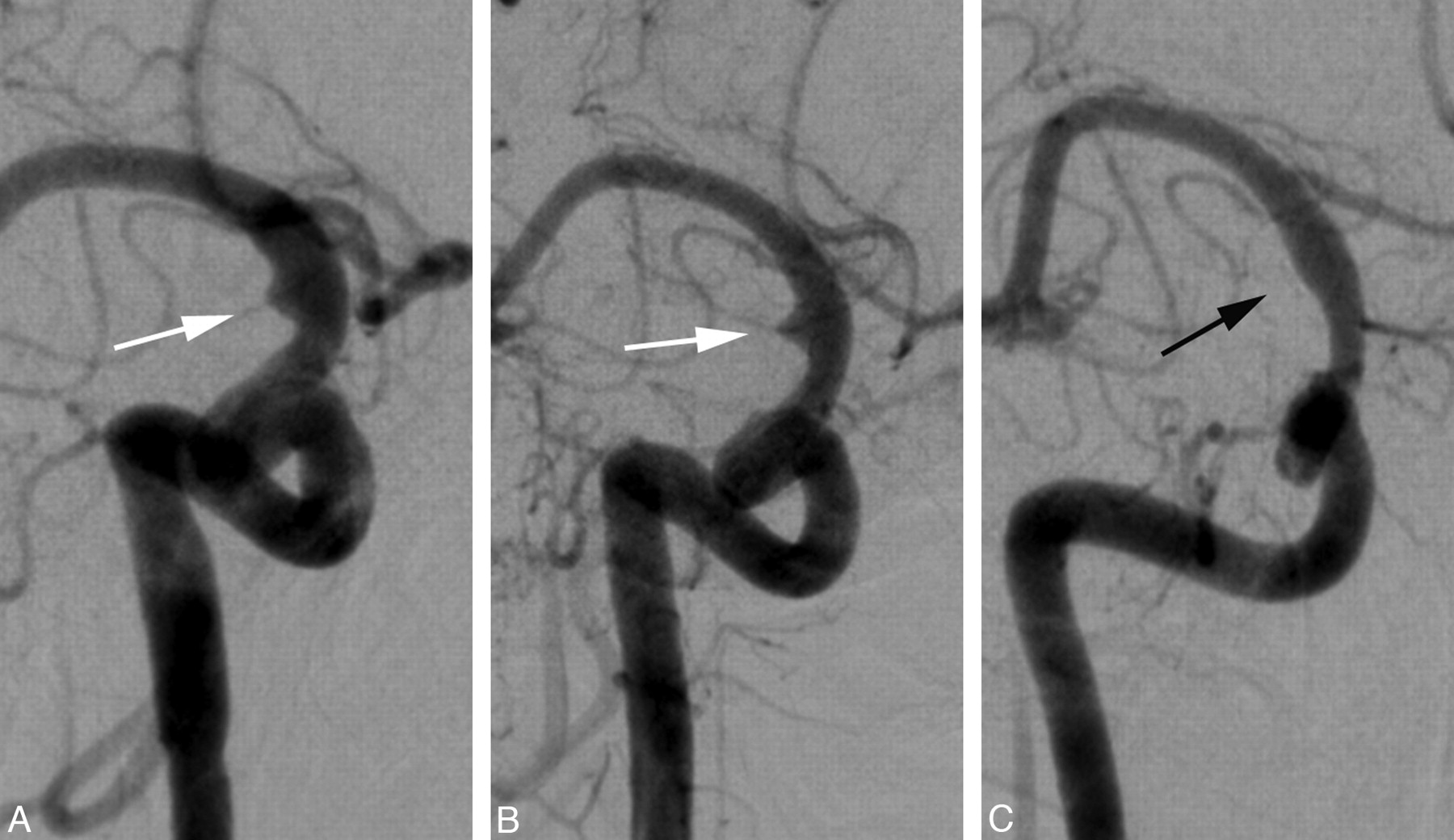
Left supraclinoid ICA blister aneurysm. Preprocedure DSA (A) shows a shallow outpouching (white arrow) arising from a nonbranching site along the medial wall of the supraclinoid segment of the left ICA, consistent with a blister aneurysm. Postprocedural DSA (B) immediately following stent deployment (stent-in-stent technique) again shows the blister aneurysm (white arrow), with diminished contrast opacification of the aneurysm sac. One-month (C) and 5-month (D) follow-up DSA show wide patency of the stents with no residual filling of the aneurysm sac.
One of the patients with progressive disease showed slight enlargement of the aneurysm on follow-up DSA performed 7 days following stent placement. A single HydroSoft 2 mm × 1 cm coil (MicroVention Terumo, Aliso Viejo, California) was then placed into the aneurysm sac, with sluggish filling of the aneurysm on the immediate postprocedural runs. Subsequent follow-up imaging at 2 weeks, 3 months, and 6 months showed no residual or recurrent aneurysm (Fig 2).
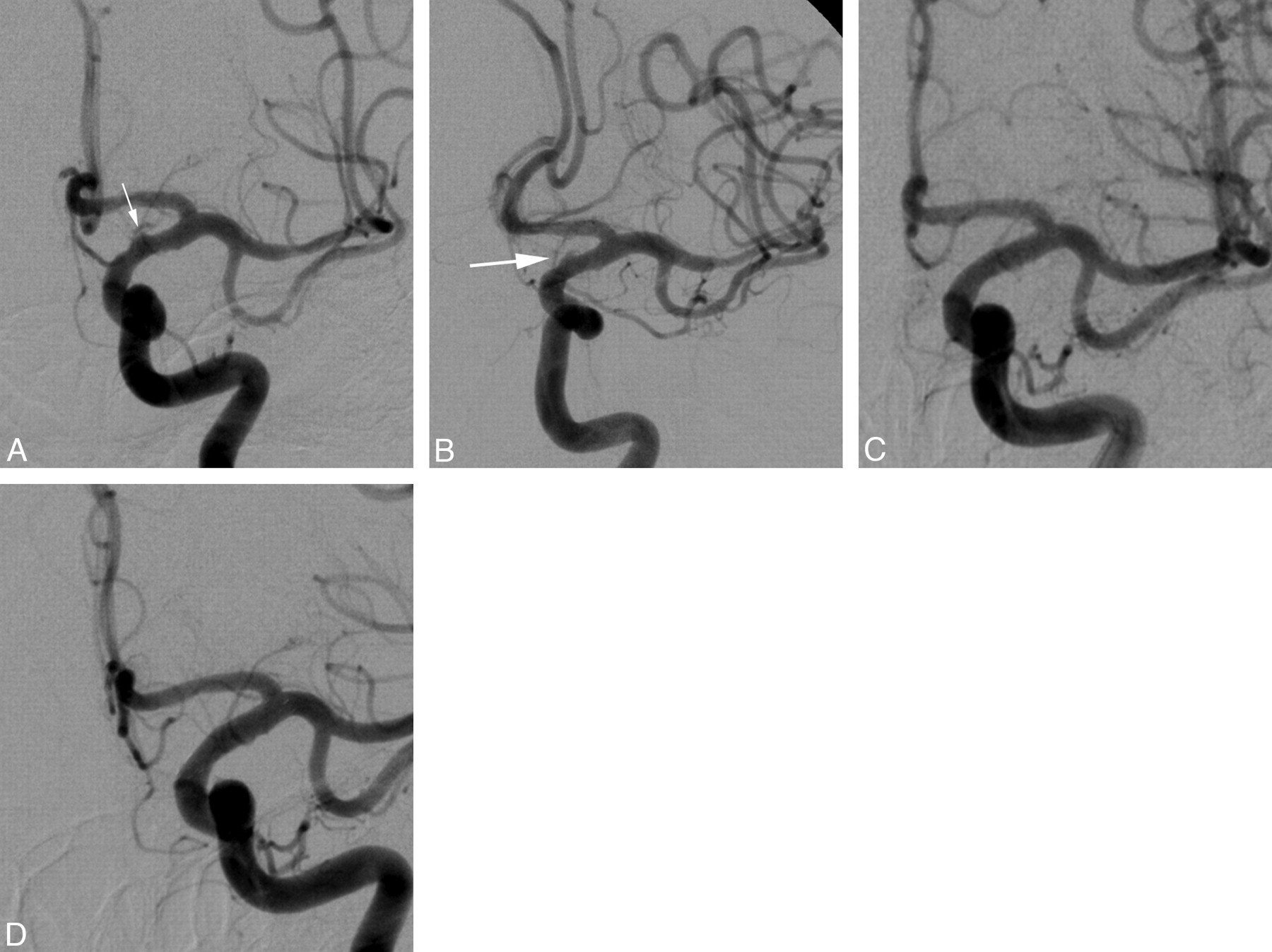
Right supraclinoid ICA blister aneurysm. Preprocedural DSA (A) shows a shallow outpouching (white arrow) arising from a nonbranching site along the anterolateral wall of the supraclinoid segment of the right ICA, consistent with a blister aneurysm. One-week follow-up DSA (B) following endovascular stent placement again shows the blister aneurysm (white arrow), now slightly increased in size. The patient subsequently underwent adjunct coil embolization with a single coil. Six-month follow-up DSA (C) following stent placement and coiling shows wide patency of the stents and no residual filling of the aneurysm sac, with note made of a single coil (black arrow) in the thrombosed aneurysm sac.
Another of the patients with progressive disease showed mild sequential enlargement and change in the configuration of the aneurysm on follow-up DSA performed 7 days, 14 days, and 30 days following stent placement. This residual aneurysm, measuring 3.4 mm, was then coil embolized by using a single HyperSoft 2 mm × 3 cm coil (MicroVention Terumo), with minimal residual filling of the aneurysm neck on the immediate postprocedural runs. However, follow-up DSA at 3 months demonstrated aneurysm recurrence (3.8 mm), which was again coil embolized with 7 coils (including a Micrus Spherical 3 mm × 5.4 cm coil [Micrus Endovascular, San Jose, California], a Compass 3.5 mm × 4.5 mm coil [MicroVention Terumo], 3 Complex 2 mm × 4 cm coils [MicroVention Terumo], and 2 HyperSoft-10 2 mm × 4 cm coils), with a small (1.7 mm) residual neck remnant. Follow-up imaging at 6 months again demonstrated aneurysm regrowth (3.3 mm), which was again coil embolized with 4 coils (including a Compass 2 mm × 4 cm coil, a HydroSoft 2 mm × 2 cm coil, and 2 HyperSoft 2 mm × 1 cm coils), with no residual aneurysm on the immediate postprocedural runs. MR angiography performed at 8 months (to evaluate for headaches) showed no evidence of aneurysm recurrence, and DSA follow-up at 9 and 12 months showed minimal filling at the base of the aneurysm with otherwise no evidence of recurrence (Fig 3).

Right supraclinoid ICA blister aneurysm. Preprocedural DSA (A) shows a shallow outpouching (white arrow) arising from a nonbranching site along the lateral wall of the supraclinoid segment of the right ICA, consistent with a blister aneurysm. One-month follow-up DSA (B) following endovascular stent placement again shows the blister aneurysm (white arrow), now increased in size and more round in shape. The patient subsequently underwent adjunct coil embolization with a single coil. Subsequent 3-month follow-up DSA (C) again shows the blister aneurysm, again increased in size and more round in shape. The patient subsequently underwent adjunct coil embolization with multiple coils. Subsequent 6-month follow-up DSA (D) again shows the blister aneurysm (white arrow), with recurrence at the aneurysm base. The patient subsequently underwent adjunct coil embolization with multiple coils. Twelve-month follow-up DSA (E) shows wide patency of the stents and minimal residual filling of the aneurysm sac (white arrow), with no filling of the coil interstices or aneurysm dome.
The final patient with progressive disease demonstrated a complicated course (Fig 4), beginning with the stent placement procedure. Overlapping Enterprise stents were deployed across the aneurysm neck, but the second stent did not deploy properly. Close examination revealed that the distal tines of the second stent were entrapped in 1 of the cells of the first stent, thus preventing the second stent from opening properly and resulting in poor apposition of the second stent to the wall of the vessel. Ten days later, the patient sustained aneurysmal rebleeding, with associated deterioration of her neurologic examination. A cerebral angiogram revealed significant enlargement of the previously treated left ICA blister aneurysm. A temporary balloon occlusion test was performed, which the patient passed. Under proximal balloon occlusion of the ICA, a single coil (GDC 360 2 mm × 4 cm; Target Therapeutics, Fremont, California) was detached in the dome of the aneurysm. This was followed by complete occlusion of the left ICA spanning from supraclinoid ICA (just proximal to the origin of the fetal posterior cerebral artery) to the cavernous ICA (just distal to the persistent trigeminal artery, which filled the basilar trunk). Postprocedurally, the left hemisphere was filling through collateral flow from the anterior communicating artery and fetal left posterior cerebral artery. A week following the ICA occlusion, the patient developed severe vasospasm, treated with medical management and multiple intra-arterial verapamil infusions, but eventually developed a large, hemorrhagic left middle cerebral artery infarction. The patient recovered during the course of the next several weeks and was discharged to an inpatient rehabilitation facility. The stroke resulted in severe, persistent neurologic deficit, including expressive aphasia, right facial palsy, and right hemiparesis.

Left supraclinoid ICA blister aneurysm. Preprocedural DSA (A) shows a blister aneurysm (white arrow) along the medial wall of the supraclinoid segment of the left ICA. Subsequent DSA (B) shows enlargement of the blister aneurysm (white arrow). Due to the repeat hemorrhage and increase in the size of the aneurysm, the patient was treated with parent vessel occlusion (C) and aneurysm trapping (after passing a temporary balloon occlusion).
Discussion
Ruptured blister aneurysms of the supraclinoid ICA present with SAH and are estimated to represent 0.9%–6.5% of ruptured intracranial aneurysms.9 Compared with saccular aneurysms, these lesions tend to have a more precipitous course, enlarging rapidly and rebleeding frequently. Morphologically, they appear as wide-necked, shallow outpouchings of nonbranching sites of the supraclinoid ICA. At surgery, they often contain a fragile, thin wall and a broad communication with the parent vessel, without an identifiable neck. Histologically, these lesions have been shown to represent focal wall defects covered with thin, fibrous tissue and adventitia, lacking the usual collagen layer, findings indicative of pseudoaneurysm.10 As such, these lesions have shown an association with arterial dissections.11
Because of their broad-based, shallow profiles, these aneurysms often represent a diagnostic challenge, both with CTA and DSA. The imaging appearance of supraclinoid ICA blister aneurysms has been described in detail elsewhere.12 In addition to an understanding of the morphology and distribution of these lesions, meticulous angiographic technique and a high index of suspicion are often both necessary to make this diagnosis. Any patient with SAH in an aneurysmal pattern and no detectable berry aneurysm should be carefully scrutinized for a blister aneurysm.
The 6 patients included in this study presented with acute SAH on unenhanced head CT and were diagnosed by CTA and/or DSA with blister aneurysms. In these patients, coil embolization was not performed at the initial treatment session because the operators deemed that the morphology of the aneurysms did not allow for safe coil deposition, even after stent placement. Instead, these patients were treated with a staged protocol: immediate treatment by using a stent-in-stent technique in an effort to temporize, preserve the artery, and redirect flow to prevent rehemorrhage. The second stage consisted of careful and rapid follow-up with coil embolization of the aneurysms that enlarged. Interestingly, in 3 patients, the aneurysms did not enlarge, and the stent-in-stent technique was curative.
Importantly, our patients also fell into 2 groups in terms of their initial imaging findings. In 1 group, we deemed the CTA and DSA to show definitive evidence of a blister aneurysm as the cause of the SAH. These patients were treated per our protocol as described. The second group of patients presented with suspicious but not definitive findings of blister aneurysms and were not treated immediately, but rather they were reevaluated with short-term follow-up DSA (within a few days). The reasoning for this decision stems from the concept that blister aneurysms tend to enlarge and change configuration rather rapidly. Three of our patients (50%) fell into the “suspicious but not definitive” category, all showing increase in the size and change in the configuration of the aneurysm on follow-up DSA. It should be noted that during DSA evaluation for endovascular treatment of blister aneurysms, consideration should be given to evaluating collateral flow to the affected circulation (cross-compression views, Alcock test) and possibly temporary balloon occlusion, as parent vessel occlusion remains the definitive endovascular treatment of ICA blister aneurysms, though the complications of parent vessel occlusion in the setting of acute subarachnoid hemorrhage are well documented.13–16
Given the fragile nature of these aneurysms, they are associated with high surgical morbidity and mortality due to intraoperative rupture, regrowth, and rehemorrhage, even after the adequate aneurysm clip application.2 Thus, the optimum treatment of these lesions remains uncertain, leading to attempts at numerous novel surgical17–20 and endovascular2–7,9,12,17 treatment techniques.
Given the histopathology of these lesions (resembling pseudoaneurysms with an incomplete wall or fibrous capsule) and the hemodynamic stress these lesions experience, it is not surprising that these aneurysms react unfavorably after coil embolization or clipping or even after stent-assisted coil embolization. The small size and shallow, wide-necked morphology of these aneurysms makes safe deposition of coil material unlikely, due to the risk of further aneurysm rupture or coil migration. Additionally, the lack of a true aneurysm wall allows for frequent aneurysm regrowth.
Our treatment strategy in the acute setting, that is, endovascular treatment with a stent-in-stent approach, attempts to diminish the flow into and hemodynamic stresses placed upon these lesions through flow diversion. Theoretically, the overlapping stents at the aneurysm neck would create better flow diversion than a single stent. In fact, in our experience, overlapping stents markedly decrease the flow into these aneurysms, at least to visual inspection on the angiogram. Postprocedural DSA following overlapping stent placement demonstrated uniformly decreased flow within the aneurysm sacs of our cohort. Indeed, this treatment alone proved effective in 3 of our patients (50%), who have not needed further treatment or shown aneurysm regrowth at intermediate follow-up. Note that the stent-in-stent protocol should always include a plan for immediate follow-up and coiling of aneurysms that enlarge.
In our small cohort, 3 of 6 aneurysms enlarged (as expected) and underwent subsequent coil embolization as part of our planned staged procedure. In 1 patient, this goal was achieved with 1 coiling session, by using a single coil. In another patient, this entailed 3 coiling sessions and several coils. This treatment has proved effective in both of these patients, who have not shown aneurysm regrowth at intermediate follow-up.
Our final patient and only complication warrants some discussion. This patient had rebleeding from the blister aneurysm following overlapping stent deployment, necessitating eventual parent vessel occlusion. The treatment was complicated by initial maldeployment of 1 of the stents (the second stent in our stent-in-stent procedure). In retrospect, we think that this was an avoidable technical complication. As the second stent was being placed, the operator, in an effort to position it properly, maintained significant forward pressure on the stent, inadvertently forcing it forward in such a way that as it deployed, the distal end of the stent became trapped in 1 of the cells of the previously placed stent, preventing the second stent from opening properly, and resulting in poor apposition of the second stent to the wall of the vessel. We hypothesize that the poor treatment response at least partially related to abnormal flow alterations in the ICA and blister aneurysm. The second stent was at least ineffective at remodeling the artery and directing flow away from the aneurysm, and it may even have directed flow into the neck of the aneurysm, unfavorably altering the hemodynamic stresses on the wall of the aneurysm.
Park et al7 published a case series comparing coil embolization (with or without stent assistance or balloon remodeling) with aneurysm trapping and parent vessel occlusion, reporting much better clinical outcomes in patients undergoing parent vessel occlusion. The results of our study resemble those of their aneurysm trapping group. Given the similarity in clinical outcomes, we think that these treatment strategies should be considered as salvage procedures when initial treatments with endovascular stent placement with or without subsequent coiling have failed. This approach allows for parent vessel preservation, with concomitant reduction in the risk of subsequent stroke development. Proximal occlusion of the ICA as a definitive treatment for ruptured blister aneurysm, despite good collateral flow, has a potential of exaggerated short- and long-term cerebral ischemia, particularly in the event that these patients experience cerebral vasospasm. This point is illustrated in our patient treated with proximal ICA occlusion, who subsequently developed severe cerebral vasospasm, which severely diminished the flow to her occluded territory. This resulted in a large hemorrhagic middle cerebral artery infarction, despite aggressive medical management and intra-arterial infusion of verapamil.
The limitations of our study should be noted, including its retrospective evaluation, small study size, and lack of a comparative control group. Although our case series suggests a beneficial treatment outcome from the treatment algorithm studied, further investigation with larger patient populations and prospective evaluation (if possible) is clearly warranted to test the validity of these results.
Conclusions
Our case series suggests that staged endovascular treatment entailing the use of a stent-in-stent technique, augmented with subsequent coil embolization as necessary for progressive disease, is a viable endovascular option for treating ruptured supraclinoid blister aneurysms, allowing for parent artery preservation. These patients must be followed carefully to detect persistent filling or regrowth of the aneurysm so that additional treatment can be performed as necessary.
References
- Received June 2, 2009.
- Accepted after revision November 20, 2009.
- Copyright © American Society of Neuroradiology
In this issue


Jump to section
Related Articles
Cited By...
- John Allcock and a brief history of Allcocks test
- Flow diversion with Pipeline Embolic Device as treatment of subarachnoid hemorrhage secondary to blister aneurysms: dual-center experience and review of the literature
- Endovascular management of intracranial blister aneurysms: spectrum and limitations of contemporary techniques
- Endovascular Treatment of Ruptured Blister-Like Aneurysms: A Systematic Review and Meta-Analysis with Focus on Deconstructive versus Reconstructive and Flow-Diverter Treatments
- Pipeline Embolization Device as primary treatment for blister aneurysms and iatrogenic pseudoaneurysms of the internal carotid artery
- Treatment of ruptured blood blister-like aneurysms with flow diverter SILK stents
- Early angiographic occlusion of ruptured blister aneurysms of the internal carotid artery using the Pipeline Embolization Device as a primary treatment option
- Multiple overlapping stents as monotherapy in the treatment of 'blister' pseudoaneurysms arising from the supraclinoid internal carotid artery: a single institution series and review of the literature
- Pipeline Embolization Device in Aneurysmal Subarachnoid Hemorrhage
- Treatment of two blood blister-like aneurysms with flow diverter stenting
- Endovascular Treatment Using Predominantly Stent-Assisted Coil Embolization and Antiplatelet and Anticoagulation Management of Ruptured Blood Blister-Like Aneurysms