Advertisement

Main menu

  • Home
  • Content
    • Current Issue
    • Accepted Manuscripts
    • Article Preview
    • Past Issue Archive
    • Video Articles
    • AJNR Case Collection
    • Case of the Week Archive
    • Case of the Month Archive
    • Classic Case Archive
  • Special Collections
    • AJNR Awards
    • ASNR Foundation Special Collection
    • Most Impactful AJNR Articles
    • Photon-Counting CT
    • Spinal CSF Leak Articles (Jan 2020-June 2024)
  • Multimedia
    • AJNR Podcasts
    • AJNR SCANtastic
    • Trainee Corner
    • MRI Safety Corner
    • Imaging Protocols
  • For Authors
    • Submit a Manuscript
    • Submit a Video Article
    • Submit an eLetter to the Editor/Response
    • Manuscript Submission Guidelines
    • Statistical Tips
    • Fast Publishing of Accepted Manuscripts
    • Graphical Abstract Preparation
    • Imaging Protocol Submission
    • Author Policies
  • About Us
    • About AJNR
    • Editorial Board
    • Editorial Board Alumni
  • More
    • Become a Reviewer/Academy of Reviewers
    • Subscribers
    • Permissions
    • Alerts
    • Feedback
    • Advertisers
    • ASNR Home

User menu

  • Alerts
  • Log in

Search

  • Advanced search
American Journal of Neuroradiology

American Journal of Neuroradiology

ASHNR American Society of Functional Neuroradiology ASHNR American Society of Pediatric Neuroradiology ASSR
  • Alerts
  • Log in

Advanced Search

  • Home
  • Content
    • Current Issue
    • Accepted Manuscripts
    • Article Preview
    • Past Issue Archive
    • Video Articles
    • AJNR Case Collection
    • Case of the Week Archive
    • Case of the Month Archive
    • Classic Case Archive
  • Special Collections
    • AJNR Awards
    • ASNR Foundation Special Collection
    • Most Impactful AJNR Articles
    • Photon-Counting CT
    • Spinal CSF Leak Articles (Jan 2020-June 2024)
  • Multimedia
    • AJNR Podcasts
    • AJNR SCANtastic
    • Trainee Corner
    • MRI Safety Corner
    • Imaging Protocols
  • For Authors
    • Submit a Manuscript
    • Submit a Video Article
    • Submit an eLetter to the Editor/Response
    • Manuscript Submission Guidelines
    • Statistical Tips
    • Fast Publishing of Accepted Manuscripts
    • Graphical Abstract Preparation
    • Imaging Protocol Submission
    • Author Policies
  • About Us
    • About AJNR
    • Editorial Board
    • Editorial Board Alumni
  • More
    • Become a Reviewer/Academy of Reviewers
    • Subscribers
    • Permissions
    • Alerts
    • Feedback
    • Advertisers
    • ASNR Home
  • Follow AJNR on Twitter
  • Visit AJNR on Facebook
  • Follow AJNR on Instagram
  • Join AJNR on LinkedIn
  • RSS Feeds

AJNR is seeking candidates for the AJNR Podcast Editor. Read the position description.

Case ReportCase Report

Subacute Thyroiditis Presenting as a Focal Lesion on [18F] Fluorodeoxyglucose Whole-Body Positron-Emission Tomography/CT

S.H. Yeo, S.K. Lee, I. Hwang and E.J. Ahn
American Journal of Neuroradiology April 2011, 32 (4) E58-E60; DOI: https://doi.org/10.3174/ajnr.A2017
S.H. Yeo
  • Find this author on Google Scholar
  • Find this author on PubMed
  • Search for this author on this site
S.K. Lee
  • Find this author on Google Scholar
  • Find this author on PubMed
  • Search for this author on this site
I. Hwang
  • Find this author on Google Scholar
  • Find this author on PubMed
  • Search for this author on this site
E.J. Ahn
  • Find this author on Google Scholar
  • Find this author on PubMed
  • Search for this author on this site
  • Article
  • Figures & Data
  • Info & Metrics
  • Responses
  • References
  • PDF
Loading

Abstract

SUMMARY: A 52-year-old man presented with subacute thyroiditis and showed an intensely hypermetabolic thyroid lesion on 18F-FDG PET/CT. The patient also had coexisting chronic lymphocytic thyroiditis. The lesion of subacute thyroiditis revealed an SUVmax of 9.1 g/mL and was ill-defined, heterogeneous, markedly hypoechoic, and hypovascular on US. Biopsy demonstrated histologic findings of subacute thyroiditis, which should be included in the differential diagnosis of an intensely hypermetabolic thyroid lesion on 18F-FDG PET/CT.

Abbreviations

18F-FDG PET/CT
[18F] fluorodeoxyglucose whole-body positron-emission tomography and CT
FDG
fluorodeoxyglucose
US
sonography
PD
power Doppler
PTC
papillary thyroid carcinoma
SUVmax
maximum standardized uptake value
US-FNAB
sonography-guided fine-needle aspiration biopsy

Subacute thyroiditis is an uncommon inflammatory disease of the thyroid gland that occurs most often in middle-aged women. It is usually preceded by an upper respiratory tract infection and is presumed to be caused by a viral infection. A postviral inflammatory response leads to giant cell infiltration into the thyroid follicles, ultimately resulting in follicular swelling, which in turn results in stretching of the thyroid capsule with subsequent pain and tenderness on palpation, symptoms that are characteristic clinical features of this illness.1 It is a self-limited illness, which often resolves spontaneously. Thyroid US is useful for the initial diagnosis and follow-up of patients with subacute thyroiditis.2,3

Thyroid lesions identified by combined 18F-FDG PET/CT can create a dilemma for both patients and physicians; the probability of malignancy in thyroid focal uptake on 18F-FDG PET/CT ranges from 14% to 63%.4,5 Diffuse FDG uptake can be an indicator of chronic lymphocytic thyroiditis,6 which can also present as focal FDG uptake, mimicking thyroid malignancy.7 Cases of subacute thyroiditis presenting as diffuse FDG uptake have also been reported8,9; however, focal FDG uptake in the case of subacute thyroiditis has not appeared in the English literature, to our knowledge.

We report a case of subacute thyroiditis presenting as an intensely hypermetabolic thyroid lesion on 18F-FDG PET/CT, along with findings at US and US-FNAB that mimicked a malignancy of the thyroid gland, in a patient with coexisting chronic lymphocytic thyroiditis.

Case Report

A 52-year-old man was referred to our department for further evaluation of an incidentally identified hypermetabolic mass in the right thyroid lobe on 18F-FDG PET/CT. Physical examination was unremarkable except for a tender palpable mass at the right anterior neck. Approximately 17 days prior, he was admitted to our medical center for 2 days for general aches and mild febrile sensation. Findings of his thyroid function test and serum radioimmunoassay were normal except for elevated antithyroglobulin antibodies (153.7 IU/mL [normal, <70 IU/mL]). An 18F-FDG PET/CT was performed for cancer screening, which showed an intensely hypermetabolic mass with an SUVmax of 9.1 g/mL in the right thyroid lobe (Fig 1), which had not been present on the previous 18F-FDG PET/CT performed 20 months earlier. Background thyroid parenchyma showed mild diffuse FDG uptake in both thyroid lobes, which suggested chronic lymphocytic thyroiditis (Fig 1).

Fig 1.
  • Download figure
  • Open in new tab
  • Download powerpoint
Fig 1.

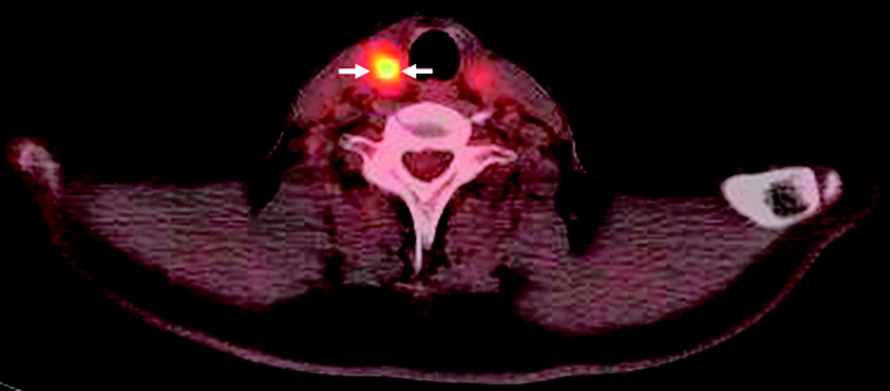
18F-FDG PET/CT image demonstrates an intensely hypermetabolic mass (SUVmax = 9.1 g/mL) in the right thyroid lobe (arrows). Background thyroid parenchyma shows mild diffuse FDG uptake.

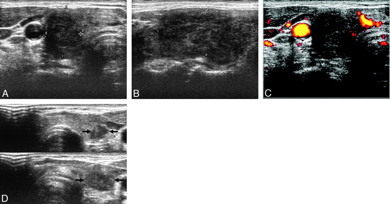
Thyroid US, performed 1 day after the 18F-FDG PET/CT examination, demonstrated an approximately 21 × 19 × 37 mm mass in the right thyroid lobe (Fig 2A−C), the location of which coincided with the hypermetabolic focus on 18F-FDG PET/CT. The mass was ill-defined, markedly hypoechoic, and heterogeneous, with scattered hyperechoic areas on gray-scale US (Fig 2A, -B), and was hypovascular compared with the background parenchyma on PD US (Fig 2C). Tenderness was noted just on compression of the mass. These sonographic findings suggested subacute thyroiditis. In addition, 2 small focal ill-defined markedly hypoechoic nodular lesions were present in the left thyroid lobe (Fig 2D). They could not be distinguished from background thyroid parenchyma that showed mild diffuse FDG uptake on 18F-FDG PET/CT. We performed US-FNAB of the right thyroid mass because the patient and referring physician were anxious about the newly developed intensely hypermetabolic focus on 18F-FDG PET/CT and because we could not definitively exclude infiltrative malignancies such as PTC or primary thyroid lymphoma.

Fig 2.
  • Download figure
  • Open in new tab
  • Download powerpoint
Fig 2.

A, Transverse gray-scale US reveals a markedly hypoechoic mass with a taller-than-wide shape in the right thyroid lobe (delineated by electronic calipers). B, A longitudinal gray-scale US shows an ill-demarcated heterogeneous mass with scattered hyperechoic areas (delineated by electronic calipers). C, A transverse PD US demonstrates hypovascularity of the mass compared with the background thyroid parenchyma. D, Split transverse gray-scale USs show 2 small focal ill-defined markedly hypoechoic nodular lesions in the left thyroid lobe (arrows).

US-FNAB was also performed on the 2 small nodular lesions of the left thyroid lobe to exclude PTC. The results of US-FNAB of the right and left thyroid lesions were consistent with subacute thyroiditis (Fig 3A) and chronic lymphocytic thyroiditis (Fig 3B), respectively.

Fig 3.
  • Download figure
  • Open in new tab
  • Download powerpoint
Fig 3.

A, Photographs of the cytologic examination of the specimen obtained by US-FNAB of the right thyroid mass show several multinucleated giant cells (arrows) with the background of smaller inflammatory cells, consistent with subacute thyroiditis (Papanicolaou, original magnification ×200). B, Photomicrograph of the cytologic examination of the specimen obtained by US-FNAB of 1 of the 2 left thyroid nodules reveals a lymphoid tangle (arrow) in the background of small lymphoid cells, suggestive of chronic lymphocytic thyroiditis (Papanicolaou, original magnification ×200).

Discussion

In subacute thyroiditis, the clinical and laboratory data are usually sufficient for making the diagnosis, and biopsy is rarely indicated. Our case presented a clinical dilemma because of unexpected focal, newly developed, intense FDG uptake of the lesion of subacute thyroiditis on 18F-FDG PET/CT, which raised clinical concern about thyroid malignancy.

Focal FDG uptake in the thyroid gland is likely caused by malignancy.10 However, focal FDG uptake may also be attributed to several benign conditions, including nodular hyperplasia, Hürthle cell and follicular neoplasms, and even chronic lymphocytic thyroiditis.7,10

Subacute thyroiditis can involve 1 or both lobes of the thyroid gland diffusely or focally.11 Several reports concerning US features of subacute thyroiditis have been published in the English literature.2,3 In a study of 27 patients with subacute thyroiditis,2 unilateral thyroid involvement was demonstrated in 23 and bilateral involvement, in 4 patients. All the lesions were ill-defined and hypoechoic. Nine patients had a hypoechoic nodular lesion with an irregular or microlobulated margin, which mimicked thyroid carcinoma. No hypervascularity was noted in any of the lesions on color or PD US. Zacharia et al3 reported the utility of color Doppler US in a case of subacute thyroiditis that mimicked thyroid carcinoma clinically. They reported that color Doppler US showed no hypervascularity at the acute stage and slightly increased vascularity at the recovery stage.

Two cases of subacute thyroiditis with diffuse hypermetabolism on 18F-FDG PET, involving a single lobe and both lobes of the thyroid gland, respectively, have been reported.8,9 However, cases of subacute thyroiditis with focal hypermetabolism have not appeared in the English literature. Our case represents the first of subacute thyroiditis that showed focal hypermetabolism on 18F-FDG PET/CT, mimicking a thyroid malignancy. In view of clinical and US features, subacute thyroiditis was suggested as a probable diagnosis. However, because the patient and referring physician were anxious about the newly developed intensely hypermetabolic focus on 18F-FDG PET/CT and because we could not definitively exclude infiltrative malignancies such as PTC and primary thyroid lymphoma, we proceeded to US-FNAB. The results of cytologic examination were consistent with subacute thyroiditis. In addition, 2 small nodular lesions of the left thyroid lobe proved to be chronic lymphocytic thyroiditis by US-FNAB.

Thus, this case represents coexistence of subacute thyroiditis and chronic lymphocytic thyroiditis. Interestingly, the lesion of subacute thyroiditis showed intense hypermetabolism, while the lesions of focal chronic lymphocytic thyroiditis were not significantly hypermetabolic compared with the background parenchyma. We hypothesized that the intense hypermetabolism of the lesion of subacute thyroiditis might be caused by active inflammation, while the lesions of focal chronic lymphocytic thyroiditis might be relatively quiescent; thus they did not show hypermetabolism on 18F-FDG PET/CT.

In conclusion, subacute thyroiditis should be included in the differential diagnosis of focal FDG uptake on 18F-FDG PET/CT. Gray-scale and PD US are useful for the characterization of this lesion, and US-FNAB can provide a definitive diagnosis of subacute thyroiditis that presents as an intensely hypermetabolic thyroid incidentaloma on 18F-FDG PET/CT.

References

  1. 1.↵
    1. Burrow GN,
    2. Oppenheimer JH,
    3. Volpé R
    1. Volpé R
    . Subacute thyroiditis. In: Burrow GN, Oppenheimer JH, Volpé R eds. Thyroid Function and Disease.Philadelphia:Saunders;1989:179–87
  2. 2.↵
    1. Park SY,
    2. Kim EK,
    3. Kim MJ,
    4. et al
    . Ultrasonographic characteristics of subacute granulomatous thyroiditis. Korean J Radiol 2006;7:229–34
    CrossRefPubMed
  3. 3.↵
    1. Zacharia TT,
    2. Perumpallichira JJ,
    3. Sindhwani V,
    4. et al
    . Gray-scale and color Doppler sonographic findings in a case of subacute granulomatous thyroiditis mimicking thyroid carcinoma. J Clin Ultrasound 2002;30:442–44
    CrossRefPubMed
  4. 4.↵
    1. King DL,
    2. Stack BC Jr.,
    3. Spring PM,
    4. et al
    . Incidence of thyroid carcinoma in fluorodeoxyglucose positron emission tomography-positive thyroid incidentalomas. Otolaryngol Head Neck Surg 2007;137:400–04
    CrossRefPubMed
  5. 5.↵
    1. Van den Bruel A,
    2. Maes A,
    3. De Potter T,
    4. et al
    . Clinical relevance of thyroid fluorodeoxyglucose-whole body positron emission tomography incidentaloma. J Clin Endocrinol Metab 2002;87:1517–20
    CrossRefPubMed
  6. 6.↵
    1. Yasuda S,
    2. Shohtsu A,
    3. Ide M,
    4. et al
    . Chronic thyroiditis: diffuse uptake of FDG at PET. Radiology 1998;207:775–78
    PubMed
  7. 7.↵
    1. Schmid DT,
    2. Kneifel S,
    3. Stoeckli SJ,
    4. et al
    . Increased 18F-FDG uptake mimicking thyroid cancer in a patient with Hashimoto's thyroiditis. Eur Radiol 2003;13:2119–21
    CrossRefPubMed
  8. 8.↵
    1. Song YS,
    2. Jang SJ,
    3. Chung JK,
    4. et al
    . F-18 fluorodeoxyglucose (FDG) positron emission tomography (PET) and Tc-99m pertechnate scan findings of a patient with unilateral subacute thyroiditis. Clin Nucl Med 2009;34:456–58
    CrossRefPubMed
  9. 9.↵
    1. Meller J,
    2. Sahlmann CO,
    3. Scheel AK.
    . 18F-FDG PET and PET/CT in fever of unknown origin. J Nucl Med 2007;48:35–45
    Abstract/FREE Full Text
  10. 10.↵
    1. Kim TY,
    2. Kim WB,
    3. Ryu JS,
    4. et al
    . 18F-fluorodeoxyglucose uptake in thyroid from positron emission tomogram (PET) for evaluation in cancer patients: high prevalence of malignancy in thyroid PET incidentaloma. Laryngoscope 2005;115:1074–78
    CrossRefPubMed
  11. 11.↵
    1. Greene JN.
    . Subacute thyroiditis. Am J Med 1971;51:97–108
    CrossRefPubMed
  • Received September 24, 2009.
  • Accepted after revision December 2, 2009.
  • Copyright © American Society of Neuroradiology
View Abstract
PreviousNext
Back to top

In this issue

American Journal of Neuroradiology: 32 (4)
American Journal of Neuroradiology
Vol. 32, Issue 4
1 Apr 2011
  • Table of Contents
  • Index by author
Advertisement
Print
Download PDF
Email Article

Thank you for your interest in spreading the word on American Journal of Neuroradiology.

NOTE: We only request your email address so that the person you are recommending the page to knows that you wanted them to see it, and that it is not junk mail. We do not capture any email address.

Enter multiple addresses on separate lines or separate them with commas.
Subacute Thyroiditis Presenting as a Focal Lesion on [18F] Fluorodeoxyglucose Whole-Body Positron-Emission Tomography/CT
(Your Name) has sent you a message from American Journal of Neuroradiology
(Your Name) thought you would like to see the American Journal of Neuroradiology web site.
CAPTCHA
This question is for testing whether or not you are a human visitor and to prevent automated spam submissions.
Cite this article
S.H. Yeo, S.K. Lee, I. Hwang, E.J. Ahn
Subacute Thyroiditis Presenting as a Focal Lesion on [18F] Fluorodeoxyglucose Whole-Body Positron-Emission Tomography/CT
American Journal of Neuroradiology Apr 2011, 32 (4) E58-E60; DOI: 10.3174/ajnr.A2017

Citation Manager Formats

  • BibTeX
  • Bookends
  • EasyBib
  • EndNote (tagged)
  • EndNote 8 (xml)
  • Medlars
  • Mendeley
  • Papers
  • RefWorks Tagged
  • Ref Manager
  • RIS
  • Zotero
0 Responses
Respond to this article
Share
Bookmark this article
Subacute Thyroiditis Presenting as a Focal Lesion on [18F] Fluorodeoxyglucose Whole-Body Positron-Emission Tomography/CT
S.H. Yeo, S.K. Lee, I. Hwang, E.J. Ahn
American Journal of Neuroradiology Apr 2011, 32 (4) E58-E60; DOI: 10.3174/ajnr.A2017
del.icio.us logo Twitter logo Facebook logo Mendeley logo
  • Tweet Widget
  • Facebook Like
  • Google Plus One
Purchase

Jump to section

  • Article
    • Abstract
    • Abbreviations
    • Case Report
    • Discussion
    • References
  • Figures & Data
  • Info & Metrics
  • Responses
  • References
  • PDF

Related Articles

  • No related articles found.
  • PubMed
  • Google Scholar

Cited By...

  • No citing articles found.
  • Crossref (9)
  • Google Scholar

This article has been cited by the following articles in journals that are participating in Crossref Cited-by Linking.

  • The Use of18F-FDG-PET/CT for Diagnosis and Treatment Monitoring of Inflammatory and Infectious Diseases
    Andor W. J. M. Glaudemans, Erik F. J. de Vries, Filippo Galli, Rudi A. J. O. Dierckx, Riemer H. J. A. Slart, Alberto Signore
    Clinical and Developmental Immunology 2013 2013
  • The Old and the New in Subacute Thyroiditis: An Integrative Review
    Nicola Lanzo, Bohdan Patera, Gaia Fazzino, Daniela Gallo, Adriana Lai, Eliana Piantanida, Silvia Ippolito, Maria Tanda
    Endocrines 2022 3 3
  • The Role of 18F-FDG PET/CT in the Management of the Autoimmune Thyroid Diseases
    Bogdan Małkowski, Zbigniew Serafin, Rafał Glonek, Szymon Suwała, Rita Łopatto, Roman Junik
    Frontiers in Endocrinology 2019 10
  • 18F-FDG PET/CT imaging of atypical subacute thyroiditis in thyrotoxicosis
    Katsuya Yoshida, Hidetaka Yokoh, Akira Toriihara, Hayahiko Fujii, Naoki Harata, Jun Isogai, Ukihide Tateishi
    Medicine 2017 96 30
  • Comparison of treatment outcome between glucocorticoids and non-steroidal anti-inflammatory drugs in subacute thyroiditis patients—a systematic review and meta-analysis
    Anqi Yuan, Jialu Wu, Hui Huang
    Frontiers in Endocrinology 2024 15
  • Benign Thyroid and Neck Lesions Mimicking Malignancy with False Positive Findings on Positron Emission Tomography-Computed Tomography
    Ye Ri Yoon, Shin Young Kim, Sang Mi Lee, Deuk Young Lee
    Journal of the Korean Society of Radiology 2017 76 2
  • Diagnostic Value of 2-[18F]FDG PET/CT in a Patient with Atypical Subacute Thyroiditis: A Case Report
    Teresa Kraus, Marcus Hacker, Werner Langsteger, Shuren Li, Raffaella Calabretta
    Life 2022 12 8
  • FDG PET/CT of Neuroendocrine Carcinoma of the Thyroid Gland
    Li-Chun Zheng, Xiang-Liu OuYang, Wen-Jun Zhang, Xiao-Ming Zhang
    Clinical Nuclear Medicine 2015 40 2
  • Subacute thyroiditis: clinical significance of diffuse 18-fluorodeoxyglucose uptake. Case report
    Nikolai A. Ognerubov, Tatiana S. Antipova, Elena E. Palkina
    Consilium Medicum 2023 25 4

More in this TOC Section

  • Atypical Diffusion-Restricted Lesion in 5-Fluorouracil Encephalopathy
  • Dural Infantile Hemangioma Masquerading as a Skull Vault Lesion
  • Multimodal Imaging of Spike Propagation: A Technical Case Report
Show more Case Reports

Similar Articles

Advertisement

Indexed Content

  • Current Issue
  • Accepted Manuscripts
  • Article Preview
  • Past Issues
  • Editorials
  • Editor's Choice
  • Fellows' Journal Club
  • Letters to the Editor
  • Video Articles

Cases

  • Case Collection
  • Archive - Case of the Week
  • Archive - Case of the Month
  • Archive - Classic Case

Special Collections

  • AJNR Awards
  • ASNR Foundation Special Collection
  • Most Impactful AJNR Articles
  • Photon-Counting CT
  • Spinal CSF Leak Articles (Jan 2020-June 2024)

More from AJNR

  • Trainee Corner
  • Imaging Protocols
  • MRI Safety Corner

Multimedia

  • AJNR Podcasts
  • AJNR Scantastics

Resources

  • Turnaround Time
  • Submit a Manuscript
  • Submit a Video Article
  • Submit an eLetter to the Editor/Response
  • Manuscript Submission Guidelines
  • Statistical Tips
  • Fast Publishing of Accepted Manuscripts
  • Graphical Abstract Preparation
  • Imaging Protocol Submission
  • Evidence-Based Medicine Level Guide
  • Publishing Checklists
  • Author Policies
  • Become a Reviewer/Academy of Reviewers
  • News and Updates

About Us

  • About AJNR
  • Editorial Board
  • Editorial Board Alumni
  • Alerts
  • Permissions
  • Not an AJNR Subscriber? Join Now
  • Advertise with Us
  • Librarian Resources
  • Feedback
  • Terms and Conditions
  • AJNR Editorial Board Alumni

American Society of Neuroradiology

  • Not an ASNR Member? Join Now

© 2025 by the American Society of Neuroradiology All rights, including for text and data mining, AI training, and similar technologies, are reserved.
Print ISSN: 0195-6108 Online ISSN: 1936-959X

Powered by HighWire