Advertisement

Main menu

  • Home
  • Content
    • Current Issue
    • Accepted Manuscripts
    • Article Preview
    • Past Issue Archive
    • Video Articles
    • AJNR Case Collection
    • Case of the Week Archive
    • Case of the Month Archive
    • Classic Case Archive
  • Special Collections
    • AJNR Awards
    • ASNR Foundation Special Collection
    • Most Impactful AJNR Articles
    • Photon-Counting CT
    • Spinal CSF Leak Articles (Jan 2020-June 2024)
  • Multimedia
    • AJNR Podcasts
    • AJNR SCANtastic
    • Trainee Corner
    • MRI Safety Corner
    • Imaging Protocols
  • For Authors
    • Submit a Manuscript
    • Submit a Video Article
    • Submit an eLetter to the Editor/Response
    • Manuscript Submission Guidelines
    • Statistical Tips
    • Fast Publishing of Accepted Manuscripts
    • Graphical Abstract Preparation
    • Imaging Protocol Submission
    • Author Policies
  • About Us
    • About AJNR
    • Editorial Board
    • Editorial Board Alumni
  • More
    • Become a Reviewer/Academy of Reviewers
    • Subscribers
    • Permissions
    • Alerts
    • Feedback
    • Advertisers
    • ASNR Home

User menu

  • Alerts
  • Log in

Search

  • Advanced search
American Journal of Neuroradiology

American Journal of Neuroradiology

ASHNR American Society of Functional Neuroradiology ASHNR American Society of Pediatric Neuroradiology ASSR
  • Alerts
  • Log in

Advanced Search

  • Home
  • Content
    • Current Issue
    • Accepted Manuscripts
    • Article Preview
    • Past Issue Archive
    • Video Articles
    • AJNR Case Collection
    • Case of the Week Archive
    • Case of the Month Archive
    • Classic Case Archive
  • Special Collections
    • AJNR Awards
    • ASNR Foundation Special Collection
    • Most Impactful AJNR Articles
    • Photon-Counting CT
    • Spinal CSF Leak Articles (Jan 2020-June 2024)
  • Multimedia
    • AJNR Podcasts
    • AJNR SCANtastic
    • Trainee Corner
    • MRI Safety Corner
    • Imaging Protocols
  • For Authors
    • Submit a Manuscript
    • Submit a Video Article
    • Submit an eLetter to the Editor/Response
    • Manuscript Submission Guidelines
    • Statistical Tips
    • Fast Publishing of Accepted Manuscripts
    • Graphical Abstract Preparation
    • Imaging Protocol Submission
    • Author Policies
  • About Us
    • About AJNR
    • Editorial Board
    • Editorial Board Alumni
  • More
    • Become a Reviewer/Academy of Reviewers
    • Subscribers
    • Permissions
    • Alerts
    • Feedback
    • Advertisers
    • ASNR Home
  • Follow AJNR on Twitter
  • Visit AJNR on Facebook
  • Follow AJNR on Instagram
  • Join AJNR on LinkedIn
  • RSS Feeds

AJNR is seeking candidates for the AJNR Podcast Editor. Read the position description.

Research ArticleTechnical Note
Open Access

Hyperintense Vessel Sign on Fluid-Attenuated Inversion Recovery MR Imaging Is Reduced by Gadolinium

K.A. Dani, L.L. Latour and S. Warach on behalf of the NINDS Natural History of Stroke Investigators
American Journal of Neuroradiology September 2012, 33 (8) E112-E114; DOI: https://doi.org/10.3174/ajnr.A2482
K.A. Dani
aFrom the Section on Stroke Diagnostics and Therapeutics, NINDS, National Institutes of Health, Bethesda, Maryland.
  • Find this author on Google Scholar
  • Find this author on PubMed
  • Search for this author on this site
L.L. Latour
aFrom the Section on Stroke Diagnostics and Therapeutics, NINDS, National Institutes of Health, Bethesda, Maryland.
  • Find this author on Google Scholar
  • Find this author on PubMed
  • Search for this author on this site
S. Warach
aFrom the Section on Stroke Diagnostics and Therapeutics, NINDS, National Institutes of Health, Bethesda, Maryland.
  • Find this author on Google Scholar
  • Find this author on PubMed
  • Search for this author on this site
  • Article
  • Figures & Data
  • Info & Metrics
  • Responses
  • References
  • PDF
Loading

Abstract

SUMMARY: The HVS on FLAIR imaging is a useful marker of acute ischemic stroke. We investigated whether prior administration of gadolinium-based contrast hindered detection of this sign on images from subjects with acute nonlacunar ischemic stroke <4.5 hours after onset. Both blinded and comparative unblinded analyses showed significantly reduced HVS detection on postcontrast images. We suggest that assessment for this sign should be performed on images acquired prior to contrast administration.

ABBREVIATIONS:

BA
blinded analysis
CA
comparative analysis
FLAIR
fluid-attenuated inversion recovery
HVS
hyperintense vessel sign
NIH
National Institutes of Health
NINDS
National Institute of Neurological Disorders and Stroke

The HVS describes hyperintensity of the cerebral arteries ipsilateral to an arterial occlusion when visualized on FLAIR imaging. It is, therefore, a useful imaging marker of acute ischemic stroke.1,2 Although precise mechanisms have not yet been elucidated, angiographic findings suggest that the pathophysiology of this sign is likely to involve either arterial occlusion or retrograde flow via collateral vessels, static blood, or slow flow.1⇓–3 At proximal locations, FLAIR HVS is likely to represent arterial occlusion, and a report from our group suggested a sensitivity of 65.9%.4 At distal locations, however, the sign may be a marker for collateral flow; a further study5 suggested that subjects with the HVS in distal vessels had milder baseline neurologic deficits and a trend toward smaller subacute lesions. Given that HVS is assessed frequently in both clinical and research settings, detection should be optimized to be as sensitive as possible. However, our experience in the acquisition of postcontrast FLAIR images suggested that their sensitivity may be less than that in such images acquired before contrast administration. We aimed to clarify this issue.

Materials and Methods

A retrospective analysis was performed on an institutional review board−approved prospectively collected dataset (Natural History of Stroke Protocol). Subject inclusion criteria for this study were the following: 1) acute nonlacunar ischemic stroke, 2) <4.5 hours from stroke onset, and 3) good quality pre- and postcontrast FLAIR images from the same examination. Some of these subjects have been included in a previous publication.5 Images were acquired from 2 hospitals affiliated with our institution, and details of image acquisition have been previously reported.5 The analysis of images was performed in 2 ways: First, 1 observer (K.A.D.) reviewed all pre- and postcontrast FLAIR images while blinded to clinical and other imaging data and without knowledge of which images were performed after contrast (BA). This analysis was performed blinded to minimize observer bias. Each FLAIR image was dichotomously graded for the presence or absence of an HVS at sites proximal and distal to the Sylvian fissure. For each site, a 2 × 2 contingency table describing the presence or absence of HVS on pre- and postcontrast images was constructed. Next, StatsDirect (Version 2.7.2, http://www.statsdirect.com) was used to perform the McNemar test with Yates correction to elicit differences in the proportion of HVS detected in precontrast images compared with postcontrast images. Next, the Fisher exact test was used to compare the proportion of cases of HVS in which contrast affected detection in proximal and in distal locations.

Next, a CA was performed to simulate clinical image reads. For this analysis, pre- and postcontrast image pairings were reviewed together, while the reviewers were unblinded to other imaging data. The presence or absence of HVS on pre- and postcontrast images was noted, in addition to recording of cases in which there was a subjective reduction in HVS intensity but without complete disappearance of the sign. In a similar manner to that in the BA, images were graded for the continued presence (persistent or reduced HVS) or absence of HVS after contrast. Statistical analysis was performed as per the BA.

Results

Of the 56 subjects who fulfilled the inclusion criteria, movement artifacts precluded analysis in 11 subjects. Of the remaining 45 subjects, BA revealed 23 (51%) subjects who demonstrated the presence of HVS on precontrast FLAIR. Of these, the HVS was present in 22 subjects proximally, in 19 subjects distally, and in both locations in 18 subjects. For the BA, disappearance of HVS after contrast was seen in 11/22 (50%) subjects proximally (P = .027) and 13/19 (68%) subjects distally (P = .003). The detection of HVS by BA in the post- but not precontrast FLAIR imaging occurred in only 2 subjects. There was no difference in the proportion of cases with HVS disappearance with respect to proximal and distal locations (P = .34). In the CA, HVS was present in 24 subjects proximally, 21 distally, and both proximally and distally in 19. HVS was undetected after contrast in 3/24 (13%) subjects proximally (P = .25) and 10/21 (48%) distally (P = .004). Although the difference between proximal and distal locations was statistically significant (P = .02), there was no difference between proximal and distal locations with respect to the proportion of cases in which contrast had any effect on the HVS (ie, a reduction in intensity or complete disappearance of the HVS): 16/24 (67%) subjects proximally and 15/21 (71%) subjects distally (P = .76, Fisher exact test). An example of HVS change with contrast is given in Fig 1.

Fig 1.
  • Download figure
  • Open in new tab
  • Download powerpoint
Fig 1.

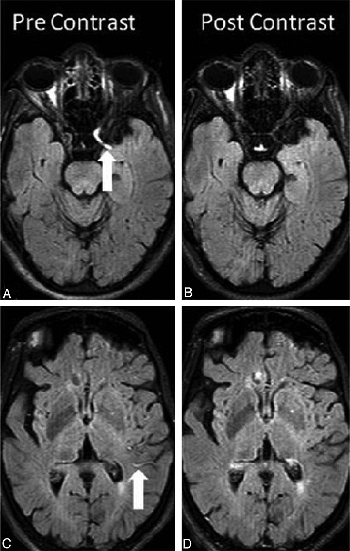
The HVS before and after gadolinium-based contrast. All images are from the same subject. A and C, Precontrast FLAIR images. The white arrow in A marks the proximal HVS, and the white arrow in C marks the distal HVS. B and D, The HVS is not detected in the postcontrast FLAIR images.

Discussion

The blinded analysis presented here demonstrates a significantly reduced rate of detection of the HVS after the administration of gadolinium-based contrast. These findings are consistent with the CA, which showed either a reduction in intensity or complete disappearance of HVS after gadolinium in most subjects. These findings are clinically important for 2 reasons: First, in concert with angiographic studies, the presence of HVS can potentially distinguish areas of slow flow from sites of arterial occlusion and, therefore, potentially highlight regions with collateral circulation, thus informing the clinician of the underlying tissue pathophysiology.5 Second, the value of postcontrast FLAIR examinations after stroke is increasingly being recognized; hyperintensity of CSF spaces (hyperintense acute reperfusion marker) seen on the postcontrast FLAIR imaging can suggest early breakdown of the blood-brain barrier. This sign is associated with hemorrhagic transformation and worse clinical outcome.6 Therefore, given the potential utility of postcontrast FLAIR images after acute ischemic stroke in clinical and research settings, neuroradiologists and neurologists should be aware of the optimal imaging conditions for detection of the HVS, which include assessment of FLAIR imaging performed before gadolinium-based contrast administration.

The cause of the postcontrast disappearance of the HVS is unclear. FLAIR images have both T1 weighting from the inversion recovery for CSF suppression and T2 weighting from a long TE. We can only speculate that the gadolinium in the vessels results in a shortening of T1 and T2 in such a way that the HVS effect is significantly decreased.

A number of limitations to this study should be acknowledged. First, we could not account for the volume or type of contrast agent used, which is likely to have varied between hospitals and across time. In addition, the FLAIR images analyzed in this study were acquired in a number of years, during which time acquisition parameters varied. In this study, we did not account for variations in TI, TE, and TR, which can all affect tissue contrast on FLAIR imaging. Section thickness and section gap thickness, which also vary between MR imaging stroke protocols, are likely to influence the detection of the HVS. Nonetheless, this heterogeneity reflects the range of routine clinical practice and, therefore, makes our observations all the more relevant. Furthermore, a particular strength of this study is that the pre- and postgadolinium FLAIR images were acquired contemporaneously (within a matter of minutes), by using exactly the same acquisition parameters, thus negating many of the potentially confounding effects of variations in acquisition parameters between examinations. Indeed, the aim of this study was to determine an effect of gadolinium in the detection of HVS, which we have successfully achieved. However, further analyses with larger numbers of subjects that examine concomitant MR angiographic findings are required to determine whether there is a difference in the effect of gadolinium on the HVS found due to arterial occlusion compared with that due to slow flow/collateral circulation, because this may explain the observed differences between proximal and distal locations.

Conclusions

Gadolinium reduces the detection of the HVS after ischemic stroke. We therefore suggest that if both pre- and postcontrast FLAIR examinations are acquired, the assessment of HVS is ideally performed on precontrast FLAIR, especially if comparisons between institutions are to be performed. Additional studies are required to determine the biologic significance of the “disappearing” HVS.

Acknowledgments

We thank Pat Lyall and Lizbeth Keller from the Section on Stroke Diagnostics and Therapeutics (NINDS) for assistance with data base searching.

Footnotes

  • Disclosures: Krishna Dani, Research Support (including provision of equipment and materials): Patrick Berthoud, Research Fellowship. The study was funded by the NIH. Details: The above fellowship provided salary and funds to travel to the NIH. This is a 3-year research training fellowship. Two years were spent at the University of Glasgow (UK) and 1 year at the NIH.

  • This work was supported by the Division of Intramural Research of the NIH and NINDS. Krishna Dani is supported by the Patrick Berthoud Charitable Trust.

  • Paper previously presented at: International Stroke Conference, February 23–24, 2010; San Antonio, Texas.

Indicates open access to non-subscribers at www.ajnr.org

References

  1. 1.↵
    1. Toyoda K,
    2. Ida M,
    3. Fukuda K
    . Fluid-attenuated inversion recovery intraarterial signal: an early sign of hyperacute cerebral ischemia. AJNR Am J Neuroradiol 2001; 22: 1021– 29
    Abstract/FREE Full Text
  2. 2.↵
    1. Noguchi K,
    2. Ogawa T,
    3. Inugami A,
    4. et al
    . MRI of acute cerebral infarction: a comparison of FLAIR and T2-weighted fast spin-echo imaging. Neuroradiology 1997; 39: 406– 10
    CrossRefPubMed
  3. 3.↵
    1. Cosnard G,
    2. Duprez T,
    3. Grandin C,
    4. et al
    . Fast FLAIR sequence for detecting major vascular abnormalities during the hyperacute phase of stroke: a comparison with MR angiography. Neuroradiology 1999; 41: 342– 46
    CrossRefPubMed
  4. 4.↵
    1. Schellinger PD,
    2. Chalela JA,
    3. Kang DW
    . Diagnostic and prognostic value of early MR imaging vessel signs in hyperacute stroke patients imaged <3 hours and treated with recombinant tissue plasminogen activator. AJNR Am J Neuroradiol 2005; 26: 618– 24
    Abstract/FREE Full Text
  5. 5.↵
    1. Lee KY,
    2. Latour LL,
    3. Luby M,
    4. et al
    . Distal hyperintense vessels on FLAIR: an MRI marker for collateral circulation in acute stroke? Neurology 2009; 72: 1134– 39. Epub 2009 Feb 11
    Abstract/FREE Full Text
  6. 6.↵
    1. Warach S,
    2. Latour LL,
    3. Warach S,
    4. et al
    . Evidence of reperfusion injury, exacerbated by thrombolytic therapy, in human focal brain ischemia using a novel imaging marker of early blood-brain barrier disruption. Stroke 2004; 35: 2659– 61
    Abstract/FREE Full Text
  • Received June 21, 2010.
  • Accepted after revision November 17, 2010.
  • © 2012 by American Journal of Neuroradiology
View Abstract
PreviousNext
Back to top

In this issue

American Journal of Neuroradiology: 33 (8)
American Journal of Neuroradiology
Vol. 33, Issue 8
1 Sep 2012
  • Table of Contents
  • Index by author
Advertisement
Print
Download PDF
Email Article

Thank you for your interest in spreading the word on American Journal of Neuroradiology.

NOTE: We only request your email address so that the person you are recommending the page to knows that you wanted them to see it, and that it is not junk mail. We do not capture any email address.

Enter multiple addresses on separate lines or separate them with commas.
Hyperintense Vessel Sign on Fluid-Attenuated Inversion Recovery MR Imaging Is Reduced by Gadolinium
(Your Name) has sent you a message from American Journal of Neuroradiology
(Your Name) thought you would like to see the American Journal of Neuroradiology web site.
CAPTCHA
This question is for testing whether or not you are a human visitor and to prevent automated spam submissions.
Cite this article
K.A. Dani, L.L. Latour, S. Warach
Hyperintense Vessel Sign on Fluid-Attenuated Inversion Recovery MR Imaging Is Reduced by Gadolinium
American Journal of Neuroradiology Sep 2012, 33 (8) E112-E114; DOI: 10.3174/ajnr.A2482

Citation Manager Formats

  • BibTeX
  • Bookends
  • EasyBib
  • EndNote (tagged)
  • EndNote 8 (xml)
  • Medlars
  • Mendeley
  • Papers
  • RefWorks Tagged
  • Ref Manager
  • RIS
  • Zotero
0 Responses
Respond to this article
Share
Bookmark this article
Hyperintense Vessel Sign on Fluid-Attenuated Inversion Recovery MR Imaging Is Reduced by Gadolinium
K.A. Dani, L.L. Latour, S. Warach
American Journal of Neuroradiology Sep 2012, 33 (8) E112-E114; DOI: 10.3174/ajnr.A2482
del.icio.us logo Twitter logo Facebook logo Mendeley logo
  • Tweet Widget
  • Facebook Like
  • Google Plus One
Purchase

Jump to section

  • Article
    • Abstract
    • ABBREVIATIONS:
    • Materials and Methods
    • Results
    • Discussion
    • Conclusions
    • Acknowledgments
    • Footnotes
    • References
  • Figures & Data
  • Info & Metrics
  • Responses
  • References
  • PDF

Related Articles

  • No related articles found.
  • PubMed
  • Google Scholar

Cited By...

  • No citing articles found.
  • Crossref (6)
  • Google Scholar

This article has been cited by the following articles in journals that are participating in Crossref Cited-by Linking.

  • Imaging Ischemic and Hemorrhagic Disease of the Brain in Dogs
    Susan A. Arnold, Simon R. Platt, Karine P. Gendron, Franklin D. West
    Frontiers in Veterinary Science 2020 7
  • Mechanism of Stroke According to the Severity and Location of Atherosclerotic Middle Cerebral Artery Disease
    Sang Hee Ha, Jun Young Chang, Sang Hun Lee, Kyung Mi Lee, Sung Hyuk Heo, Dae-il Chang, Bum Joon Kim
    Journal of Stroke and Cerebrovascular Diseases 2021 30 2
  • Interpretation of fluid-attenuated inversion recovery vascular hyperintensity in stroke
    Kyung-Yul Lee, Jin Woo Kim, Mina Park, Sang Hyun Suh, Sung Jun Ahn
    Journal of Neuroradiology 2022 49 3
  • Fluid-attenuated inversion recovery vascular hyperintensities are not visible using 3D CUBE FLAIR sequence
    Jérôme Hodel, Xavier Leclerc, Mathieu Rodallec, Sophie Gerber, Raphael Blanc, Olivier Outteryck, Samir Benadjaoud, Cécile Rabrait, Mathieu Zuber, Jean-Pierre Pruvo, Marc Zins
    European Radiology 2013 23 7
  • Analysis of blood hydrodynamic characteristics
    Zhongchang Wang, Sen Zhang
    Journal of Complexity in Health Sciences 2018 1 2
  • FLAIR vascular hyperintensity is associated with functional outcome in patients with ischemic stroke receiving endovascular treatment: a meta-analysis
    Chunyan Wang, Chuanliu Wang, Yongjun Ni
    Frontiers in Neurology 2024 15

More in this TOC Section

  • 3D Fast Spin-Echo T1 Black-Blood Imaging for the Diagnosis of Cervical Artery Dissection
  • The Kety-Schmidt Technique for Quantitative Perfusion and Oxygen Metabolism Measurements in the MR Imaging Environment
  • A Swine Model to Analyze Arterial Structural Changes Induced by Mechanical Thrombectomy
Show more Technical Note

Similar Articles

Advertisement

Indexed Content

  • Current Issue
  • Accepted Manuscripts
  • Article Preview
  • Past Issues
  • Editorials
  • Editor's Choice
  • Fellows' Journal Club
  • Letters to the Editor
  • Video Articles

Cases

  • Case Collection
  • Archive - Case of the Week
  • Archive - Case of the Month
  • Archive - Classic Case

Special Collections

  • AJNR Awards
  • ASNR Foundation Special Collection
  • Most Impactful AJNR Articles
  • Photon-Counting CT
  • Spinal CSF Leak Articles (Jan 2020-June 2024)

More from AJNR

  • Trainee Corner
  • Imaging Protocols
  • MRI Safety Corner

Multimedia

  • AJNR Podcasts
  • AJNR Scantastics

Resources

  • Turnaround Time
  • Submit a Manuscript
  • Submit a Video Article
  • Submit an eLetter to the Editor/Response
  • Manuscript Submission Guidelines
  • Statistical Tips
  • Fast Publishing of Accepted Manuscripts
  • Graphical Abstract Preparation
  • Imaging Protocol Submission
  • Evidence-Based Medicine Level Guide
  • Publishing Checklists
  • Author Policies
  • Become a Reviewer/Academy of Reviewers
  • News and Updates

About Us

  • About AJNR
  • Editorial Board
  • Editorial Board Alumni
  • Alerts
  • Permissions
  • Not an AJNR Subscriber? Join Now
  • Advertise with Us
  • Librarian Resources
  • Feedback
  • Terms and Conditions
  • AJNR Editorial Board Alumni

American Society of Neuroradiology

  • Not an ASNR Member? Join Now

© 2025 by the American Society of Neuroradiology All rights, including for text and data mining, AI training, and similar technologies, are reserved.
Print ISSN: 0195-6108 Online ISSN: 1936-959X

Powered by HighWire