Advertisement

Main menu

  • Home
  • Content
    • Current Issue
    • Accepted Manuscripts
    • Article Preview
    • Past Issue Archive
    • Video Articles
    • AJNR Case Collection
    • Case of the Week Archive
    • Case of the Month Archive
    • Classic Case Archive
  • Special Collections
    • AJNR Awards
    • ASNR Foundation Special Collection
    • Most Impactful AJNR Articles
    • Photon-Counting CT
    • Spinal CSF Leak Articles (Jan 2020-June 2024)
  • Multimedia
    • AJNR Podcasts
    • AJNR SCANtastic
    • Trainee Corner
    • MRI Safety Corner
    • Imaging Protocols
  • For Authors
    • Submit a Manuscript
    • Submit a Video Article
    • Submit an eLetter to the Editor/Response
    • Manuscript Submission Guidelines
    • Statistical Tips
    • Fast Publishing of Accepted Manuscripts
    • Graphical Abstract Preparation
    • Imaging Protocol Submission
    • Author Policies
  • About Us
    • About AJNR
    • Editorial Board
    • Editorial Board Alumni
  • More
    • Become a Reviewer/Academy of Reviewers
    • Subscribers
    • Permissions
    • Alerts
    • Feedback
    • Advertisers
    • ASNR Home

User menu

  • Alerts
  • Log in

Search

  • Advanced search
American Journal of Neuroradiology

American Journal of Neuroradiology

ASHNR American Society of Functional Neuroradiology ASHNR American Society of Pediatric Neuroradiology ASSR
  • Alerts
  • Log in

Advanced Search

  • Home
  • Content
    • Current Issue
    • Accepted Manuscripts
    • Article Preview
    • Past Issue Archive
    • Video Articles
    • AJNR Case Collection
    • Case of the Week Archive
    • Case of the Month Archive
    • Classic Case Archive
  • Special Collections
    • AJNR Awards
    • ASNR Foundation Special Collection
    • Most Impactful AJNR Articles
    • Photon-Counting CT
    • Spinal CSF Leak Articles (Jan 2020-June 2024)
  • Multimedia
    • AJNR Podcasts
    • AJNR SCANtastic
    • Trainee Corner
    • MRI Safety Corner
    • Imaging Protocols
  • For Authors
    • Submit a Manuscript
    • Submit a Video Article
    • Submit an eLetter to the Editor/Response
    • Manuscript Submission Guidelines
    • Statistical Tips
    • Fast Publishing of Accepted Manuscripts
    • Graphical Abstract Preparation
    • Imaging Protocol Submission
    • Author Policies
  • About Us
    • About AJNR
    • Editorial Board
    • Editorial Board Alumni
  • More
    • Become a Reviewer/Academy of Reviewers
    • Subscribers
    • Permissions
    • Alerts
    • Feedback
    • Advertisers
    • ASNR Home
  • Follow AJNR on Twitter
  • Visit AJNR on Facebook
  • Follow AJNR on Instagram
  • Join AJNR on LinkedIn
  • RSS Feeds

AJNR is seeking candidates for the AJNR Podcast Editor. Read the position description.

Research ArticleADULT BRAIN
Open Access

Ataxia Severity Correlates with White Matter Degeneration in Spinocerebellar Ataxia Type 7

C.R. Hernandez-Castillo, I. Vaca-Palomares, F. Barrios, L. Martinez, M.-C. Boll and J. Fernandez-Ruiz
American Journal of Neuroradiology November 2016, 37 (11) 2050-2054; DOI: https://doi.org/10.3174/ajnr.A4903
C.R. Hernandez-Castillo
aFrom the Consejo Nacional de Ciencia y Tecnologia–Instituto de Neuroetologia (C.R.H.-C.), Universidad Veracruzana, Veracruz, Mexico
  • Find this author on Google Scholar
  • Find this author on PubMed
  • Search for this author on this site
  • ORCID record for C.R. Hernandez-Castillo
I. Vaca-Palomares
bDepartamento de Fisiologia (I.V.-P., J.F.-R.), Facultad de Medicina
  • Find this author on Google Scholar
  • Find this author on PubMed
  • Search for this author on this site
  • ORCID record for I. Vaca-Palomares
F. Barrios
cInstituto de Neurobiologia (F.B.), Universidad Nacional Autonoma de Mexico, Mexico City, Mexico
  • Find this author on Google Scholar
  • Find this author on PubMed
  • Search for this author on this site
  • ORCID record for F. Barrios
L. Martinez
dDepartamento de Neurogenética y Biologıa Molecular (L.M.)
  • Find this author on Google Scholar
  • Find this author on PubMed
  • Search for this author on this site
  • ORCID record for L. Martinez
M.-C. Boll
eLaboratorio de Investigacion Clinica (M.-C.B.), Instituto Nacional de Neurología y Neurocirugía, “Manuel Velasco Suárez,” Mexico City, México.
  • Find this author on Google Scholar
  • Find this author on PubMed
  • Search for this author on this site
  • ORCID record for M.-C. Boll
J. Fernandez-Ruiz
bDepartamento de Fisiologia (I.V.-P., J.F.-R.), Facultad de Medicina
  • Find this author on Google Scholar
  • Find this author on PubMed
  • Search for this author on this site
  • ORCID record for J. Fernandez-Ruiz
  • Article
  • Figures & Data
  • Info & Metrics
  • Responses
  • References
  • PDF
Loading

Abstract

BACKGROUND AND PURPOSE: There is a scarcity of information on the effect of white matter degeneration in patients with spinocerebellar ataxia type 7. Therefore, we investigated the WM integrity in a large group of patients with spinocerebellar ataxia type 7 by using Tract-Based Spatial Statistics.

MATERIALS AND METHODS: Thirty-three patients with a molecular diagnosis of spinocerebellar ataxia type 7 and their age- and sex-matched healthy controls participated in this study. The patients' ataxia severity was evaluated with the Scale for the Assessment and Rating of Ataxia. Voxelwise analyses of diffusion metrics, including fractional anisotropy and mean diffusivity, were performed with Tract-Based Spatial Statistics. The correlation between WM abnormalities and ataxia severity was then calculated.

RESULTS: Tract-Based Spatial Statistics analysis revealed WM abnormalities in the cerebellum and the cerebellar peduncles, as well as in other major cortical and subcortical pathways. Further analysis between the Scale for the Assessment and Rating of Ataxia score and WM mean diffusivity showed significant associations only in key areas related to motor control and visuospatial processing, including the cerebellar WM, the middle occipital WM, the superior cerebellar peduncle, and bilateral anterior thalamic radiation. No significant associations between fractional anisotropy and the Scale for the Assessment and Rating of Ataxia were found.

CONCLUSIONS: These results suggest a significant contribution of local cerebellar and cerebellar-midbrain connections to ataxic impairment in spinocerebellar ataxia type 7. The results also suggest an involvement of cortical WM abnormalities including tracts within the occipital and frontal cortices. These findings contribute to a more comprehensive view of the clinical impact of the white matter degeneration in spinocerebellar ataxia type 7.

ABBREVIATIONS:

FA
fractional anisotropy
MD
mean diffusivity
SARA
Scale for the Assessment and Rating of Ataxia
SCA
spinocerebellar ataxia
SCA7
spinocerebellar ataxia type 7
TBSS
Tract-Based Spatial Statistics

Spinocerebellar ataxia type 7 (SCA7) is an autosomal dominant cerebellar ataxia caused by a mutation consisting in the expansion of the cytosine-adenine-guanine trinucleotide in the codon region of the chromosome 3p21, encoding the protein ataxin 7.1 SCA7 is considered one of the rarest forms of genetic autosomal dominant cerebellar ataxia.2 Clinically, SCA7 is characterized by a combination of cerebellar ataxia and macular degeneration and is the only spinocerebellar ataxia that manifests in permanent blindness.3,4 Furthermore, patients may eventually develop other neurologic deficits, including loss of manual dexterity, speech dysarthria, dysphagia, and eye movement abnormalities.2 A number of neuropathologic studies have documented the anatomic consequences of the neurodegenerative process. These include severe degeneration of the cerebellar cortex and other cortical regions.5,6

MR imaging techniques such as diffusion tensor imaging can produce high-resolution structural images of white matter tracts. On the basis of the measurement of water diffusion properties, DTI allows quantifying the tissue microstructure and infering its integrity.7 This MR imaging technique also enables mapping of white matter tract changes across the life span and alterations in neurologic disorders, becoming an important tool in the study of neurodegenerative diseases.8,9 Diffusion properties include mean diffusivity (MD) (also referred to as apparent diffusion coefficient) and fractional anisotropy (FA). In neurodegenerative diseases including spinocerebellar ataxias (SCAs), both MD and FA have gained widespread acceptance as sensitive indicators for quantifying microstructural damage to white matter, showing the associations between the white matter integrity and behavioral impairment in some cases.6,10⇓⇓–13 Among several studies using diffusion measurements, only a preliminary study has explored the white matter integrity in a small group of patients with SCA7,6 reporting significant FA decreases that correlate with the number of years passed from the onset of symptoms. However, the contribution of the degeneration of projection fibers to the patients' impairments was not explored, possibly due to the small number of subjects. To test this hypothesis, we explored the relationship between ataxia severity and WM metrics that result from the statistical analysis of diffusion tensor imaging in a larger cohort of patients with SCA7.

Materials and Methods

Subjects

Thirty-three patients with a molecular diagnosis of SCA7 were invited to participate in this study (15 right-handed women; mean age, 39.0 ± 14.6 years). The Scale for the Assessment and Rating of Ataxia (SARA)14 was used as a semiquantitative valuation of movement impairment, comprising 8 items related to gait, stance, sitting, speech, finger-chase test, nose-finger test, fast alternating movements, and the heel-shin test.15 Extended information about the SCA7 group can be found in Table 1. Thirty-three age- and sex-matched controls (15 right-handed women; mean age, 41.7 ± 13.8 years) participated in the study. The control group declared that they did not have any history of neurologic or psychiatric disorders. All procedures were in accordance with the ethical standards of the Declaration of Helsinki of 1975 and the applicable revisions at the time of the investigation. Therefore, the committees on human experimentation of the Universidad Nacional Autónoma de México specifically approved this study. All participants gave their written informed consent before entering the study.

View this table:
  • View inline
  • View popup
Table 1:

Demographic information of the SCA7 group

Image Acquisition

Images were acquired by using a 3T Achieva MR imaging scanner (Philips Healthcare, Best, the Netherlands). The study included the acquisition of a structural high-resolution volume and diffusion tensor imaging. For detailed acquisition parameters, please see Hernandez-Castillo et al.11

Diffusion Tensor Preprocessing

The FSL software (http://www.fmrib.ox.ac.uk/fsl) was used to process and analyze the raw DTI data.16 First, we corrected the eddy current effects; second, the eddy-corrected diffusion-weighted images were spatially normalized by using a rigid-body transformation. Last, the diffusion tensor model was adjusted to generate the fractional anisotropy maps for each participant.

Tract-Based Spatial Statistics

The statistical analysis was performed in a voxelwise manner by using the standard Tract-Based Spatial Statistics (TBSS; http://fsl.fmrib.ox.ac.uk/fsl/fslwiki/TBSS) methodology reported elsewhere.17 The TBSS procedure had the following steps: All subjects' FA data were aligned into a 1-mm isotropic FA target image in standard space (FMRIB58_FA standard space image; http://fsl.fmrib.ox.ac.uk/fsl/fslwiki/FMRIB58_FA) by using the FMRIB Nonlinear Registration Tool (FNIRT; http://fsl.fmrib.ox.ac.uk/fsl/fslwiki/FNIRT).18 Next, the mean FA image was calculated and thinned to create a mean FA skeleton, which represented the centers of all tracts common to the group. This process had 2 steps: 1) averaging the warped FA images, and then 2) thresholding at FA > 0.2. Each subject's aligned FA data were then projected onto this skeleton. Using the same nonlinear registration derived from the FA analysis, we projected MD data onto the skeleton before voxelwise statistical analysis across subjects.17

Differences in DTI (FA, MD) parameters between patients with SCA7 and healthy controls were assessed by using a permutation-based nonparametric independent 2-sample t test (FSL Randomize tool; http://fsl.fmrib.ox.ac.uk/fsl/fslwiki/Randomise/).19 Age was included in the design matrix as a covariate of no interest. Correction for multiple comparisons was assessed by using threshold-free cluster enhancement.20 We generated 5000 permutations of the data, producing uncorrected and family-wise error–corrected statistical maps. Only those voxels surviving this correction at a P value < .05 showed a significant group difference. In a second analysis, we explored the correlation between the ataxia severity and WM measurements (FA and MD). We performed a 1-sample t test of the SCA7 group for FA and MD independently, which included the SARA score in the design matrix.

Results

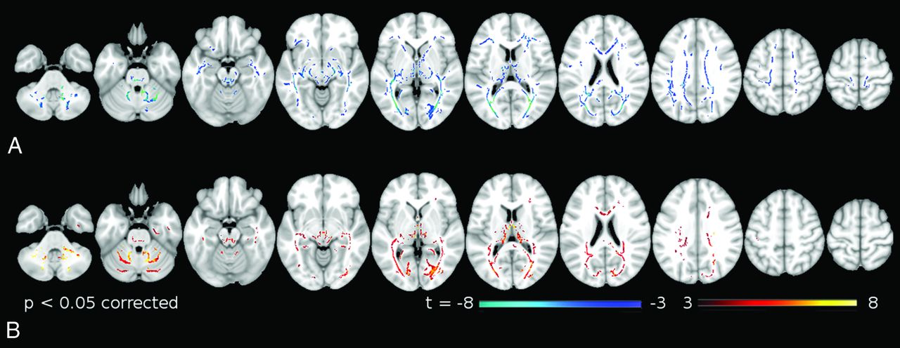
TBSS group comparison revealed significant FA decreases in patients with SCA7 (Fig 1A and Table 2) in the white matter tracts, including the inferior/middle/superior cerebellar peduncles, the bilateral internal/external capsule, the bilateral corona radiata, the bilateral optical radiation, and the occipital/temporal/frontal white matter.

Fig 1.
  • Download figure
  • Open in new tab
  • Download powerpoint
Fig 1.

TBSS significant differences in diffusion measurements between patients with SCA7 and healthy controls. A, Fractional anisotropy. B, Mean diffusivity. Warm and cold colors indicate an increase and decrease in these measures in patients with SCA7, respectively.

View this table:
  • View inline
  • View popup
Table 2:

Significant group differences in fractional anisotropy between patients with SCA7 and healthy controlsa

TBSS group comparison revealed significant MD increases in patients with SCA7 (Fig 1B and Table 3) in the cerebellar WM, including the medial lemniscus, the middle cerebellar peduncle, the optical radiations, the bilateral corona radiata, the posterior limbs of internal capsule, and the corticospinal tract.

View this table:
  • View inline
  • View popup
Table 3:

Significant group differences in mean diffusivity between patients with SCA7 and healthy controlsa

We found associations between the patients' SARA scores and mean diffusivity in several abnormal WM tracts. Specifically, we found SARA associations with MD in the right middle occipital WM, stria terminalis, superior cerebellar peduncle, anterior cerebellar WM, superior longitudinal fasciculus, and the anterior thalamic radiation (Fig 2 and Table 4). No significant association was found between the SARA scores and FA.

Fig 2.
  • Download figure
  • Open in new tab
  • Download powerpoint
Fig 2.

White matter regions where MD correlates with the SARA score in the SCA7 group. Warm colors indicate significant correlations between MD values and SARA scores.

View this table:
  • View inline
  • View popup
Table 4:

White matter regions showing significant correlation between MD and the SARA score in the SCA7 groupa

Discussion

In this study, we used Tract-Based Spatial Statistics to assess the white matter abnormalities in a group of patients with SCA7 compared with matched healthy controls and the relationship between the white matter integrity and the ataxia severity in the patient group. Our results showed significant differences in FA and MD when comparing patients with SCA7 with healthy controls and a correlation between the ataxia severity and MD changes in the patient group.

Previous neuropathologic reports have shown loss of myelinated fibers in the cerebellar white matter, corpus callosum, red nucleus capsule, oculomotor nerve, lateral lemniscus, mesencephalic trigeminal tract, abducens nerve, trapezoid body, pontocerebellar fibers, pyramidal tract, internal arcuate and olivocerebellar fibers, cuneate, and gracile fascicles, as well as the spinocerebellar tracts.5,6 Our results support those findings but also extend them by showing abnormalities in the occipital WM, the stria terminalis, and the thalamic radiations not previously reported. As expected, the cerebellar WM and the cerebellar peduncles showed a decrease in FA and an increase of MD, implying SCA7-related microstructural changes in the afferent and efferent projections of the cerebellum. The combination of white matter degeneration and gray matter loss in the cerebellum results in a variety of clinical motor impairments, including ataxia and extrapyramidal signs.21 In the same way, the degeneration of the thalamic radiations affects the information flow between the cerebellum and the motor and frontal cortices, which might result in the loss of coordination and dexterity in this set of patients.

The most relevant finding of this study is the association of the white matter mean diffusivity and the ataxia severity in this group of patients. The higher correlation between MD and SARA was found in the white matter of the middle occipital gyrus, which is involved in visual and spatial processing.22,23 Similarly, the superior longitudinal fasciculus showed significant correlation, which suggests a malfunction in the projections between the occipital-to-frontal cortices, including the premotor areas. A failure of these pathways could affect the planning and action of visuospatial tasks, especially in this disease because it has been reported to show decreases in the functional connectivity between the occipital and motor cortices.24,25 Future studies, including neuropsychological evaluation focused on visuospatial performance combined with ophthalmologic data, should be helpful in corroborating this hypothesis.

Other white matter regions showing significant correlation between the MD and SARA score were the superior cerebellar peduncles and the white matter in the anterior cerebellum. On the basis of the distribution of the neuropathologic changes in SCA7, this result was expected.5,6 Several reports have shown that lesions in these regions can lead to motor incoordination and loss of movement dexterity.26 Both the cerebellar peduncles and the anterior cerebellum have been reported as degenerated in previous MR imaging studies in SCA2.6,24 However, in previous studies, no significant correlations were found between the changes in the water diffusion properties and SARA scores, probably due to the small number of participants recruited.6

No significant correlations between FA and SARA were found. FA and MD are not equivalent measurements27,28; and as expected in our group comparison, FA and MD maps led to different results (Fig 1). In addition to MD and FA, other water diffusion properties such as axial diffusivity and radial diffusivity have been reported in previous studies, including SCA1, SCA2, and Friedreich ataxia.10,29 In this work, we focused on the analyses of FA and MD because the other measurements are subcomponents of the apparent diffusion coefficient. Furthermore, several studies of patients with neurodegenerative diseases have suggested that MD is more useful and sensitive to neurodegeneration than the other measurements.8,9,30

Conclusions

Our results show that specific changes in the diffusion properties of white matter resulting from the SCA7 mutation are associated with the severity of the ataxia. The distribution of the mean diffusivity abnormalities and its association with SARA scores suggest a disruption of information flow between motor-, visual-, and sensory-integration areas. Overall, these findings contribute to a better understanding of the neural basis of the symptomatology of patients with SCA7.

Footnotes

  • This work was supported by Consejo Nacional de Ciencia y Tecnologia grant 220871 and Universidad Nacional Autonoma de Mexico grant PAPIIT IN214716 to J.F.-R.

Indicates open access to non-subscribers at www.ajnr.org

References

  1. 1.↵
    1. Garden GA,
    2. La Spada AR
    . Molecular pathogenesis and cellular pathology of spinocerebellar ataxia type 7 neurodegeneration. Cerebellum 2008;7:138–49 doi:10.1007/s12311-008-0027-y pmid:18418675
    CrossRefPubMed
  2. 2.↵
    1. Hugosson T,
    2. Gränse L,
    3. Ponjavic V, et al
    . Macular dysfunction and morphology in spinocerebellar ataxia type 7 (SCA 7). Ophthalmic Genet 2009;30:1–6 doi:10.1080/13816810802454081 pmid:19172503
    CrossRefPubMed
  3. 3.↵
    1. Miller RC,
    2. Tewari A,
    3. Miller JA, et al
    . Neuro-ophthalmologic features of spinocerebellar ataxia type 7. J Neuroophthalmol 2009;29:180–86 doi:10.1097/WNO.0b013e3181b1b3f8 pmid:19726938
    CrossRefPubMed
  4. 4.↵
    1. Michalik A,
    2. Martin JJ,
    3. Van Broeckhoven C
    . Spinocerebellar ataxia type 7 associated with pigmentary retinal dystrophy. Eur J Hum Genet 2004;12:2–15 doi:10.1038/sj.ejhg.5201108 pmid:14571264
    CrossRefPubMed
  5. 5.↵
    1. Masciullo M,
    2. Modoni A,
    3. Pomponi MG, et al
    . Evidence of white matter involvement in SCA 7. J Neurol 2007;254:536–38 doi:10.1007/s00415-006-0274-0 pmid:16988791
    CrossRefPubMed
  6. 6.↵
    1. Alcauter S,
    2. Barrios FA,
    3. Díaz R, et al
    . Gray and white matter alterations in spinocerebellar ataxia type 7: an in vivo DTI and VBM study. Neuroimage 2011;55:1–7 doi:10.1016/j.neuroimage.2010.12.014 pmid:21147232
    CrossRefPubMed
  7. 7.↵
    1. Mori S,
    2. Zhang J
    . Principles of diffusion tensor imaging and its applications to basic neuroscience research. Neuron 2006;51:527–39 doi:10.1016/j.neuron.2006.08.012 pmid:16950152
    CrossRefPubMed
  8. 8.↵
    1. Acosta-Cabronero J,
    2. Williams GB,
    3. Pengas G, et al
    . Absolute diffusivities define the landscape of white matter degeneration in Alzheimer's disease. Brain 2010;133:529–39 doi:10.1093/brain/awp257 pmid:19914928
    Abstract/FREE Full Text
  9. 9.↵
    1. Della Nave R,
    2. Ginestroni A,
    3. Tessa C, et al
    . Regional distribution and clinical correlates of white matter structural damage in Huntington disease: a tract-based spatial statistics study. AJNR Am J Neuroradiol 2010;31:1675–81 doi:10.3174/ajnr.A2128 pmid:20488902
    Abstract/FREE Full Text
  10. 10.↵
    1. Della Nave R,
    2. Ginestroni A,
    3. Tessa C, et al
    . Brain white matter damage in SCA1 and SCA2: an in vivo study using voxel-based morphometry, histogram analysis of mean diffusivity and tract-based spatial statistics. Neuroimage 2008;43:10–19 doi:10.1016/j.neuroimage.2008.06.036 pmid:18672073
    CrossRefPubMed
  11. 11.↵
    1. Hernandez-Castillo CR,
    2. Galvez V,
    3. Mercadillo R, et al
    . Extensive white matter alterations and its correlations with ataxia severity in SCA 2 patients. PLoS One 2015;10:e0135449 doi:10.1371/journal.pone.0135449 pmid:26263162
    CrossRefPubMed
  12. 12.↵
    1. Hernandez-Castillo CR,
    2. Vaca-Palomares I,
    3. Galvez V, et al
    . Cognitive deficits correlate with white matter deterioration in spinocerebellar ataxia type 2. J Int Neuropsychol Soc 2016;22:486–91 doi:10.1017/S1355617716000084 pmid:26888086
    CrossRefPubMed
  13. 13.↵
    1. Li H,
    2. Ma J,
    3. Zhang X
    . Diffusion tensor imaging of spinocerebellar ataxia type 12. Med Sci Monit 2014;20:1783–91 doi:10.12659/MSM.891104 pmid:25274186
    CrossRefPubMed
  14. 14.↵
    1. Schmitz-Hübsch T,
    2. du Montcel ST,
    3. Baliko L, et al
    . Scale for the assessment and rating of ataxia: development of a new clinical scale. Neurology 2006;66:1717–20 doi:10.1212/01.wnl.0000219042.60538.92 pmid:16769946
    Abstract/FREE Full Text
  15. 15.↵
    1. Weyer A,
    2. Abele M,
    3. Schmitz-Hübsch T, et al
    . Reliability and validity of the scale for the assessment and rating of ataxia: a study in 64 ataxia patients. Mov Disord 2007;22:1633–37 doi:10.1002/mds.21544 pmid:17516493
    CrossRefPubMed
  16. 16.↵
    1. Smith SM,
    2. Jenkinson M,
    3. Woolrich MW, et al
    . Advances in functional and structural MR image analysis and implementation as FSL. Neuroimage 2004;23:S208–19 doi:10.1016/j.neuroimage.2004.07.051 pmid:15501092
    CrossRefPubMed
  17. 17.↵
    1. Smith SM,
    2. Jenkinson M,
    3. Johansen-Berg H, et al
    . Tract-based spatial statistics: voxelwise analysis of multi-subject diffusion data. Neuroimage 2006;31:1487–505 doi:10.1016/j.neuroimage.2006.02.024 pmid:16624579
    CrossRefPubMed
  18. 18.↵
    1. Jenkinson M,
    2. Bannister P,
    3. Brady M, et al
    . Improved optimization for the robust and accurate linear registration and motion correction of brain images. Neuroimage 2002;17:825–41 doi:10.1006/nimg.2002.1132 pmid:12377157
    CrossRefPubMed
  19. 19.↵
    1. Nichols TE,
    2. Holmes AP
    . Nonparametric permutation tests for functional neuroimaging: a primer with examples. Hum Brain Mapp 2002;15:1–25 doi:10.1002/hbm.1058 pmid:11747097
    CrossRefPubMed
  20. 20.↵
    1. Winkler AM,
    2. Ridgway GR,
    3. Webster MA, et al
    . Permutation inference for the general linear model. Neuroimage 2014;92:381–97 doi:10.1016/j.neuroimage.2014.01.060 pmid:24530839
    CrossRefPubMed
  21. 21.↵
    1. Hernandez-Castillo CR,
    2. Galvez V,
    3. Diaz R, et al
    . Specific cerebellar and cortical degeneration correlates with ataxia severity in spinocerebellar ataxia type 7. Brain Imaging Behav 2016;10:252–57 doi:10.1007/s11682-015-9389-1 pmid:25917872
    CrossRefPubMed
  22. 22.↵
    1. Renier LA,
    2. Anurova I,
    3. De Volder AG, et al
    . Preserved functional specialization for spatial processing in the middle occipital gyrus of the early blind. Neuron 2010;68:138–48 doi:10.1016/j.neuron.2010.09.021 pmid:20920797
    CrossRefPubMed
  23. 23.↵
    1. Martínez A,
    2. Anllo-Vento L,
    3. Sereno MI, et al
    . Involvement of striate and extrastriate visual cortical areas in spatial attention. Nat Neurosci 1999;2:364–69 doi:10.1038/7274 pmid:10204544
    CrossRefPubMed
  24. 24.↵
    1. Hernandez-Castillo CR,
    2. Alcauter S,
    3. Galvez V, et al
    . Disruption of visual and motor connectivity in spinocerebellar ataxia type 7. Mov Disord 2013;28:1708–16 doi:10.1002/mds.25618 pmid:23926060
    CrossRefPubMed
  25. 25.↵
    1. Hernandez-Castillo CR,
    2. Galvez V,
    3. Morgado-Valle C, et al
    . Whole-brain connectivity analysis and classification of spinocerebellar ataxia type 7 by functional MRI. Cerebellum Ataxias 2014;1:2 doi:10.1186/2053-8871-1-2 pmid:26331026
    CrossRefPubMed
  26. 26.↵
    1. Schmahmann JD
    . Disorders of the cerebellum: ataxia, dysmetria of thought, and the cerebellar cognitive affective syndrome. J Neuropsychiatry Clin Neurosci 2004;16:367–78 doi:10.1176/jnp.16.3.367 pmid:15377747
    CrossRefPubMed
  27. 27.↵
    1. Cosottini M,
    2. Giannelli M,
    3. Siciliano G, et al
    . Diffusion-tensor MR imaging of corticospinal tract in amyotrophic lateral sclerosis and progressive muscular atrophy. Radiology 2005;237:258–64 doi:10.1148/radiol.2371041506 pmid:16183935
    CrossRefPubMed
  28. 28.↵
    1. Pierpaoli C,
    2. Barnett A,
    3. Pajevic S, et al
    . Water diffusion changes in Wallerian degeneration and their dependence on white matter architecture. Neuroimage 2001;13(6 pt 1):1174–85 doi:10.1006/nimg.2001.0765 pmid:11352623
    CrossRefPubMed
  29. 29.↵
    1. Mandelli ML,
    2. De Simone T,
    3. Minati L, et al
    . Diffusion tensor imaging of spinocerebellar ataxias types 1 and 2. AJNR Am J Neuroradiol 2007;28:1996–2000 doi:10.3174/ajnr.A0716 pmid:17998418
    Abstract/FREE Full Text
  30. 30.↵
    1. Vos SB,
    2. Jones DK,
    3. Jeurissen B, et al
    . The influence of complex white matter architecture on the mean diffusivity in diffusion tensor MRI of the human brain. Neuroimage 2012;59:2208–16 doi:10.1016/j.neuroimage.2011.09.086 pmid:22005591
    CrossRefPubMed
  • Received March 28, 2016.
  • Accepted after revision May 26, 2016.
  • © 2016 by American Journal of Neuroradiology
View Abstract
PreviousNext
Back to top

In this issue

American Journal of Neuroradiology: 37 (11)
American Journal of Neuroradiology
Vol. 37, Issue 11
1 Nov 2016
  • Table of Contents
  • Index by author
  • Complete Issue (PDF)
Advertisement
Print
Download PDF
Email Article

Thank you for your interest in spreading the word on American Journal of Neuroradiology.

NOTE: We only request your email address so that the person you are recommending the page to knows that you wanted them to see it, and that it is not junk mail. We do not capture any email address.

Enter multiple addresses on separate lines or separate them with commas.
Ataxia Severity Correlates with White Matter Degeneration in Spinocerebellar Ataxia Type 7
(Your Name) has sent you a message from American Journal of Neuroradiology
(Your Name) thought you would like to see the American Journal of Neuroradiology web site.
CAPTCHA
This question is for testing whether or not you are a human visitor and to prevent automated spam submissions.
Cite this article
C.R. Hernandez-Castillo, I. Vaca-Palomares, F. Barrios, L. Martinez, M.-C. Boll, J. Fernandez-Ruiz
Ataxia Severity Correlates with White Matter Degeneration in Spinocerebellar Ataxia Type 7
American Journal of Neuroradiology Nov 2016, 37 (11) 2050-2054; DOI: 10.3174/ajnr.A4903

Citation Manager Formats

  • BibTeX
  • Bookends
  • EasyBib
  • EndNote (tagged)
  • EndNote 8 (xml)
  • Medlars
  • Mendeley
  • Papers
  • RefWorks Tagged
  • Ref Manager
  • RIS
  • Zotero
0 Responses
Respond to this article
Share
Bookmark this article
Ataxia Severity Correlates with White Matter Degeneration in Spinocerebellar Ataxia Type 7
C.R. Hernandez-Castillo, I. Vaca-Palomares, F. Barrios, L. Martinez, M.-C. Boll, J. Fernandez-Ruiz
American Journal of Neuroradiology Nov 2016, 37 (11) 2050-2054; DOI: 10.3174/ajnr.A4903
del.icio.us logo Twitter logo Facebook logo Mendeley logo
  • Tweet Widget
  • Facebook Like
  • Google Plus One
Purchase

Jump to section

  • Article
    • Abstract
    • ABBREVIATIONS:
    • Materials and Methods
    • Results
    • Discussion
    • Conclusions
    • Footnotes
    • References
  • Figures & Data
  • Info & Metrics
  • Responses
  • References
  • PDF

Related Articles

  • No related articles found.
  • PubMed
  • Google Scholar

Cited By...

  • In vivo assessment of neurodegeneration in Spinocerebellar Ataxia type 7
  • Crossref (10)
  • Google Scholar

This article has been cited by the following articles in journals that are participating in Crossref Cited-by Linking.

  • Cerebellar Astrocytes: Much More Than Passive Bystanders In Ataxia Pathophysiology
    Valentina Cerrato
    Journal of Clinical Medicine 2020 9 3
  • Founder Effects of Spinocerebellar Ataxias in the American Continents and the Caribbean
    Roberto Rodríguez-Labrada, Ana Carolina Martins, Jonathan J. Magaña, Yaimeé Vazquez-Mojena, Jacqueline Medrano-Montero, Juan Fernandez-Ruíz, Bulmaro Cisneros, Helio Teive, Karen N. McFarland, Maria Luiza Saraiva-Pereira, César M. Cerecedo-Zapata, Christopher M. Gomez, Tetsuo Ashizawa, Luis Velázquez-Pérez, Laura Bannach Jardim
    The Cerebellum 2020 19 3
  • Myelinating Glia: Potential Therapeutic Targets in Polyglutamine Spinocerebellar Ataxias
    Alexandra F. Putka, Juan P. Mato, Hayley S. McLoughlin
    Cells 2023 12 4
  • A Proposal for Classification of Retinal Degeneration in Spinocerebellar Ataxia Type 7
    Bruna Ferraço Marianelli, Flávio Moura Rezende Filho, Mariana Vallim Salles, João Brainer Clares de Andrade, José Luiz Pedroso, Juliana Maria Ferraz Sallum, Orlando Graziani P. Barsottini
    The Cerebellum 2021 20 3
  • Cerebellar Abnormalities on Proton MR Spectroscopy and Imaging in Patients With Gluten Ataxia: A Pilot Study
    Vishwa Rawat, Ritu Tyagi, Inder Singh, Prasenjit Das, Achal Kumar Srivastava, Govind K. Makharia, Uma Sharma
    Frontiers in Human Neuroscience 2022 16
  • In vivo assessment of neurodegeneration in Spinocerebellar Ataxia type 7
    Jacob A. Parker, Shabbir H. Merchant, Sanaz Attaripour-Isfahani, Hyun Joo Cho, Patrick McGurrin, Brian P. Brooks, Albert R. La Spada, Mark Hallett, Laryssa A. Huryn, Silvina G. Horovitz
    NeuroImage: Clinical 2021 29
  • Longitudinal MRI and 1H-MRS study of SCA7 mouse forebrain reveals progressive multiregional atrophy and early brain metabolite changes indicating early neuronal and glial dysfunction
    Jean-Baptiste Pérot, Anna Niewiadomska-Cimicka, Emmanuel Brouillet, Yvon Trottier, Julien Flament, Stephen D. Ginsberg
    PLOS ONE 2024 19 1
  • Deep multimodal saliency parcellation of cerebellar pathways: Linking microstructure and individual function through explainable multitask learning
    Ari Tchetchenian, Leo Zekelman, Yuqian Chen, Jarrett Rushmore, Fan Zhang, Edward H. Yeterian, Nikos Makris, Yogesh Rathi, Erik Meijering, Yang Song, Lauren J. O'Donnell
    Human Brain Mapping 2024 45 12
  • Genetic and Clinical Predictors of Ataxia in Pediatric Primary Mitochondrial Disorders
    Juan Sebastian Martin-Saavedra, Sara Reis Teixeira, Cesar Augusto Pinheiro Ferreira Alves, Fabrício Guimarães Gonçalves, Luis Octavio Tierradentro-García, Martin Kidd, Colleen Muraresku, Amy Goldstein, Arastoo Vossough
    The Cerebellum 2022 21 1
  • Neuroglia
    Alexei Verkhratsky, Arthur M. Butt
    2023

More in this TOC Section

  • Diagnostic Neuroradiology of Monoclonal Antibodies
  • Clinical Outcomes After Chiari I Decompression
  • Segmentation of Brain Metastases with BLAST
Show more Adult Brain

Similar Articles

Advertisement

Indexed Content

  • Current Issue
  • Accepted Manuscripts
  • Article Preview
  • Past Issues
  • Editorials
  • Editor's Choice
  • Fellows' Journal Club
  • Letters to the Editor
  • Video Articles

Cases

  • Case Collection
  • Archive - Case of the Week
  • Archive - Case of the Month
  • Archive - Classic Case

Special Collections

  • AJNR Awards
  • ASNR Foundation Special Collection
  • Most Impactful AJNR Articles
  • Photon-Counting CT
  • Spinal CSF Leak Articles (Jan 2020-June 2024)

More from AJNR

  • Trainee Corner
  • Imaging Protocols
  • MRI Safety Corner

Multimedia

  • AJNR Podcasts
  • AJNR Scantastics

Resources

  • Turnaround Time
  • Submit a Manuscript
  • Submit a Video Article
  • Submit an eLetter to the Editor/Response
  • Manuscript Submission Guidelines
  • Statistical Tips
  • Fast Publishing of Accepted Manuscripts
  • Graphical Abstract Preparation
  • Imaging Protocol Submission
  • Evidence-Based Medicine Level Guide
  • Publishing Checklists
  • Author Policies
  • Become a Reviewer/Academy of Reviewers
  • News and Updates

About Us

  • About AJNR
  • Editorial Board
  • Editorial Board Alumni
  • Alerts
  • Permissions
  • Not an AJNR Subscriber? Join Now
  • Advertise with Us
  • Librarian Resources
  • Feedback
  • Terms and Conditions
  • AJNR Editorial Board Alumni

American Society of Neuroradiology

  • Not an ASNR Member? Join Now

© 2025 by the American Society of Neuroradiology All rights, including for text and data mining, AI training, and similar technologies, are reserved.
Print ISSN: 0195-6108 Online ISSN: 1936-959X

Powered by HighWire