Advertisement

Main menu

  • Home
  • Content
    • Current Issue
    • Accepted Manuscripts
    • Article Preview
    • Past Issue Archive
    • Video Articles
    • AJNR Case Collection
    • Case of the Week Archive
    • Case of the Month Archive
    • Classic Case Archive
  • Special Collections
    • AJNR Awards
    • ASNR Foundation Special Collection
    • Most Impactful AJNR Articles
    • Photon-Counting CT
    • Spinal CSF Leak Articles (Jan 2020-June 2024)
  • Multimedia
    • AJNR Podcasts
    • AJNR SCANtastic
    • Trainee Corner
    • MRI Safety Corner
    • Imaging Protocols
  • For Authors
    • Submit a Manuscript
    • Submit a Video Article
    • Submit an eLetter to the Editor/Response
    • Manuscript Submission Guidelines
    • Statistical Tips
    • Fast Publishing of Accepted Manuscripts
    • Graphical Abstract Preparation
    • Imaging Protocol Submission
    • Author Policies
  • About Us
    • About AJNR
    • Editorial Board
    • Editorial Board Alumni
  • More
    • Become a Reviewer/Academy of Reviewers
    • Subscribers
    • Permissions
    • Alerts
    • Feedback
    • Advertisers
    • ASNR Home

User menu

  • Alerts
  • Log in

Search

  • Advanced search
American Journal of Neuroradiology

American Journal of Neuroradiology

ASHNR American Society of Functional Neuroradiology ASHNR American Society of Pediatric Neuroradiology ASSR
  • Alerts
  • Log in

Advanced Search

  • Home
  • Content
    • Current Issue
    • Accepted Manuscripts
    • Article Preview
    • Past Issue Archive
    • Video Articles
    • AJNR Case Collection
    • Case of the Week Archive
    • Case of the Month Archive
    • Classic Case Archive
  • Special Collections
    • AJNR Awards
    • ASNR Foundation Special Collection
    • Most Impactful AJNR Articles
    • Photon-Counting CT
    • Spinal CSF Leak Articles (Jan 2020-June 2024)
  • Multimedia
    • AJNR Podcasts
    • AJNR SCANtastic
    • Trainee Corner
    • MRI Safety Corner
    • Imaging Protocols
  • For Authors
    • Submit a Manuscript
    • Submit a Video Article
    • Submit an eLetter to the Editor/Response
    • Manuscript Submission Guidelines
    • Statistical Tips
    • Fast Publishing of Accepted Manuscripts
    • Graphical Abstract Preparation
    • Imaging Protocol Submission
    • Author Policies
  • About Us
    • About AJNR
    • Editorial Board
    • Editorial Board Alumni
  • More
    • Become a Reviewer/Academy of Reviewers
    • Subscribers
    • Permissions
    • Alerts
    • Feedback
    • Advertisers
    • ASNR Home
  • Follow AJNR on Twitter
  • Visit AJNR on Facebook
  • Follow AJNR on Instagram
  • Join AJNR on LinkedIn
  • RSS Feeds

AJNR is seeking candidates for the AJNR Podcast Editor. Read the position description.

Research ArticleHEAD AND NECK

Perineural Spread in a Case of Sinonasal Sarcoidosis: Case Report

Silvio Mazziotti, Michele Gaeta, Alfredo Blandino, Sergio Vinci and Ignazio Pandolfo
American Journal of Neuroradiology June 2001, 22 (6) 1207-1208;
Silvio Mazziotti
  • Find this author on Google Scholar
  • Find this author on PubMed
  • Search for this author on this site
Michele Gaeta
  • Find this author on Google Scholar
  • Find this author on PubMed
  • Search for this author on this site
Alfredo Blandino
  • Find this author on Google Scholar
  • Find this author on PubMed
  • Search for this author on this site
Sergio Vinci
  • Find this author on Google Scholar
  • Find this author on PubMed
  • Search for this author on this site
Ignazio Pandolfo
  • Find this author on Google Scholar
  • Find this author on PubMed
  • Search for this author on this site
  • Article
  • Figures & Data
  • Info & Metrics
  • Responses
  • References
  • PDF
Loading

Abstract

Summary: We report a case of sinonasal sarcoidosis with perineural spread along the trigeminal and vidian nerves in which primarily MR imaging but also CT allowed excellent visualization of this infiltration.

Sarcoidosis is an idiopathic multisystemic granulomatous disease characterized by the association of clinical signs with histopathologic evidence of noncaseating granulomas (1). While sarcoidosis is mainly a pneumologic disease, extrapulmonary locations (eg, peripheral lymph nodes, skin, liver, eye, spleen, brain, bones) are also seen, though less frequently (1). Severe sinonasal involvement occurs in 12% to 27% of patients, whereas CNS involvement occurs in only about 5% of patients (2–7).

We describe the CT and MR imaging findings of a patient with sinonasal sarcoidosis that spread in a retrograde fashion along the trigeminal and vidian nerves. To our knowledge, no previous report has documented the spread of sinonasal sarcoidosis or involvement of the vidian nerve.

Case Report

A 48 year-old man had undergone surgery for sinonasal polyposis associated with a frontal mucocele. Six months later he presented with deep facial pain, nasal obstruction, anosmia, and rhinorrhea.

CT scans showed postsurgical changes and abnormal soft tissue in the maxillary sinuses, in the residual ethmoidal cavities, in the sphenoidal sinus, and in the pterygopalatine fossae bilaterally. Coronal scans showed enlargement of the left pterygoid canal and foramen rotundum, while the infraorbital canals appeared enlarged bilaterally (Fig 1A–C).

fig 1.
  • Download figure
  • Open in new tab
  • Download powerpoint
fig 1.

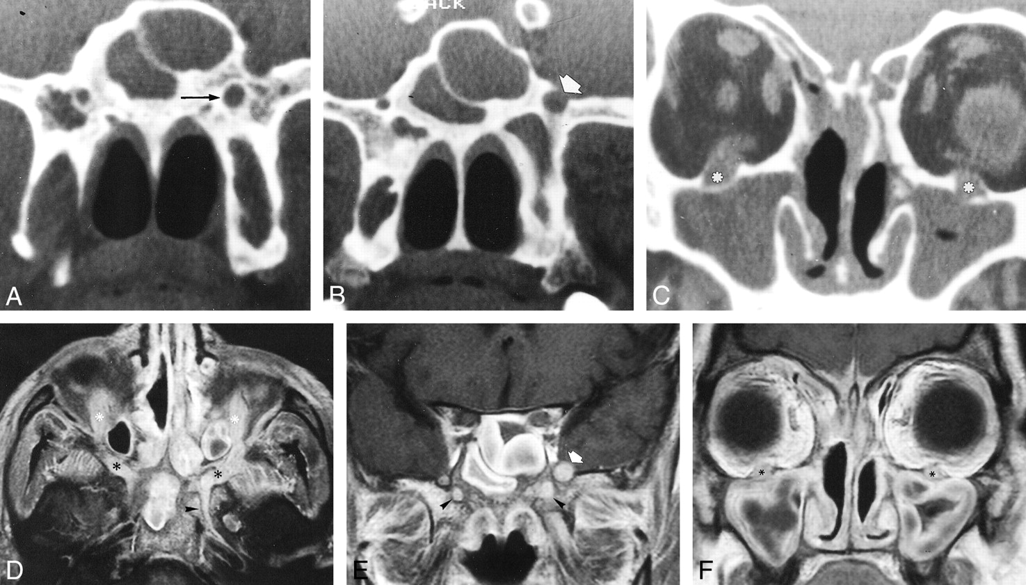
48-year-old man with deep facial pain, nasal obstruction, anosmia, and rhinorrhea 6 months after surgery for sinonasal polyposis associated with a frontal mucocele.

A–B, Coronal CT scans through the pterygoid process show enlargement of the left pterygoid canal (arrow, A) as well as enlargement of the left foramen rotundum (arrow, B).

C, Coronal CT scan more anterior than A and B shows widening and erosion of both infraorbital canals (asterisks). Note the presence of abnormal soft tissue in the maxillary and sphenoidal sinuses.

D, Contrast-enhanced axial T1-weighted MR image (700/12/6 [TR/TE/excitations]) through the base of the skull, obtained with a fat-saturation technique, shows abnormal enhancing tissue in the right and left upper pterygopalatine fossae (black asterisks). Enlargement and enhancement of the infraorbital nerves (white asterisks) and the left pterygoid nerve (arrowhead) can also be seen.

E, Contrast-enhanced coronal T1-weighted MR image (700/12/6) shows enhancement of the right and left vidian nerves (arrowheads), enlargement of the left vidian nerve, and left-sided maxillary division of the trigeminal nerve (arrow).

F, Contrast-enhanced coronal T1-weighted MR image (700/12/6) through the anterior part of the maxillary sinus shows enlargement and enhancement of both infraorbital nerves (asterisks). Abnormal enhancing tissue infiltrates through the skull base, involving the meninges in the anterior cranial fossa. Thickening of the sinus mucosa is visible on all images.

MR images obtained before and after intravenous administration of contrast material showed diffuse thickening and enhancement of the mucosa in the nasal cavities and in the maxillary, ethmoidal, and sphenoidal sinuses as well as abnormal tissue in the pterygopalatine fossa (Fig 1D–F). The residual lumen of the maxillary and sphenoidal sinus was filled with fluid. Abnormal tissue was spread along the infraorbital nerves, which were enlarged. On the left, perineural infiltration involved the pterygopalatine fossa, vidian nerve, and maxillary nerve, up to the gasserian ganglion. On the right, the pterygopalatine fossa and, to a lesser extent than on the left, the vidian nerve appeared involved, but not the maxillary nerve. Abnormal tissue infiltrated through the skull base, with involvement of the dura in the anterior cranial fossa (Fig 1F).

A biopsy sample of the nasal mucosa revealed multiple noncaseating granulomas, indicative of sarcoidosis. An increase in the erythrosedimentation rate and serum angiotensin-converting enzyme, together with radiographic findings of hilar adenopathy, confirmed the diagnosis of sarcoidosis.

Discussion

Sarcoidosis is an idiopathic, systemic, granulomatous disease whose onset is usually unifocal, but with a strong predilection for systemic involvement, characterized by alternating periods of remission and relapse (1). Sinonasal involvement is a relatively rare occurrence, with a frequency of 12% to 27% (3).

Several other diseases involving the sinonasal region are characterized by granulomatous lesions. Thus, the diagnosis of sinonasal sarcoidosis is often difficult, and is made by correlating clinical, radiologic, histologic, and laboratory findings (8, 9). Although cranial nerve involvement occurs frequently in neurosarcoidosis, the trigeminal nerve is rarely involved, and, to our knowledge, no report has documented involvement of the vidian nerve or perineural infiltration secondary to sinonasal sarcoidosis.

In our case, sarcoidosis had spread in a perineural fashion along the infraorbital and vidian nerves bilaterally as well as along the right maxillary nerve. This process resulted in enlargement of the bony canals and erosion of the cortical wall (10). MR imaging showed enhancement and enlargement of the involved nerves, providing direct evidence of neural spread and representing an advantage over CT in that it allowed complete evaluation of the stage of illness (10–12). It is important to remember, however, that the findings described are not specific, as they may also be seen in perineural spread of neoplasm (10–16).

Conclusion

The findings in our case enabled us to reach the following conclusions. 1) Perineural spread is a possible complication of sinonasal sarcoidosis; in fact, in our case, the process began presumably in the paranasal sinuses and spread upward along the cranial nerves, as suggested by the absence of neural involvement on the presurgical imaging studies. This is in accordance with the higher prevalence of sinonasal sarcoidosis as compared with CNS involvement (2–7). 2) MR imaging, but also CT, allows excellent visualization of this process. 3) Perineural spread of sarcoidosis along the trigeminal and vidian nerves may simulate perineural involvement by head and neck cancer, in particular when sarcoidosis is not confirmed by histologic studies.

Footnotes

  • 1 Address reprint requests to Silvio Mazziotti, MD, Via Consolare Pompea, 45 (ex 23), 98168 Messina, Italy.

References

  1. ↵
    James DJ, Jones Williams W. Sarcoidosis and Other Granulomatous Diseases.. Philadelphia: Saunders; 1984
  2. ↵
    Pickuth D, Spielmann RP, Heywang-Kobrunner SH. Role of radiology in the diagnosis of neurosarcoidosis. Eur Radiol 2000;10:941-944
    CrossRefPubMed
  3. ↵
    Page JR, Seth SM. Sarcoidosis of the upper respiratory tract. J Laryngol 1969;83:1005-1011
  4. Zouaoui A, Maillard JC, Dormont D, Chiras J, Marsault C. MRI in neurosarcoidosis. J Neuroradiol 1992;19:271-284
    PubMed
  5. Oksanen V. Neurosarcoidosis. Sarcoidosis 1994;11:76-79
    PubMed
  6. Scott TF. Neurosarcoidosis: progress and clinical aspects. Neurology 1993;43:8-12
    FREE Full Text
  7. Lexa FJ, Grossman RI. MR of sarcoidosis in the head and spine: spectrum of manifestations and radiographic response to steroid therapy. AJNR Am J Neuroradiol 1994;15:973-982
    Abstract/FREE Full Text
  8. ↵
    Maillard Alberto AJ, Goepfert H. Nasal and paranasal sarcoidosis. Arch Otolaryngol 1978;104:197-201
    CrossRefPubMed
  9. Trachtenberg SB, Wilkinson EE, Jacobson G. Sarcoidosis of the nose and paranasal sinuses. Radiology 1974;113:619-620
    CrossRefPubMed
  10. ↵
    Blandino A, Gaeta M, Minutoli F, Pandolfo I. CT and MR findings in neoplastic perineural spread along the vidian nerve. Eur Radiol 2000;10:521-526
    CrossRefPubMed
  11. Pandolfo I, Gaeta M, Blandino A, Salvi L, Longo M. MR imaging of perineural metastasis along the vidian nerve. J Comput Assist Tomogr 1989;13:498-500
    CrossRefPubMed
  12. Carmody RF, Mafee MF, Goodwin JA, Small K, Haery C. Orbital and optic pathway sarcoidosis: MR findings. AJNR Am J Neuroradiol 1994;15:775-783
    Abstract/FREE Full Text
  13. Dodd GD, Dolan PA, Ballantyne AJ, Ibanez ML, Chan P. The dissemination of tumors of the head and neck via the cranial nerves. Radiol Clin North Am 1970;8:445-461
    PubMed
  14. Pandolfo I, Gaeta M, Blandino A, Longo M, Faranda C. Perineural spread of nasopharyngeal carcinoma: radiological and CT demonstration. Eur J Radiol 1988;8:231-235
    PubMed
  15. Curtin HD, Williams R, Johnson J. CT of perineural tumor extension: pterygopalatine fossa. AJR Am J Roentgenol 1985;144:163-169
    PubMed
  16. Ginsberg LE, Eicher SA. Great auricular nerve: anatomy and imaging in a case of perineural tumor spread. AJNR Am J Neuroradiol 2000;21:568-571
    Abstract/FREE Full Text
  • Received October 26, 2000.
  • Accepted after revision February 6, 2001.
  • Copyright © American Society of Neuroradiology
View Abstract
PreviousNext
Back to top

In this issue

American Journal of Neuroradiology: 22 (6)
American Journal of Neuroradiology
Vol. 22, Issue 6
1 Jun 2001
  • Table of Contents
  • Index by author
Advertisement
Print
Download PDF
Email Article

Thank you for your interest in spreading the word on American Journal of Neuroradiology.

NOTE: We only request your email address so that the person you are recommending the page to knows that you wanted them to see it, and that it is not junk mail. We do not capture any email address.

Enter multiple addresses on separate lines or separate them with commas.
Perineural Spread in a Case of Sinonasal Sarcoidosis: Case Report
(Your Name) has sent you a message from American Journal of Neuroradiology
(Your Name) thought you would like to see the American Journal of Neuroradiology web site.
CAPTCHA
This question is for testing whether or not you are a human visitor and to prevent automated spam submissions.
Cite this article
Silvio Mazziotti, Michele Gaeta, Alfredo Blandino, Sergio Vinci, Ignazio Pandolfo
Perineural Spread in a Case of Sinonasal Sarcoidosis: Case Report
American Journal of Neuroradiology Jun 2001, 22 (6) 1207-1208;

Citation Manager Formats

  • BibTeX
  • Bookends
  • EasyBib
  • EndNote (tagged)
  • EndNote 8 (xml)
  • Medlars
  • Mendeley
  • Papers
  • RefWorks Tagged
  • Ref Manager
  • RIS
  • Zotero
0 Responses
Respond to this article
Share
Bookmark this article
Perineural Spread in a Case of Sinonasal Sarcoidosis: Case Report
Silvio Mazziotti, Michele Gaeta, Alfredo Blandino, Sergio Vinci, Ignazio Pandolfo
American Journal of Neuroradiology Jun 2001, 22 (6) 1207-1208;
del.icio.us logo Twitter logo Facebook logo Mendeley logo
  • Tweet Widget
  • Facebook Like
  • Google Plus One

Jump to section

  • Article
    • Abstract
    • Case Report
    • Discussion
    • Conclusion
    • Footnotes
    • References
  • Figures & Data
  • Info & Metrics
  • Responses
  • References
  • PDF

Related Articles

  • No related articles found.
  • PubMed
  • Google Scholar

Cited By...

  • No citing articles found.
  • Crossref
  • Google Scholar

This article has not yet been cited by articles in journals that are participating in Crossref Cited-by Linking.

More in this TOC Section

  • Correlation of Apparent Diffusion Coefficient at 3T with Prognostic Parameters of Retinoblastoma
  • Parathyroid Lesions: Characterization with Dual-Phase Arterial and Venous Enhanced CT of the Neck
  • Efficacy of Diffusion-Weighted Imaging for the Differentiation between Lymphomas and Carcinomas of the Nasopharynx and Oropharynx: Correlations of Apparent Diffusion Coefficients and Histologic Features
Show more HEAD AND NECK

Similar Articles

Advertisement

Indexed Content

  • Current Issue
  • Accepted Manuscripts
  • Article Preview
  • Past Issues
  • Editorials
  • Editor's Choice
  • Fellows' Journal Club
  • Letters to the Editor
  • Video Articles

Cases

  • Case Collection
  • Archive - Case of the Week
  • Archive - Case of the Month
  • Archive - Classic Case

Special Collections

  • AJNR Awards
  • ASNR Foundation Special Collection
  • Most Impactful AJNR Articles
  • Photon-Counting CT
  • Spinal CSF Leak Articles (Jan 2020-June 2024)

More from AJNR

  • Trainee Corner
  • Imaging Protocols
  • MRI Safety Corner

Multimedia

  • AJNR Podcasts
  • AJNR Scantastics

Resources

  • Turnaround Time
  • Submit a Manuscript
  • Submit a Video Article
  • Submit an eLetter to the Editor/Response
  • Manuscript Submission Guidelines
  • Statistical Tips
  • Fast Publishing of Accepted Manuscripts
  • Graphical Abstract Preparation
  • Imaging Protocol Submission
  • Evidence-Based Medicine Level Guide
  • Publishing Checklists
  • Author Policies
  • Become a Reviewer/Academy of Reviewers
  • News and Updates

About Us

  • About AJNR
  • Editorial Board
  • Editorial Board Alumni
  • Alerts
  • Permissions
  • Not an AJNR Subscriber? Join Now
  • Advertise with Us
  • Librarian Resources
  • Feedback
  • Terms and Conditions
  • AJNR Editorial Board Alumni

American Society of Neuroradiology

  • Not an ASNR Member? Join Now

© 2025 by the American Society of Neuroradiology All rights, including for text and data mining, AI training, and similar technologies, are reserved.
Print ISSN: 0195-6108 Online ISSN: 1936-959X

Powered by HighWire