Advertisement

Main menu

  • Home
  • Content
    • Current Issue
    • Accepted Manuscripts
    • Article Preview
    • Past Issue Archive
    • Video Articles
    • AJNR Case Collection
    • Case of the Week Archive
    • Case of the Month Archive
    • Classic Case Archive
  • Special Collections
    • AJNR Awards
    • ASNR Foundation Special Collection
    • Most Impactful AJNR Articles
    • Photon-Counting CT
    • Spinal CSF Leak Articles (Jan 2020-June 2024)
  • Multimedia
    • AJNR Podcasts
    • AJNR SCANtastic
    • Trainee Corner
    • MRI Safety Corner
    • Imaging Protocols
  • For Authors
    • Submit a Manuscript
    • Submit a Video Article
    • Submit an eLetter to the Editor/Response
    • Manuscript Submission Guidelines
    • Statistical Tips
    • Fast Publishing of Accepted Manuscripts
    • Graphical Abstract Preparation
    • Imaging Protocol Submission
    • Author Policies
  • About Us
    • About AJNR
    • Editorial Board
    • Editorial Board Alumni
  • More
    • Become a Reviewer/Academy of Reviewers
    • Subscribers
    • Permissions
    • Alerts
    • Feedback
    • Advertisers
    • ASNR Home

User menu

  • Alerts
  • Log in

Search

  • Advanced search
American Journal of Neuroradiology

American Journal of Neuroradiology

ASHNR American Society of Functional Neuroradiology ASHNR American Society of Pediatric Neuroradiology ASSR
  • Alerts
  • Log in

Advanced Search

  • Home
  • Content
    • Current Issue
    • Accepted Manuscripts
    • Article Preview
    • Past Issue Archive
    • Video Articles
    • AJNR Case Collection
    • Case of the Week Archive
    • Case of the Month Archive
    • Classic Case Archive
  • Special Collections
    • AJNR Awards
    • ASNR Foundation Special Collection
    • Most Impactful AJNR Articles
    • Photon-Counting CT
    • Spinal CSF Leak Articles (Jan 2020-June 2024)
  • Multimedia
    • AJNR Podcasts
    • AJNR SCANtastic
    • Trainee Corner
    • MRI Safety Corner
    • Imaging Protocols
  • For Authors
    • Submit a Manuscript
    • Submit a Video Article
    • Submit an eLetter to the Editor/Response
    • Manuscript Submission Guidelines
    • Statistical Tips
    • Fast Publishing of Accepted Manuscripts
    • Graphical Abstract Preparation
    • Imaging Protocol Submission
    • Author Policies
  • About Us
    • About AJNR
    • Editorial Board
    • Editorial Board Alumni
  • More
    • Become a Reviewer/Academy of Reviewers
    • Subscribers
    • Permissions
    • Alerts
    • Feedback
    • Advertisers
    • ASNR Home
  • Follow AJNR on Twitter
  • Visit AJNR on Facebook
  • Follow AJNR on Instagram
  • Join AJNR on LinkedIn
  • RSS Feeds

AJNR is seeking candidates for the AJNR Podcast Editor. Read the position description.

Research ArticlePEDIATRICS

MR Imaging Characteristics of Pilomyxoid Astrocytomas

Atilla Arslanoglu, Bayram Cirak, Alena Horska, James Okoh, Tarik Tihan, Leslie Aronson, Anthony M. Avellino, Peter C. Burger and David M. Yousem
American Journal of Neuroradiology October 2003, 24 (9) 1906-1908;
Atilla Arslanoglu
  • Find this author on Google Scholar
  • Find this author on PubMed
  • Search for this author on this site
Bayram Cirak
  • Find this author on Google Scholar
  • Find this author on PubMed
  • Search for this author on this site
Alena Horska
  • Find this author on Google Scholar
  • Find this author on PubMed
  • Search for this author on this site
James Okoh
  • Find this author on Google Scholar
  • Find this author on PubMed
  • Search for this author on this site
Tarik Tihan
  • Find this author on Google Scholar
  • Find this author on PubMed
  • Search for this author on this site
Leslie Aronson
  • Find this author on Google Scholar
  • Find this author on PubMed
  • Search for this author on this site
Anthony M. Avellino
  • Find this author on Google Scholar
  • Find this author on PubMed
  • Search for this author on this site
Peter C. Burger
  • Find this author on Google Scholar
  • Find this author on PubMed
  • Search for this author on this site
David M. Yousem
  • Find this author on Google Scholar
  • Find this author on PubMed
  • Search for this author on this site
  • Article
  • Figures & Data
  • Info & Metrics
  • Responses
  • References
  • PDF
Loading

Abstract

BACKGROUND AND PURPOSE: Pilomyxoid astrocytoma (PMA) is a recently described tumor that typically occurs in the chiasmatic-hypothalamic region in young children and has unique histopathologic and clinical characteristics. These tumors have been previously diagnosed as pilocytic astrocytoma (PA). PMA appears to have a higher rate of recurrence and CSF dissemination than typical PA.

METHODS: We analyzed MR findings in four patients with PMA and compared them with those of typical chiasmatic-hypothalamic PA.

RESULTS: MR findings of PMA were chiasmatic or hypothalamic enhancing solid tumor with hydrocephalus, highly homogeneous T2 signal intensity that extended into the deep white and gray matter, and CSF dissemination.

CONCLUSION: Larger series are needed before the MR imaging findings of chiasmatic or hypothalamic enhancing solid tumor with hydrocephalus, highly homogeneous T2 signal intensity extending into the deep white and gray matter, and CSF dissemination can be used in the differential diagnosis of such tumors.

Pilomyxoid astrocytoma (PMA) is a recently described astrocytic tumor with unique histopathologic and clinical characteristics (1). Some authors have defined these tumors as a pilocytic astrocytoma (PA) variant and named them an infantile type of PA, because they often occur very early in life (2). Others have described them as a separate entity or an atypical variant of PA (3, 4). The most pertinent features of PMA that differentiate it from typical PA are a higher rate of recurrence and CSF dissemination (1, 5). MR imaging has a major role in preoperative assessment of PMA for an appropriate therapeutic algorithm. In this article, we evaluate the MR imaging findings of PMA and compare them with the characteristic features of typical chiasmatic-hypothalamic PA.

Description of Cases

We analyzed the imaging findings in four patients with the diagnosis of PMA (three male, one female; age range, 5 months to 2 years [mean age at the presentation, 15.8 months]). Patients presented with failure to thrive (two cases), feeding difficulties, and developmental delay. MR imaging of the brain was performed in all patients, and two underwent MR imaging of the spine. All MR studies were performed on a 1.5-T GE Signa MR system (General Electric Medical Systems, Milwaukee, WI) by using the standard quadrature birdcage head and spine coil. In all four patients, gadopentetate dimeglumine (0.1 mmol/kg) was administered.

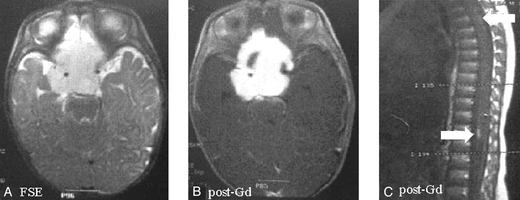
MR images were reviewed to localize lesions as well as to analyze composition (ie, solid versus cystic) and enhancement patterns of PMAs. Images were also reviewed to determine whether metastasis to surrounding tissues existed. Typical localization was determined in the areas of the optic chiasm and hypothalamus. The composition of PMAs was 100% solid in two patients, >90% solid in one patient, and >70% solid in one patient. Histopathologic analysis revealed that two of the solid tumors had small, central necrotic areas. Mass effect and encasement of surrounding vessels was extensive in two patients and moderate in two patients. Basilar and spinal meningeal enhancement was present in three and two patients, respectively. Extension of tumor to deep gray matter and white matter was found in two patients. Two patients had mild to moderate hydrocephalus. MR imaging findings and clinical features of our patients were compared with those of typical chiasmatic-hypothalamic PA (Table 1). A sample of our PMA case with spinal seeding via CSF is shown in Figure 1 .

Fig 1.
  • Download figure
  • Open in new tab
  • Download powerpoint
Fig 1.

Suprasellar pilomyxoid astrocytoma in a 5-month-old boy (patient 2). The mass, surrounding the circle of Willis, shows hyperintense signal on fast spin-echo images (A) and homogeneous enhancement after contrast material administration except for right anterior small cystic component (B). Peripheral pial enhancement of spinal cord on postcontrast sagittal T1 spin-echo images was detected (C).

View this table:
  • View inline
  • View popup

Clinical and Imaging Findings in Four Patients with the Diagnosis of PmA Compared with that of Chiasmatic/Hypothalamic PA

Three of the patients reported in this study were included in a previously published report (1). All tumors consisted of monomorphic and piloid cells in a prominent myxoid background. All had a compact architecture with marked vascularity and an angiocentric pattern. The angiocentric arrangement of cells was reminiscent of perivascular pseudorosettes. There were no eosinophilic granular bodies or Rosenthal fibers. Tumor necrosis was seen in two patients. None of the specimens had glomeruloid vascular proliferation, calcification, or ossification. These histologic features mirror those described in the previous report (1).

All patients had undergone surgical excision of tumor. Two of the patients had rapid regrowth of the tumor, and each had undergone a decompressive reoperation. Chemotherapy was instituted for all four patients. None of the patients showed diencephalic syndrome. Two patients were alive with disease 30 and 36 months after initial surgery. One patient died of tumor after 61 months. The other patient was lost to follow-up 26 months after initial surgery.

Discussion

PMA can be defined as either a variant of PA or a separate entity with monomorphous pilomyxoid characteristics (1, 5). We have elsewhere demonstrated the monomorphous cellular composition and a higher rate of recurrence compared with typical PA (1). Progression-free survival at 1 year was 38.7% in PMA and 69.2% in PA (1).

PA is an indolent tumor typically located in the cerebellum, optic nerve, and hypothalamic-chiasmatic region. Although anaplastic evolution or CSF dissemination was reported for PA, this has been extremely rare (5, 6). The postoperative survival rate for PA is 86–100% at 5 years, 83% at 10 years, and 70% at 20 years (6). Survival rate with gross total tumor resection is 100%. Long-term survival is expected even in subtotally resected tumors. A small number of PAs tend to be aggressive (7).

There are definite histologic differences between PMA and PA. Eosinophilic granular bodies, Rosenthal fibers, plump “protoplasmic” cells, and a biphasic pattern are extremely rare in PMA but common in PA. PMA has a monomorphous myxoid background and angiocentric pattern, which are not common in PA. Both tumor types have abundant piloid cells (1).

The radiologic appearance of chiasmatic hypothalamic PA is characteristic (7–9). In general, the tumors do not have peritumoral edema. They may have a solid nodular and cystic part. Contrast enhancement is variable. The walls of PA consist of nonneoplastic-compressed brain parenchyma that typically does not enhance, although occasionally some cases will show mural enhancement. Obstructive hydrocephalus occurs late (usually mild to moderate), and 10% of PAs have calcification (8). PA may be associated with neurofibromatosis and involve the optic pathways (10, 11). With this typical radiologic appearance and age of the patient, the diagnosis of typical PA can be made with confidence (8).

PMA is a relatively newly described entity; there are no reports on imaging findings of these unusual tumors. In our study, the most distinguishing radiologic features suggestive of PMA as opposed to a typical PA are predominantly solid tumor, homogeneous enhancement, hydrocephalus, extension of T2 signal intensity abnormality into the deep white mater and gray matter, and CSF dissemination.

Reports of atypical PA with more aggressive and worse clinical outcome have been clearly documented in the past. We have identified a substantial number of the PMA patients in our original study from such cases. It may be equally possible that the incidence of PMA may be much higher than that perceived among such aggressive tumors.

Treatment of PAs is mainly surgical debulking, after which the tumors remain stable for many years. Despite slow growth of the solid component, multiple cystic recurrences can be observed, and they can be treated by stereotactic surgical drainage, intracystic irradiation with P32, or craniotomy (5). By contrast, PMAs are more likely to recur and disseminate in CSF, and recurrence may be more solid than cystic. PMAs usually require adjuvant treatment (radiation therapy or chemotherapy) to control the disease. There is no specific adjuvant therapy protocol for PMA (1, 5).

Conclusion

The findings of a chiasmatic or hypothalamic enhancing solid tumor with hydrocephalus, highly homogeneous T2 signal intensity, extension of T2 signal intensity abnormality into the deep white and gray matter, and CSF dissemination characterize the radiologic features of PMA. Larger series are needed before these MR imaging findings can be used in the differential diagnosis from typical PAs.

References

  1. ↵
    Tihan T, Fisher PG, Kepner JL, et al. Pediatric astrocytomas with monomorphous pilomyxoid features and a less favorable outcome. J Neuropathol Exp Neurol 1999;58:1061–1068
    CrossRefPubMed
  2. ↵
    Fuller CE, Frankel B, Smith M, et al. Suprasellar monomorphous pilomyxoid neoplasm: an ultrastructural analysis. Clin Neuropathol 2001;20:256–262
    PubMed
  3. ↵
    Burger P. Pathology of brain stem astrocytomas. Pediatr Neurosurg 1996;24:35–40
    PubMed
  4. ↵
    Krieger MD, Gonzales-Gomez I, Levy ML, McComb JG. Recurrence patterns and anaplastic change in a long term study of pilocytic astrocytomas. Pediatr Neurosurg 1997;27:1–11
    PubMed
  5. ↵
    Burger PC, Cohen KJ, Rosenblum MK, Tihan T. Pathology of diencephalic astrocytomas. Pediatr Neurosurg 2000;32:214–219
    CrossRefPubMed
  6. ↵
    Obana WG, Cogen PH, Davis RL, Edwards MSB. Metastatic juvenile pilocytic astrocytoma. J Neurosurg 1991;75:972–975
    PubMed
  7. ↵
    Strong JA, Hatten HP Jr, Brown MT, et al. Pilocytic astrocytoma: correlation between the initial imaging features and clinical aggressiveness. AJR Am J Roentgenol 1993;161:369–372
    PubMed
  8. ↵
    Lee YY, Van Tassel P, Bruner JM, et al. Juvenile pilocytic astrocytomas: CT and MR characteristics. AJR Am J Roentgenol 1989;152:1263–1270
    PubMed
  9. ↵
    Kulkarni AV, Amstrong DC, Drake JM. MR characteristics of malignant spinal cord astrocytomas in children. Can J Neurol Sci 1999;26:290–293
    PubMed
  10. ↵
    Blah J, Jaffe R, Deutsch M, Adkins JC. Neurofibromatosis and childhood tumors. Cancer 1986;57:1225–1229
    CrossRefPubMed
  11. ↵
    Jacoby CG, Go RT, Beren RA. Cranial CT of neurofibromatosis. AJR Am J Roentgenol 1980;135:553–557
    PubMed
  • Received January 13, 2003.
  • Accepted after revision April 28, 2003.
  • Copyright © American Society of Neuroradiology
View Abstract
PreviousNext
Back to top

In this issue

American Journal of Neuroradiology: 24 (9)
American Journal of Neuroradiology
Vol. 24, Issue 9
1 Oct 2003
  • Table of Contents
  • Index by author
Advertisement
Print
Download PDF
Email Article

Thank you for your interest in spreading the word on American Journal of Neuroradiology.

NOTE: We only request your email address so that the person you are recommending the page to knows that you wanted them to see it, and that it is not junk mail. We do not capture any email address.

Enter multiple addresses on separate lines or separate them with commas.
MR Imaging Characteristics of Pilomyxoid Astrocytomas
(Your Name) has sent you a message from American Journal of Neuroradiology
(Your Name) thought you would like to see the American Journal of Neuroradiology web site.
CAPTCHA
This question is for testing whether or not you are a human visitor and to prevent automated spam submissions.
Cite this article
Atilla Arslanoglu, Bayram Cirak, Alena Horska, James Okoh, Tarik Tihan, Leslie Aronson, Anthony M. Avellino, Peter C. Burger, David M. Yousem
MR Imaging Characteristics of Pilomyxoid Astrocytomas
American Journal of Neuroradiology Oct 2003, 24 (9) 1906-1908;

Citation Manager Formats

  • BibTeX
  • Bookends
  • EasyBib
  • EndNote (tagged)
  • EndNote 8 (xml)
  • Medlars
  • Mendeley
  • Papers
  • RefWorks Tagged
  • Ref Manager
  • RIS
  • Zotero
0 Responses
Respond to this article
Share
Bookmark this article
MR Imaging Characteristics of Pilomyxoid Astrocytomas
Atilla Arslanoglu, Bayram Cirak, Alena Horska, James Okoh, Tarik Tihan, Leslie Aronson, Anthony M. Avellino, Peter C. Burger, David M. Yousem
American Journal of Neuroradiology Oct 2003, 24 (9) 1906-1908;
del.icio.us logo Twitter logo Facebook logo Mendeley logo
  • Tweet Widget
  • Facebook Like
  • Google Plus One

Jump to section

  • Article
    • Abstract
    • Description of Cases
    • Discussion
    • Conclusion
    • References
  • Figures & Data
  • Info & Metrics
  • Responses
  • References
  • PDF

Related Articles

  • No related articles found.
  • PubMed
  • Google Scholar

Cited By...

  • Hypothalamic Pilomyxoid Astrocytoma in a Child with Lipodystrophy
  • Management of Pilomyxoid Astrocytomas: Our Experience
  • Crossref
  • Google Scholar

This article has not yet been cited by articles in journals that are participating in Crossref Cited-by Linking.

More in this TOC Section

  • SyMRI & MR Fingerprinting in Brainstem Myelination
  • Comparison of Image Quality and Radiation Dose in Pediatric Temporal Bone CT Using Photon-Counting Detector CT and Energy-Integrating Detector CT
  • Dandy-Walker Phenotype with Brainstem Involvement
Show more Pediatrics

Similar Articles

Advertisement

Indexed Content

  • Current Issue
  • Accepted Manuscripts
  • Article Preview
  • Past Issues
  • Editorials
  • Editor's Choice
  • Fellows' Journal Club
  • Letters to the Editor
  • Video Articles

Cases

  • Case Collection
  • Archive - Case of the Week
  • Archive - Case of the Month
  • Archive - Classic Case

Special Collections

  • AJNR Awards
  • ASNR Foundation Special Collection
  • Most Impactful AJNR Articles
  • Photon-Counting CT
  • Spinal CSF Leak Articles (Jan 2020-June 2024)

More from AJNR

  • Trainee Corner
  • Imaging Protocols
  • MRI Safety Corner

Multimedia

  • AJNR Podcasts
  • AJNR Scantastics

Resources

  • Turnaround Time
  • Submit a Manuscript
  • Submit a Video Article
  • Submit an eLetter to the Editor/Response
  • Manuscript Submission Guidelines
  • Statistical Tips
  • Fast Publishing of Accepted Manuscripts
  • Graphical Abstract Preparation
  • Imaging Protocol Submission
  • Evidence-Based Medicine Level Guide
  • Publishing Checklists
  • Author Policies
  • Become a Reviewer/Academy of Reviewers
  • News and Updates

About Us

  • About AJNR
  • Editorial Board
  • Editorial Board Alumni
  • Alerts
  • Permissions
  • Not an AJNR Subscriber? Join Now
  • Advertise with Us
  • Librarian Resources
  • Feedback
  • Terms and Conditions
  • AJNR Editorial Board Alumni

American Society of Neuroradiology

  • Not an ASNR Member? Join Now

© 2025 by the American Society of Neuroradiology All rights, including for text and data mining, AI training, and similar technologies, are reserved.
Print ISSN: 0195-6108 Online ISSN: 1936-959X

Powered by HighWire