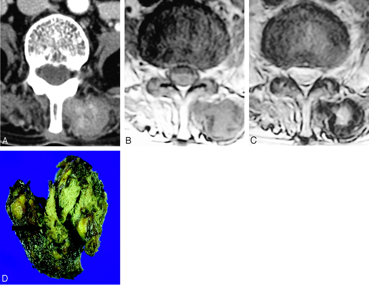
Advertisement

Main menu

  • Home
  • Content
    • Current Issue
    • Accepted Manuscripts
    • Article Preview
    • Past Issue Archive
    • Video Articles
    • AJNR Case Collection
    • Case of the Week Archive
    • Case of the Month Archive
    • Classic Case Archive
  • Special Collections
    • AJNR Awards
    • ASNR Foundation Special Collection
    • Most Impactful AJNR Articles
    • Photon-Counting CT
    • Spinal CSF Leak Articles (Jan 2020-June 2024)
  • Multimedia
    • AJNR Podcasts
    • AJNR SCANtastic
    • Trainee Corner
    • MRI Safety Corner
    • Imaging Protocols
  • For Authors
    • Submit a Manuscript
    • Submit a Video Article
    • Submit an eLetter to the Editor/Response
    • Manuscript Submission Guidelines
    • Statistical Tips
    • Fast Publishing of Accepted Manuscripts
    • Graphical Abstract Preparation
    • Imaging Protocol Submission
    • Author Policies
  • About Us
    • About AJNR
    • Editorial Board
    • Editorial Board Alumni
  • More
    • Become a Reviewer/Academy of Reviewers
    • Subscribers
    • Permissions
    • Alerts
    • Feedback
    • Advertisers
    • ASNR Home

User menu

  • Alerts
  • Log in

Search

  • Advanced search
American Journal of Neuroradiology

American Journal of Neuroradiology

ASHNR American Society of Functional Neuroradiology ASHNR American Society of Pediatric Neuroradiology ASSR
  • Alerts
  • Log in

Advanced Search

  • Home
  • Content
    • Current Issue
    • Accepted Manuscripts
    • Article Preview
    • Past Issue Archive
    • Video Articles
    • AJNR Case Collection
    • Case of the Week Archive
    • Case of the Month Archive
    • Classic Case Archive
  • Special Collections
    • AJNR Awards
    • ASNR Foundation Special Collection
    • Most Impactful AJNR Articles
    • Photon-Counting CT
    • Spinal CSF Leak Articles (Jan 2020-June 2024)
  • Multimedia
    • AJNR Podcasts
    • AJNR SCANtastic
    • Trainee Corner
    • MRI Safety Corner
    • Imaging Protocols
  • For Authors
    • Submit a Manuscript
    • Submit a Video Article
    • Submit an eLetter to the Editor/Response
    • Manuscript Submission Guidelines
    • Statistical Tips
    • Fast Publishing of Accepted Manuscripts
    • Graphical Abstract Preparation
    • Imaging Protocol Submission
    • Author Policies
  • About Us
    • About AJNR
    • Editorial Board
    • Editorial Board Alumni
  • More
    • Become a Reviewer/Academy of Reviewers
    • Subscribers
    • Permissions
    • Alerts
    • Feedback
    • Advertisers
    • ASNR Home
  • Follow AJNR on Twitter
  • Visit AJNR on Facebook
  • Follow AJNR on Instagram
  • Join AJNR on LinkedIn
  • RSS Feeds

AJNR is seeking candidates for the AJNR Podcast Editor. Read the position description.

Research ArticleSPINE

MR Imaging Findings of Paravertebral Gossypiboma

H.S. Kim, T.-S. Chung, S.H. Suh and S.Y. Kim
American Journal of Neuroradiology April 2007, 28 (4) 709-713;
H.S. Kim
  • Find this author on Google Scholar
  • Find this author on PubMed
  • Search for this author on this site
T.-S. Chung
  • Find this author on Google Scholar
  • Find this author on PubMed
  • Search for this author on this site
S.H. Suh
  • Find this author on Google Scholar
  • Find this author on PubMed
  • Search for this author on this site
S.Y. Kim
  • Find this author on Google Scholar
  • Find this author on PubMed
  • Search for this author on this site
  • Article
  • Figures & Data
  • Info & Metrics
  • Responses
  • References
  • PDF
Loading
  1. Fig 1.
    • Download figure
    • Open in new tab
    • Download powerpoint
    Fig 1.

    A 59-year-old woman (patient 1) who underwent partial laminectomy 8 months before the MR imaging examination. The CT scan (A) shows a lobulating contoured isoattenuated paravertebral mass with a slightly hyperattenuated center. The axial T1-weighted image (B) shows the paravertebral mass with an intermediate signal intensity. The axial T2-weighted image (C) shows the paravertebral mass with a high signal intensity in the center and a low signal intensity at the periphery. Initial radiologic differential diagnosis was abscess. The pathologic specimen (D) of the mass shows a surgical gauze (arrow) at the center of the mass.
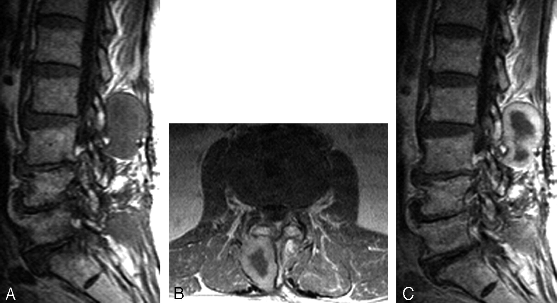
  2. Fig 2.
    • Download figure
    • Open in new tab
    • Download powerpoint
    Fig 2.

    A 34-year-old man (patient 2) who underwent partial laminectomy and diskectomy 4 months before the MR imaging examination. Sagittal T2-weighted image (A) shows a paravertebral mass at the S1–2 level (arrow) with a high signal intensity in the center and a low signal intensity at the periphery. The axial T1-weighted image (B) with contrast enhancement shows bilateral posterior paravertebral masses with peripheral rim enhancement at the same location as in A. Initial radiologic differential diagnosis was abscess and gossypiboma. Small surgical gauzes were removed from the bilateral masses.

  3. Fig 3.
    • Download figure
    • Open in new tab
    • Download powerpoint
    Fig 3.

    An 86-year-old man (patient 5) who underwent laminectomy and diskectomy 5 years before the MR imaging examination. Sagittal T1-weighted image (A) shows a posterior paravertebral mass with an intermediate signal intensity at the L2–3 level. Axial and sagittal T1-weighted images with contrast enhancement (B and C) show a right posterior paravertebral mass with peripheral rim enhancement. Initial radiologic differential diagnosis was abscess and gossypiboma. The mass was proved to contain a small surgical gauze surrounded by a thick fibrous capsule.

  4. Fig 4.
    • Download figure
    • Open in new tab
    • Download powerpoint
    Fig 4.

    A 39-year-old man (patient 7) who underwent total laminectomy 7 years before the MR imaging examination. Sagittal T2-weighted image (A) shows a posterior paravertebral mass with low-signal-intensity and multifocal high-signal-intensity spots at the L4–5 level. Axial and sagittal T1-weighted images with contrast enhancement (B and C) show a mass with heterogeneous strong enhancement. Initial radiologic differential diagnosis was abscess, neurogenic tumor, and gossypiboma. Grossly, the mass did not contain a surgical gauze, but histopathologically, the cut surface of the mass contained suture threads surrounded by attenuated fibrosis related to the foreign body reaction.

  • Copyright © American Society of Neuroradiology
PreviousNext
Back to top

In this issue

American Journal of Neuroradiology: 28 (4)
American Journal of Neuroradiology
Vol. 28, Issue 4
April 2007
  • Table of Contents
  • Index by author
Advertisement
Print
Download PDF
Email Article

Thank you for your interest in spreading the word on American Journal of Neuroradiology.

NOTE: We only request your email address so that the person you are recommending the page to knows that you wanted them to see it, and that it is not junk mail. We do not capture any email address.

Enter multiple addresses on separate lines or separate them with commas.
MR Imaging Findings of Paravertebral Gossypiboma
(Your Name) has sent you a message from American Journal of Neuroradiology
(Your Name) thought you would like to see the American Journal of Neuroradiology web site.
CAPTCHA
This question is for testing whether or not you are a human visitor and to prevent automated spam submissions.
Cite this article
H.S. Kim, T.-S. Chung, S.H. Suh, S.Y. Kim
MR Imaging Findings of Paravertebral Gossypiboma
American Journal of Neuroradiology Apr 2007, 28 (4) 709-713;

Citation Manager Formats

  • BibTeX
  • Bookends
  • EasyBib
  • EndNote (tagged)
  • EndNote 8 (xml)
  • Medlars
  • Mendeley
  • Papers
  • RefWorks Tagged
  • Ref Manager
  • RIS
  • Zotero
0 Responses
Respond to this article
Share
Bookmark this article
MR Imaging Findings of Paravertebral Gossypiboma
H.S. Kim, T.-S. Chung, S.H. Suh, S.Y. Kim
American Journal of Neuroradiology Apr 2007, 28 (4) 709-713;
del.icio.us logo Twitter logo Facebook logo Mendeley logo
  • Tweet Widget
  • Facebook Like
  • Google Plus One

Jump to section

  • Article
    • Abstract
    • Materials and Methods
    • Results
    • Discussion
    • Conclusion
    • References
  • Figures & Data
  • Info & Metrics
  • Responses
  • References
  • PDF

Related Articles

  • No related articles found.
  • PubMed
  • Google Scholar

Cited By...

  • Asymptomatic gossypiboma with complete intramural migration and ileoileal fistula
  • Imaging Retained Surgical Instruments
  • Crossref
  • Google Scholar

This article has not yet been cited by articles in journals that are participating in Crossref Cited-by Linking.

More in this TOC Section

  • MP2RAGE 7T in MS Lesions of the Cervical Spine
  • Bern Score Validity for SIH
  • Resisted Inspiration for CSF-Venous Fistula
Show more Spine

Similar Articles

Advertisement

Indexed Content

  • Current Issue
  • Accepted Manuscripts
  • Article Preview
  • Past Issues
  • Editorials
  • Editor's Choice
  • Fellows' Journal Club
  • Letters to the Editor
  • Video Articles

Cases

  • Case Collection
  • Archive - Case of the Week
  • Archive - Case of the Month
  • Archive - Classic Case

Special Collections

  • AJNR Awards
  • ASNR Foundation Special Collection
  • Most Impactful AJNR Articles
  • Photon-Counting CT
  • Spinal CSF Leak Articles (Jan 2020-June 2024)

More from AJNR

  • Trainee Corner
  • Imaging Protocols
  • MRI Safety Corner

Multimedia

  • AJNR Podcasts
  • AJNR Scantastics

Resources

  • Turnaround Time
  • Submit a Manuscript
  • Submit a Video Article
  • Submit an eLetter to the Editor/Response
  • Manuscript Submission Guidelines
  • Statistical Tips
  • Fast Publishing of Accepted Manuscripts
  • Graphical Abstract Preparation
  • Imaging Protocol Submission
  • Evidence-Based Medicine Level Guide
  • Publishing Checklists
  • Author Policies
  • Become a Reviewer/Academy of Reviewers
  • News and Updates

About Us

  • About AJNR
  • Editorial Board
  • Editorial Board Alumni
  • Alerts
  • Permissions
  • Not an AJNR Subscriber? Join Now
  • Advertise with Us
  • Librarian Resources
  • Feedback
  • Terms and Conditions
  • AJNR Editorial Board Alumni

American Society of Neuroradiology

  • Not an ASNR Member? Join Now

© 2025 by the American Society of Neuroradiology All rights, including for text and data mining, AI training, and similar technologies, are reserved.
Print ISSN: 0195-6108 Online ISSN: 1936-959X

Powered by HighWire