Advertisement

Main menu

  • Home
  • Content
    • Current Issue
    • Accepted Manuscripts
    • Article Preview
    • Past Issue Archive
    • Video Articles
    • AJNR Case Collection
    • Case of the Week Archive
    • Case of the Month Archive
    • Classic Case Archive
  • Special Collections
    • AJNR Awards
    • ASNR Foundation Special Collection
    • Most Impactful AJNR Articles
    • Photon-Counting CT
    • Spinal CSF Leak Articles (Jan 2020-June 2024)
  • Multimedia
    • AJNR Podcasts
    • AJNR SCANtastic
    • Trainee Corner
    • MRI Safety Corner
    • Imaging Protocols
  • For Authors
    • Submit a Manuscript
    • Submit a Video Article
    • Submit an eLetter to the Editor/Response
    • Manuscript Submission Guidelines
    • Statistical Tips
    • Fast Publishing of Accepted Manuscripts
    • Graphical Abstract Preparation
    • Imaging Protocol Submission
    • Author Policies
  • About Us
    • About AJNR
    • Editorial Board
    • Editorial Board Alumni
  • More
    • Become a Reviewer/Academy of Reviewers
    • Subscribers
    • Permissions
    • Alerts
    • Feedback
    • Advertisers
    • ASNR Home

User menu

  • Alerts
  • Log in

Search

  • Advanced search
American Journal of Neuroradiology

American Journal of Neuroradiology

ASHNR American Society of Functional Neuroradiology ASHNR American Society of Pediatric Neuroradiology ASSR
  • Alerts
  • Log in

Advanced Search

  • Home
  • Content
    • Current Issue
    • Accepted Manuscripts
    • Article Preview
    • Past Issue Archive
    • Video Articles
    • AJNR Case Collection
    • Case of the Week Archive
    • Case of the Month Archive
    • Classic Case Archive
  • Special Collections
    • AJNR Awards
    • ASNR Foundation Special Collection
    • Most Impactful AJNR Articles
    • Photon-Counting CT
    • Spinal CSF Leak Articles (Jan 2020-June 2024)
  • Multimedia
    • AJNR Podcasts
    • AJNR SCANtastic
    • Trainee Corner
    • MRI Safety Corner
    • Imaging Protocols
  • For Authors
    • Submit a Manuscript
    • Submit a Video Article
    • Submit an eLetter to the Editor/Response
    • Manuscript Submission Guidelines
    • Statistical Tips
    • Fast Publishing of Accepted Manuscripts
    • Graphical Abstract Preparation
    • Imaging Protocol Submission
    • Author Policies
  • About Us
    • About AJNR
    • Editorial Board
    • Editorial Board Alumni
  • More
    • Become a Reviewer/Academy of Reviewers
    • Subscribers
    • Permissions
    • Alerts
    • Feedback
    • Advertisers
    • ASNR Home
  • Follow AJNR on Twitter
  • Visit AJNR on Facebook
  • Follow AJNR on Instagram
  • Join AJNR on LinkedIn
  • RSS Feeds

AJNR is seeking candidates for the AJNR Podcast Editor. Read the position description.

Case ReportPediatrics

Syndrome of Megalencephaly, Polydactyly, and Polymicrogyria Lacking Frank Hydrocephalus, with Associated MR Imaging Findings

H.G. Tore, A.M. McKinney, V.A. Nagar, B. Lohman, C.L. Truwit and C. Raybaud
American Journal of Neuroradiology September 2009, 30 (8) 1620-1622; DOI: https://doi.org/10.3174/ajnr.A1566
H.G. Tore
  • Find this author on Google Scholar
  • Find this author on PubMed
  • Search for this author on this site
A.M. McKinney
  • Find this author on Google Scholar
  • Find this author on PubMed
  • Search for this author on this site
V.A. Nagar
  • Find this author on Google Scholar
  • Find this author on PubMed
  • Search for this author on this site
B. Lohman
  • Find this author on Google Scholar
  • Find this author on PubMed
  • Search for this author on this site
C.L. Truwit
  • Find this author on Google Scholar
  • Find this author on PubMed
  • Search for this author on this site
C. Raybaud
  • Find this author on Google Scholar
  • Find this author on PubMed
  • Search for this author on this site
  • Article
  • Figures & Data
  • Info & Metrics
  • Responses
  • References
  • PDF
Loading

SUMMARY:

Megalencephaly, polymicrogyria, polydactyly, and hydrocephalus (MPPH) syndrome has been recently recognized and is very rare. Each case reported so far has demonstrated hydrocephalus to varying degrees. We report an infant with MPPH syndrome, but lacking frank hydrocephalus. The additional finding of an abnormally elongated pituitary infundibulum has not been described in this syndrome and, along with the presence of a regressing cystic cavum septum pellucidum, suggests that chronic underlying hydrocephalus may have been present.

Megalencephaly, polymicrogyria, polydactyly, and hydrocephalus (MPPH) syndrome was first described in 2004, with only 9 cases reported so far; hence, the prevalence has not yet been determined.1,2 Hydrocephalus has been considered a mandatory component of this syndrome.1—4 We report a patient with macrocephaly, polymicrogyria, and polydactyly, but without overt hydrocephalus. We also report an elongated pituitary infundibulum along with a regressing cystic cavum septum pellucidum (CSP); cystic CSP has been described previously in MPPH, but without reports of regression.2,5,6 We discuss how to distinguish MPPH from other syndromes presenting with polymicrogyria or megalencephaly.

Case Report

A 1-day-old boy was born to a healthy nonconsanguineous family at 39-weeks’ gestation. Findings of prenatal sonography had suggested mildly dilated lateral and third ventricles, but with an otherwise unremarkable prenatal follow-up. After initially low Apgar scores but subsequent improvement with bag-mask ventilation, the patient was admitted to the neonatal intensive care unit with normal breathing. On physical examination, he was macrocephalic (head circumference >3.5 SDs above normal) with normal body length and weight, but with bilateral sixth digits along the ulnar aspects of the hands on plain films (Fig 1A). He had low-set ears, a high arching palate without cleft or cyst, and esotropia. No hypotonia/hypertonia was present, and deep tendon reflexes were normal. Results of blood biochemistry, cultures, and investigations for metabolic disease were unremarkable. Findings of cytogenetic analysis were unrevealing. Kidney and cardiac ultrasound findings were unremarkable.

Fig 1.
  • Download figure
  • Open in new tab
  • Download powerpoint
Fig 1.

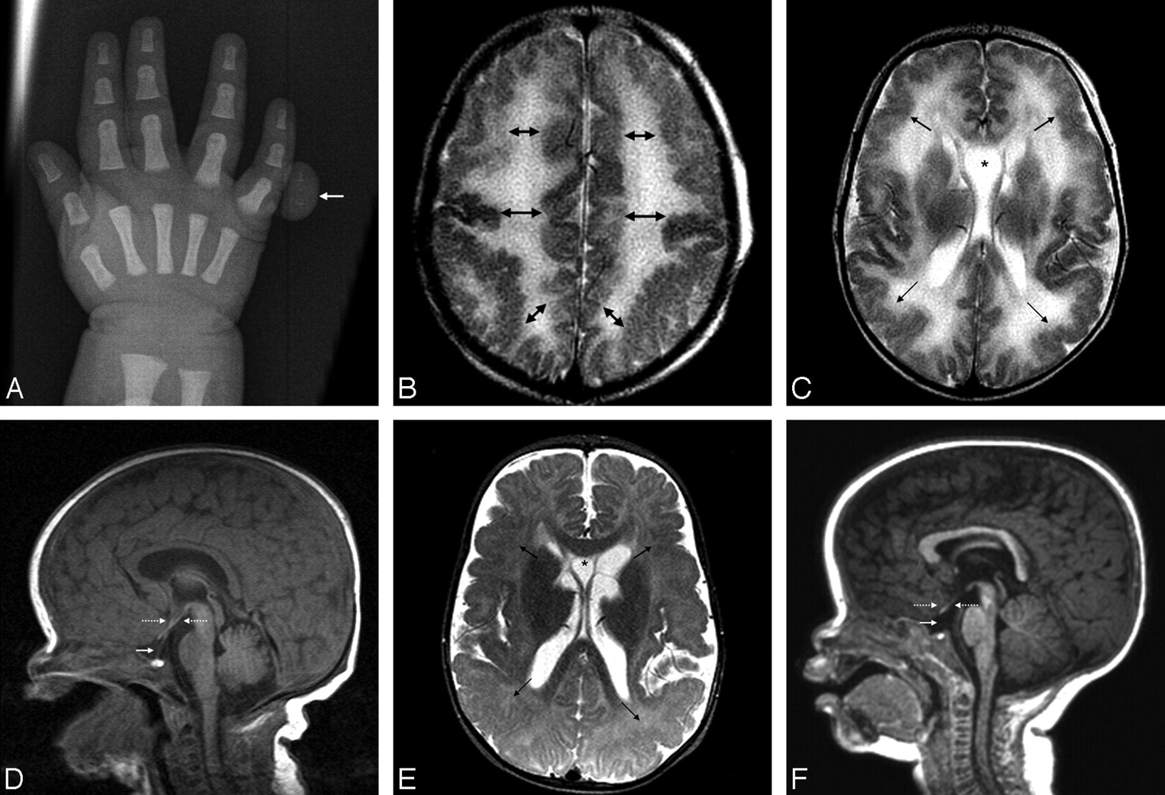
A−C, A neonate with macrocephaly and a sixth digit on plain film (arrow, A), who underwent MR imaging demonstrating diffuse polymicrogyria on T2WI (arrows, B and C). A prominent cystic CSP bows the cyst walls laterally (asterisk, C). D, The temporal horns are not overtly dilated on T2WI images (not shown), though the pituitary infundibulum is elongated (arrow), without dilated anterior third ventricular recesses, arguing against frank hydrocephalus (dotted arrows). E, At 8 months of age, the cystic CSP becomes much smaller (asterisk), but with enlarged subarachnoid spaces over the cerebral convexities. The arrows denote polymicrogyria in the midst of progressing myelination. F, The infundibulum is even more difficult to visualize, whereas the brain stem appears slightly small.

Cranial sonography was then performed, which suggested subtle lateral ventricular dilation. Thereafter, MR imaging demonstrated diffuse polymicrogyria bilaterally, favoring the posterior frontal lobes and perisylvian regions (Fig 1B, -C). Additional findings included an elongated pituitary infundibulum without other sellar abnormalities. A cystic CSP was present, measuring 1.5-cm maximum transverse diameter (Fig 1C). The myelination pattern appeared normal. There was mild colpocephaly of the lateral ventricles, but without enlargement of the anterior recesses of the third ventricle or of the temporal horns of the lateral ventricles (Fig 1D). The midbrain and corpus callosum appeared normal.

At 7 months, the patient developed seizures, which were controlled by medication. An electroencephalogram indicated seizure activity in the right occipital region spreading to the temporal lobe. Follow-up MR imaging at 8 months age demonstrated that the pituitary stalk remained elongated and even more difficult to visualize, still without overt ventricular enlargement (Fig 1E, -F). The brain- stem at this point appeared somewhat small, but not frankly hypoplastic. The cystic CSP decreased to 7 mm, whereas the subarachnoid spaces over the convexities had increased. There were no overt abnormalities in myelination, with appropriate myelination for age of the callosal genu and splenium on T2-weighted images (T2WI), though there was no repeat MR imaging in the second year of life to exclude entirely abnormalities in myelination. At this point, the patient has been followed for more than 1 year and has reached age-appropriate developmental milestones.

Discussion

MPPH syndrome was originally described in 2004 in 5 patients with megalencephaly, polymicrogyria, polydactyly, and hydrocephalus, along with severe psychomotor retardation, blindness, hypotonia, epilepsy, and facial dysmorphism.1 Subsequent reports have described phenotypic variation with facial dysmorphism of varying degrees (present in our patient); syndactyly, callosal, and midbrain abnormalities have also been described but were not present in our patient, who also had a later onset of epilepsy.1—4 Hence, he may have a milder phenotypic variant, given his later onset of seizures and appropriate psychomotor development to this point.

The finding of colpocephaly without overt hydrocephalus raises the question as to whether frank hydrocephalus is mandatory in MPPH. In the small number of patients reported with MPPH, hydrocephalus has been consistently described, along with macrocephaly, polymicrogyria, and polydactyly. The lack of definite hydrocephalus in our patient also suggests that he represents a milder variant. However, the infundibular elongation raises the question of chronic underlying hydrocephalus, possibly extending from an enlarged suprasellar cistern; in such a situation, the ventricles may not dilate due to the elasticity of the infant skull with extracerebral communication of the hydrocephalus. However, it is difficult to discern whether overt hydrocephalus will ultimately develop without years of follow-up.

We noted 2 MR imaging findings in our patient that, to our knowledge, have not been described previously in MPPH syndrome: pituitary infundibular elongation and decreasing size of a cystic CSP. The elongation of the infundibulum is nonspecific and again could relate to chronic low-level increased intracranial pressure. Regarding the cystic CSP, some reports in patients without MPPH have described intermittent hydrocephalus from its enlargement, with improvement or resolution of symptoms following treatment.6 In MPPH syndrome, a cystic CSP has been described in all except 1 patient, a higher occurrence rate than that in the general infant population of 10%–40%.2,5,7,8 Hence, the cystic CSP in our patient could have related to chronic low-level hydrocephalus, with a decreased size from extraventricular decompression outside the ventricles, because the subarachnoid spaces along the convexities were more prominent on follow-up MR imaging. However, we cannot exclude the possibility that the cystic CSP is a developmental defect associated with MPPH.

Notably, given the polymicrogyria, other causes of seizures should also be investigated on MR imaging because the differential would include the symmetric perisylvian polymicrogyria syndromes, megalencephaly-polymicrogyria-mega-corpus callosum (MPMCC) syndrome, and macrocephaly-cutis-marmorata telangiectatica congenita syndrome (MCMTC). The presence of polydactyly and megalencephaly in our patient excluded the isolated symmetric perisylvian syndromes.9 Regarding MPMCC, a thickened corpus callosum is an essential feature, but was not present in our patient.10 Regarding MCMTC, which can have polydactyly, the absence of cutaneous hemangiomata and the presence of polymicrogyria excluded this syndrome.11

No genetic alterations in MPPH have been found, and accordingly, our patient had no chromosomal abnormality. However, we did not perform a high-resolution cytogenetic examination, a potential limitation of this report. The presence of congenital metabolic disorders was largely excluded by negative findings on an extensive work-up via laboratory screening.

Footnotes

  • Paper previously presented at: Annual Meeting of the American Society of Neuroradiology and the American Society of Pediatric Neuroradiology, June 3, 2008; New Orleans, La.

References

  1. ↵
    1. Mirzaa G,
    2. Dodge NN,
    3. Glass I,
    4. et al
    . Megalencephaly and perisylvian polymicrogyria with postaxial polydactyly and hydrocephalus: a rare brain malformation syndrome associated with mental retardation and seizures. Neuropediatrics 2004; 35: 353– 59
    CrossRefPubMed
  2. ↵
    1. Pisano T,
    2. Meloni M,
    3. Cianchetti C,
    4. et al
    . Megalencephaly, polymicrogyria, and hydrocephalus (MPPH) syndrome: a new case with syndactyly. J Child Neurol 2008; 23: 916– 18
    Abstract/FREE Full Text
  3. ↵
    1. Tohyama J,
    2. Akasaka N,
    3. Saito N,
    4. et al
    . Megalencephaly and polymicrogyria with polydactyly syndrome. Pediatr Neurol 2007; 37: 148– 51
    CrossRefPubMed
  4. ↵
    1. Garavelli L,
    2. Guareschi E,
    3. Errico S,
    4. et al
    . Megalencephaly and perisylvian polymicrogyria with postaxial polydactyly and hydrocephalus (MPPH): report of a case. Neuropediatrics 2007; 38: 200– 03
    CrossRefPubMed
  5. ↵
    1. Colombani M,
    2. Chouchane M,
    3. Pitelet G,
    4. et al
    . A new case of megalencephaly and perisylvian polymicrogyria with post-axial polydactyly and hydrocephalus: MPPH syndrome. Eur J Med Genet 2006; 49: 466– 71
    CrossRefPubMed
  6. ↵
    1. Silbert PL,
    2. Gubbay SS,
    3. Vaughan RJ
    . Cavum septum pellucidum and obstructive hydrocephalus. J Neurol Neurosurg Psychiatry 1993; 56: 820– 22
    Abstract/FREE Full Text
  7. ↵
    1. Farruggia S,
    2. Babcock DS
    . The cavum septi pellucidi: its appearance and incidence with cranial ultrasonography in infancy. Radiology 1981; 139: 147– 50
    PubMed
  8. ↵
    1. Nakano S,
    2. Hojo H,
    3. Kataoka K,
    4. et al
    . Age related incidence of cavum septi pellucidi and cavum vergae on CT scans of pediatric patients. J Comput Assist Tomogr 1981; 5: 348– 49
    PubMed
  9. ↵
    1. Barkovich AJ,
    2. Hevner R,
    3. Guerrini R
    . Syndromes of bilateral symmetrical polymicrogyria. AJNR Am J Neuroradiol 1999; 20: 1814– 21
    Abstract/FREE Full Text
  10. ↵
    1. Göchlich-Ratmann G,
    2. Baethman M,
    3. Lorenz P,
    4. et al
    . Megalencephaly, mega corpus callosum, and complete lack of motor development: a previously undescribed syndrome. Am J Med Genet 1998; 79: 161– 67
    CrossRefPubMed
  11. ↵
    1. Franceschini P,
    2. Licata D,
    3. Di Cara G,
    4. et al
    . Macrocephaly-cutis marmorata telangiectatica congenita without cutis marmorata? Am J Med Genet 2000; 90: 265– 69
    CrossRefPubMed
  • Received January 1, 2009.
  • Accepted after revision January 27, 2009.
  • Copyright © American Society of Neuroradiology
View Abstract
PreviousNext
Back to top

In this issue

American Journal of Neuroradiology: 30 (8)
American Journal of Neuroradiology
Vol. 30, Issue 8
1 Sep 2009
  • Table of Contents
  • Index by author
Advertisement
Print
Download PDF
Email Article

Thank you for your interest in spreading the word on American Journal of Neuroradiology.

NOTE: We only request your email address so that the person you are recommending the page to knows that you wanted them to see it, and that it is not junk mail. We do not capture any email address.

Enter multiple addresses on separate lines or separate them with commas.
Syndrome of Megalencephaly, Polydactyly, and Polymicrogyria Lacking Frank Hydrocephalus, with Associated MR Imaging Findings
(Your Name) has sent you a message from American Journal of Neuroradiology
(Your Name) thought you would like to see the American Journal of Neuroradiology web site.
CAPTCHA
This question is for testing whether or not you are a human visitor and to prevent automated spam submissions.
Cite this article
H.G. Tore, A.M. McKinney, V.A. Nagar, B. Lohman, C.L. Truwit, C. Raybaud
Syndrome of Megalencephaly, Polydactyly, and Polymicrogyria Lacking Frank Hydrocephalus, with Associated MR Imaging Findings
American Journal of Neuroradiology Sep 2009, 30 (8) 1620-1622; DOI: 10.3174/ajnr.A1566

Citation Manager Formats

  • BibTeX
  • Bookends
  • EasyBib
  • EndNote (tagged)
  • EndNote 8 (xml)
  • Medlars
  • Mendeley
  • Papers
  • RefWorks Tagged
  • Ref Manager
  • RIS
  • Zotero
0 Responses
Respond to this article
Share
Bookmark this article
Syndrome of Megalencephaly, Polydactyly, and Polymicrogyria Lacking Frank Hydrocephalus, with Associated MR Imaging Findings
H.G. Tore, A.M. McKinney, V.A. Nagar, B. Lohman, C.L. Truwit, C. Raybaud
American Journal of Neuroradiology Sep 2009, 30 (8) 1620-1622; DOI: 10.3174/ajnr.A1566
del.icio.us logo Twitter logo Facebook logo Mendeley logo
  • Tweet Widget
  • Facebook Like
  • Google Plus One
Purchase

Jump to section

  • Article
    • SUMMARY:
    • Case Report
    • Discussion
    • Footnotes
    • References
  • Figures & Data
  • Info & Metrics
  • Responses
  • References
  • PDF

Related Articles

  • No related articles found.
  • PubMed
  • Google Scholar

Cited By...

  • Cerebral cortex expansion and folding: what have we learned?
  • Crossref (12)
  • Google Scholar

This article has been cited by the following articles in journals that are participating in Crossref Cited-by Linking.

  • A developmental and genetic classification for malformations of cortical development: update 2012
    A. James Barkovich, Renzo Guerrini, Ruben I. Kuzniecky, Graeme D. Jackson, William B. Dobyns
    Brain 2012 135 5
  • Cerebral cortex expansion and folding: what have we learned?
    Virginia Fernández, Cristina Llinares‐Benadero, Víctor Borrell
    The EMBO Journal 2016 35 10
  • Megalencephaly‐capillary malformation (MCAP) and megalencephaly‐polydactyly‐polymicrogyria‐hydrocephalus (MPPH) syndromes: Two closely related disorders of brain overgrowth and abnormal brain and body morphogenesis
    Ghayda M. Mirzaa, Robert L. Conway, Karen W. Gripp, Tally Lerman‐Sagie, Dawn H. Siegel, Linda S. deVries, Dorit Lev, Nancy Kramer, Elizabeth Hopkins, John M. Graham, William B. Dobyns
    American Journal of Medical Genetics Part A 2012 158A 2
  • Megalencephaly Syndromes and Activating Mutations in the PI3K‐AKT Pathway: MPPH and MCAP
    GHAYDA M. MIRZAA, JEAN‐BAPTISTE RIVIÈRE, WILLIAM B. DOBYNS
    American Journal of Medical Genetics Part C: Seminars in Medical Genetics 2013 163 2
  • MPPH Syndrome: Two New Cases
    Wendy L. Osterling, Richard S. Boyer, Gary L. Hedlund, James F. Bale
    Pediatric Neurology 2011 44 5
  • Prenatal diagnosis of MPPH syndrome
    Bart De Keersmaecker, Hilde Van Esch, Dominique Van Schoubroeck, Filip Claus, Philippe Moerman, Luc De Catte
    Prenatal Diagnosis 2013 33 3
  • Megalencephaly-Polymicrogyria-Polydactyly-Hydrocephalus Syndrome
    Ariana Kariminejad, Farid Radmanesh, Ali-reza Rezayi, Seyed-Hasan Tonekaboni, Joseph G. Gleeson
    Journal of Child Neurology 2013 28 5
  • Systematic Approach to Pediatric Macrocephaly
    Jennifer Huang, Asha Sarma, Stephen Little, Sumit Pruthi
    RadioGraphics 2023 43 5
  • Medulloblastoma in the setting of megalencephaly polymicrogyria polydactyly hydrocephalus
    Muhamed Hadzipasic, Madeline B. Karsten, Heather Olson, Lance Rodan, Hart Lidov, Sanjay P. Prabhu, Karen Wright, Katie Pricola Fehnel
    American Journal of Medical Genetics Part A 2021 185 5
  • Four-year follow-up of megalencephaly, polymicrogyria, postaxial polydactyly and hydrocephalus (MPPH) syndrome
    Tara G Zamora, Kari D Roberts
    BMJ Case Reports 2013 2013

More in this TOC Section

  • SyMRI & MR Fingerprinting in Brainstem Myelination
  • Comparison of Image Quality and Radiation Dose in Pediatric Temporal Bone CT Using Photon-Counting Detector CT and Energy-Integrating Detector CT
  • Pons&Vermis Localization on Fetal MRI Using U-Net
Show more Pediatrics

Similar Articles

Advertisement

Indexed Content

  • Current Issue
  • Accepted Manuscripts
  • Article Preview
  • Past Issues
  • Editorials
  • Editor's Choice
  • Fellows' Journal Club
  • Letters to the Editor
  • Video Articles

Cases

  • Case Collection
  • Archive - Case of the Week
  • Archive - Case of the Month
  • Archive - Classic Case

Special Collections

  • AJNR Awards
  • ASNR Foundation Special Collection
  • Most Impactful AJNR Articles
  • Photon-Counting CT
  • Spinal CSF Leak Articles (Jan 2020-June 2024)

More from AJNR

  • Trainee Corner
  • Imaging Protocols
  • MRI Safety Corner

Multimedia

  • AJNR Podcasts
  • AJNR Scantastics

Resources

  • Turnaround Time
  • Submit a Manuscript
  • Submit a Video Article
  • Submit an eLetter to the Editor/Response
  • Manuscript Submission Guidelines
  • Statistical Tips
  • Fast Publishing of Accepted Manuscripts
  • Graphical Abstract Preparation
  • Imaging Protocol Submission
  • Evidence-Based Medicine Level Guide
  • Publishing Checklists
  • Author Policies
  • Become a Reviewer/Academy of Reviewers
  • News and Updates

About Us

  • About AJNR
  • Editorial Board
  • Editorial Board Alumni
  • Alerts
  • Permissions
  • Not an AJNR Subscriber? Join Now
  • Advertise with Us
  • Librarian Resources
  • Feedback
  • Terms and Conditions
  • AJNR Editorial Board Alumni

American Society of Neuroradiology

  • Not an ASNR Member? Join Now

© 2025 by the American Society of Neuroradiology All rights, including for text and data mining, AI training, and similar technologies, are reserved.
Print ISSN: 0195-6108 Online ISSN: 1936-959X

Powered by HighWire