Advertisement

Main menu

  • Home
  • Content
    • Current Issue
    • Accepted Manuscripts
    • Article Preview
    • Past Issue Archive
    • Video Articles
    • AJNR Case Collection
    • Case of the Week Archive
    • Case of the Month Archive
    • Classic Case Archive
  • Special Collections
    • AJNR Awards
    • ASNR Foundation Special Collection
    • Most Impactful AJNR Articles
    • Photon-Counting CT
    • Spinal CSF Leak Articles (Jan 2020-June 2024)
  • Multimedia
    • AJNR Podcasts
    • AJNR SCANtastic
    • Trainee Corner
    • MRI Safety Corner
    • Imaging Protocols
  • For Authors
    • Submit a Manuscript
    • Submit a Video Article
    • Submit an eLetter to the Editor/Response
    • Manuscript Submission Guidelines
    • Statistical Tips
    • Fast Publishing of Accepted Manuscripts
    • Graphical Abstract Preparation
    • Imaging Protocol Submission
    • Author Policies
  • About Us
    • About AJNR
    • Editorial Board
    • Editorial Board Alumni
  • More
    • Become a Reviewer/Academy of Reviewers
    • Subscribers
    • Permissions
    • Alerts
    • Feedback
    • Advertisers
    • ASNR Home

User menu

  • Alerts
  • Log in

Search

  • Advanced search
American Journal of Neuroradiology

American Journal of Neuroradiology

ASHNR American Society of Functional Neuroradiology ASHNR American Society of Pediatric Neuroradiology ASSR
  • Alerts
  • Log in

Advanced Search

  • Home
  • Content
    • Current Issue
    • Accepted Manuscripts
    • Article Preview
    • Past Issue Archive
    • Video Articles
    • AJNR Case Collection
    • Case of the Week Archive
    • Case of the Month Archive
    • Classic Case Archive
  • Special Collections
    • AJNR Awards
    • ASNR Foundation Special Collection
    • Most Impactful AJNR Articles
    • Photon-Counting CT
    • Spinal CSF Leak Articles (Jan 2020-June 2024)
  • Multimedia
    • AJNR Podcasts
    • AJNR SCANtastic
    • Trainee Corner
    • MRI Safety Corner
    • Imaging Protocols
  • For Authors
    • Submit a Manuscript
    • Submit a Video Article
    • Submit an eLetter to the Editor/Response
    • Manuscript Submission Guidelines
    • Statistical Tips
    • Fast Publishing of Accepted Manuscripts
    • Graphical Abstract Preparation
    • Imaging Protocol Submission
    • Author Policies
  • About Us
    • About AJNR
    • Editorial Board
    • Editorial Board Alumni
  • More
    • Become a Reviewer/Academy of Reviewers
    • Subscribers
    • Permissions
    • Alerts
    • Feedback
    • Advertisers
    • ASNR Home
  • Follow AJNR on Twitter
  • Visit AJNR on Facebook
  • Follow AJNR on Instagram
  • Join AJNR on LinkedIn
  • RSS Feeds

AJNR is seeking candidates for the AJNR Podcast Editor. Read the position description.

Research ArticleHead and Neck

Recurrent Orbital Myositis Mimicking Sixth Nerve Palsy: Diagnosis with MR Imaging

M. Fischer, U. Kempkes, P. Haage and S. Isenmann
American Journal of Neuroradiology February 2010, 31 (2) 275-276; DOI: https://doi.org/10.3174/ajnr.A1751
M. Fischer
  • Find this author on Google Scholar
  • Find this author on PubMed
  • Search for this author on this site
U. Kempkes
  • Find this author on Google Scholar
  • Find this author on PubMed
  • Search for this author on this site
P. Haage
  • Find this author on Google Scholar
  • Find this author on PubMed
  • Search for this author on this site
S. Isenmann
  • Find this author on Google Scholar
  • Find this author on PubMed
  • Search for this author on this site
  • Article
  • Figures & Data
  • Info & Metrics
  • Responses
  • References
  • PDF
Loading

Abstract

SUMMARY: We present a case with recurrent orbital myositis sequentially affecting both lateral rectus muscles separately. In the first episode, the absence of the required symptoms for the diagnosis of orbital myositis led to the erroneous diagnosis of sixth nerve palsy. Eventually, the correct diagnosis was established with cerebral MR imaging. Orbital myositis should be included in the differential diagnosis of what appears clinically to be abducens palsy, and MR imaging with a focus on the orbita is mandatory in such patients.

Orbital myositis implies orbital inflammation confined to ≥1 extraocular muscle and usually begins acutely with unilateral periorbital pain, particularly with eye movement, and diplopia due to restriction of extraocular muscle movement. In addition, eyelid swelling and conjunctival injection usually are present.1 These may be related to specific inflammatory processes associated with systemic disease or restricted to the orbit.2 Typically, inflammation is unilateral and can be recurrent. Recurrences can involve different muscles and, occasionally, the other eye.2 Diagnosis can usually be made clinically due to the characteristic pain and signs of inflammation. Here, we report a young patient with 2 asynchronous bilateral orbital myositis episodes, separated by 18 months, presenting without any clinical signs of inflammation. This case emphasizes the notion that orbital myositis should be considered in the differential diagnosis of clinically apparent abducens nerve palsy.

Case Reports

A 30-year-old man presented after acute onset of diplopia on gazing to the right. He reported pain around the right eye for 3 days, especially when looking to the right. Neurologic examination revealed a discrete deficit in abduction movement of the right eye that progressed during the following days. There was no conjunctival injection, chemosis, or bulb pain present; and visual acuity, visual fields, pupillary reactions, funduscopy findings, and eyelids were normal as were the findings on the remainder of the neurologic examination. Results of thyroid function tests were normal. MR imaging of the brain showed swelling and signal-intensity hyperintensity in the right lateral rectus muscle on T2-weighted images (Fig 1) and contrast-enhancement on T1-weighted images (Fig 2), establishing the diagnosis of myositis. Symptomatic causes were excluded; steroid treatment with intravenous prednisolone, followed by oral prednisolone in a decreasing dosage, led to complete remission.

Fig 1.
  • Download figure
  • Open in new tab
  • Download powerpoint
Fig 1.

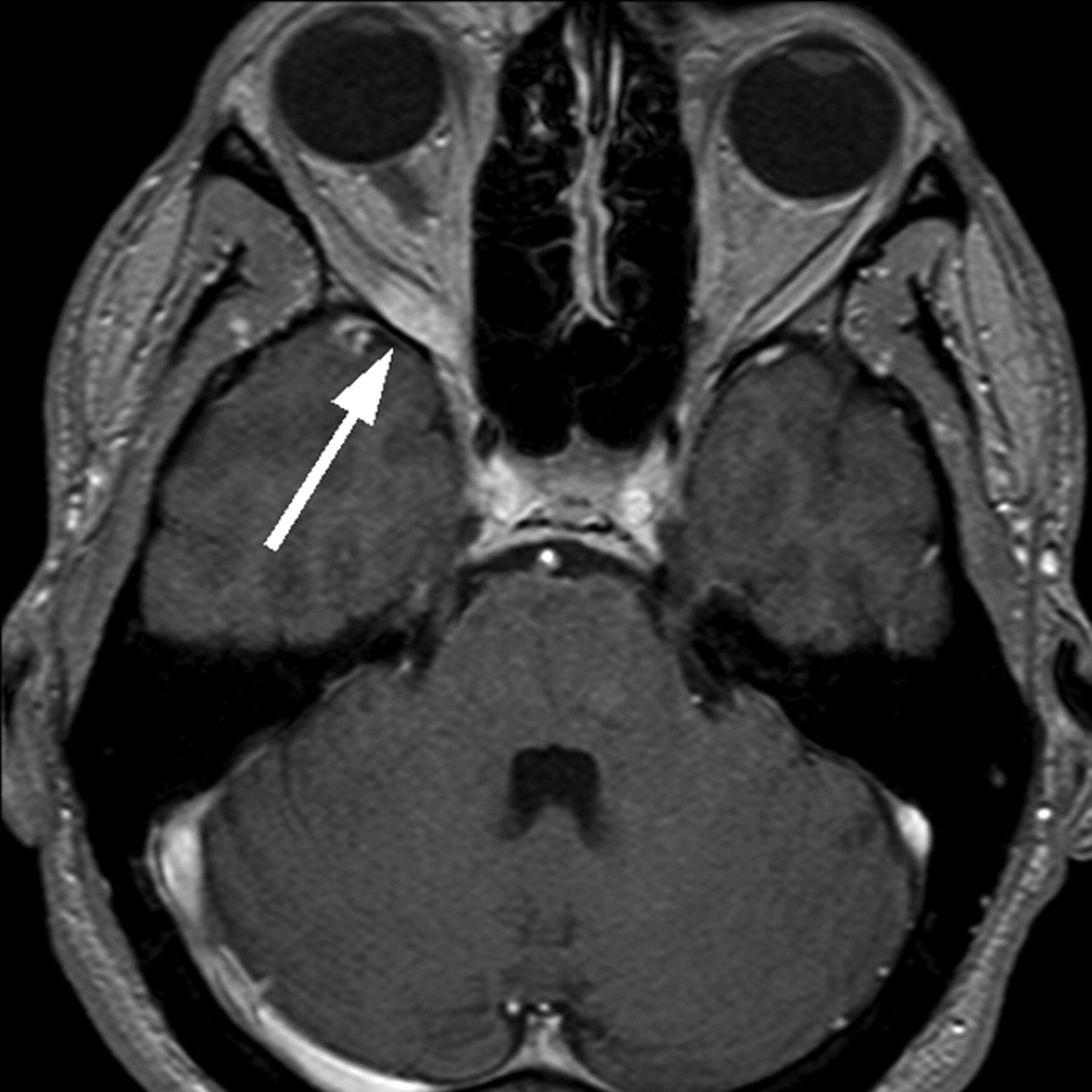
Cerebral MR image shows swelling of the right lateral rectus muscle (arrow) on a T2-weighted image. Note the position of the bulbi indicating right-sided weakness for abduction.

Fig 2.
  • Download figure
  • Open in new tab
  • Download powerpoint
Fig 2.

Cerebral MR image shows swelling of the right lateral rectus muscle (arrow) on a T1-weighted image with contrast enhancement. Note the position of the bulbi, indicating right-sided weakness for abduction.

During the stay, he reported having had similar symptoms at the contralateral (left) eye 18 months previously, with diplopia on looking to the left. According to the patient and the medical records, pain and other signs of inflammation had been absent. At that time, abducens nerve palsy had been diagnosed. Brain MR imaging and intracranial MR angiography, CSF, visual-evoked potentials, and acoustic-evoked potentials had been unremarkable on first impression. Reassessment of the MR imaging scans revealed signal-intensity hyperintensity and slight thickening of the left lateral rectus muscle on T2-weighted images (Fig 3), which had not been noticed 18 months earlier. Therefore, the diagnosis of recurrent ocular myositis was established. There had been no family history of orbital myositis.

Fig 3.
  • Download figure
  • Open in new tab
  • Download powerpoint
Fig 3.

Eighteen months previously, similar swelling was present in the left lateral rectus muscle (arrow) on a T2-weighted image. Note the position of the bulbi, indicating left-sided weakness for abduction.

Discussion

In patients 50 years of age or older, sixth nerve palsy is usually attributed to extra-axial microvascular injury associated with arteriosclerosis, hypertension, or diabetes.3 In younger patients, nerve compression by aneurysms or tumors, multiple sclerosis, and vascular brain stem lesions must be considered, yet in many cases, the cause remains elusive.4,5

Typically, orbital myositis can be differentiated from neurogenic lesions because of the characteristic clinical triad of periorbital pain, diplopia, and signs of inflammation, such as conjunctival injections or swelling. Concurrent pain is present in >95% of all cases.2,6 Nevertheless, the diagnosis in the painless cases could be established due to other signs of inflammation. Unilateral single-muscle involvement is the most common presentation; multiple or bilateral involvement can also be seen. Superior, lateral, and medial rectus muscles are involved equally frequently, and recurrences occur in ≤56% of cases.7 Multiple muscle involvement, male sex, eyelid retraction, lack of protoptosis, and lack of response to systemic corticosteroids are associated with recurrent orbital myositis.8 Thyroid function must be assessed in recurrent orbital myositis, and a familial incidence is possible. Myositis can be detected on MR images or CT scans through thickening of the affected muscle and uptake of contrast medium, and on MR imaging through signal-intensity hyperintensity in T2-weighted images. Localized inflammation can be evaluated best on fat-suppressed T2-weighted images.9 There is usually a rapid response to systemic steroid therapy. To our knowledge, isolated orbital myositis of the lateral rectus muscle mimicking abducens nerve palsy has not been reported, and recurrence of the condition affecting the contralateral eye in a second episode is unprecedented in the literature.

In approximately 5% of orbital myositis cases, pain is absent, and our case shows that other clinical signs of inflammation can also be missing. Isolated alterations in an individual rectus muscle can occur. This leads to our speculation that some patients presenting clinically with abducens palsy may in fact have inflammation of the lateral rectus muscle, which may be overlooked unless specifically assessed. Although cerebral MR imaging or CT is mandatory in the diagnostic work-up of abducens palsy in younger patients,4 our case illustrates that thickening of the lateral rectus muscle may be missed when the focus is centered on other structures. Orbital myositis should be considered in the differential diagnosis of abducens palsy, especially in younger individuals. MR images with thin sections through the orbits should be included, and special attention should be paid to the lateral rectus muscle of the affected eye. Therapy with steroids leads characteristically to a good and rapid response.7

References

  1. 1.↵
    1. Moorman C,
    2. Elston J
    . Acute orbital myositis. Eye 1995;9:96–101
    PubMed
  2. 2.↵
    1. Lacey B,
    2. Chang W,
    3. Rootman J
    . Nonthyroid causes of extraocular muscle disease. Surv Ophthalmol 1999;44:187–213
    CrossRefPubMed
  3. 3.↵
    1. Rush J,
    2. Younge B
    . Paralysis of cranial nerves III, IV and VI: cause and prognosis in 1000 cases. Arch Ophthalmol 1981;99:76–79
    CrossRefPubMed
  4. 4.↵
    1. Goodwin D
    . Differential diagnosis and management of acquired sixth cranial nerve palsy. Optometry 2006;77:534–39
    PubMed
  5. 5.↵
    1. Barr D,
    2. Kupersmith MJ,
    3. Turbin R,
    4. et al
    . Isolated sixth nerve palsy: an uncommon presenting sign of multiple sclerosis. J Neurol 2000;247:701–04
    CrossRefPubMed
  6. 6.↵
    1. Berkhoff M,
    2. Sturzenegger M,
    3. Schroth G,
    4. et al
    . Ocular myositis [in German]. Nervenarzt 1997;68:792–800
    CrossRefPubMed
  7. 7.↵
    1. Mombaerts I,
    2. Koornneef L
    . Current status in the treatment of orbital myositis. Ophthalmology 1997;104:402–08
    PubMed
  8. 8.↵
    1. Mannor G,
    2. Rose GE,
    3. Moseley I,
    4. et al
    . Outcome of orbital myositis: clinical features associated with recurrence. Ophthalmology 1997;104:409–14
    PubMed
  9. 9.↵
    1. Kubota T,
    2. Kano H
    . Assessment of inflammation in idiopathic orbital myositis with fat-suppressed T2-weighted magnetic resonance imaging. Am J Ophthalmol 2007;143:718–20
    CrossRefPubMed
  • Received April 5, 2009.
  • Accepted after revision May 17, 2009.
  • Copyright © American Society of Neuroradiology
View Abstract
PreviousNext
Back to top

In this issue

American Journal of Neuroradiology: 31 (2)
American Journal of Neuroradiology
Vol. 31, Issue 2
1 Feb 2010
  • Table of Contents
  • Index by author
Advertisement
Print
Download PDF
Email Article

Thank you for your interest in spreading the word on American Journal of Neuroradiology.

NOTE: We only request your email address so that the person you are recommending the page to knows that you wanted them to see it, and that it is not junk mail. We do not capture any email address.

Enter multiple addresses on separate lines or separate them with commas.
Recurrent Orbital Myositis Mimicking Sixth Nerve Palsy: Diagnosis with MR Imaging
(Your Name) has sent you a message from American Journal of Neuroradiology
(Your Name) thought you would like to see the American Journal of Neuroradiology web site.
CAPTCHA
This question is for testing whether or not you are a human visitor and to prevent automated spam submissions.
Cite this article
M. Fischer, U. Kempkes, P. Haage, S. Isenmann
Recurrent Orbital Myositis Mimicking Sixth Nerve Palsy: Diagnosis with MR Imaging
American Journal of Neuroradiology Feb 2010, 31 (2) 275-276; DOI: 10.3174/ajnr.A1751

Citation Manager Formats

  • BibTeX
  • Bookends
  • EasyBib
  • EndNote (tagged)
  • EndNote 8 (xml)
  • Medlars
  • Mendeley
  • Papers
  • RefWorks Tagged
  • Ref Manager
  • RIS
  • Zotero
0 Responses
Respond to this article
Share
Bookmark this article
Recurrent Orbital Myositis Mimicking Sixth Nerve Palsy: Diagnosis with MR Imaging
M. Fischer, U. Kempkes, P. Haage, S. Isenmann
American Journal of Neuroradiology Feb 2010, 31 (2) 275-276; DOI: 10.3174/ajnr.A1751
del.icio.us logo Twitter logo Facebook logo Mendeley logo
  • Tweet Widget
  • Facebook Like
  • Google Plus One
Purchase

Jump to section

  • Article
    • Abstract
    • Case Reports
    • Discussion
    • References
  • Figures & Data
  • Info & Metrics
  • Responses
  • References
  • PDF

Related Articles

  • No related articles found.
  • PubMed
  • Google Scholar

Cited By...

  • No citing articles found.
  • Crossref (11)
  • Google Scholar

This article has been cited by the following articles in journals that are participating in Crossref Cited-by Linking.

  • Clinical Features of Ocular Motility in Idiopathic Orbital Myositis
    Min Seok Kang, Hee Kyung Yang, Namju Kim, Jeong-Min Hwang
    Journal of Clinical Medicine 2020 9 4
  • Inferior rectus myositis: a rare cause of painful ophthalmoplegia and a therapeutic challenge
    Sara Gori, Cinzia Lucchesi, Rosanna Calabrese, Mirco Cosottini, Eleonora Catarsi, Antonio Tavoni, Gabriele Siciliano
    Neurological Sciences 2013 34 5
  • Neuroradiological and clinical features in ophthalmoplegia
    Stefan Weidauer, Christian Hofmann, Marlies Wagner, Elke Hattingen
    Neuroradiology 2019 61 4
  • Ocular myositis: insights into recurrence and semiological presentation
    William Alves Martins, Luiz Carlos Porcello Marrone, Ricardo Saute, Jefferson Becker, José Amadeu Almeida Vargas, Juliana Ferreira da Costa Vargas, Antonio Carlos Huf Marrone
    International Journal of Neuroscience 2015 125 9
  • Orbital myositis associated with focal active colitis in a teenage girl
    Meda Kondolot, Ekrem Unal, Gamze Poyrazoglu, Ahu Kara, Ali Yikilmaz, Sefer Kumandas, Duran Arslan
    Child's Nervous System 2012 28 4
  • Cutting the edge of idiopathic recurrent orbital myositis
    Alessandra Ariatti, Giuliana Galassi, Raffaella Rovati, Annalisa Chiari
    Open Medicine 2013 8 6
  • La tomografía computarizada y la resonancia magnética en patologías frecuentes de órbita y vía óptica. Una revisión
    Lina Marcela Acuña Bedoya, Sara Viviana Angulo Sánchez
    Ciencia y Tecnología para la Salud Visual y Ocular 2013 11 1
  • Lateral Rectus Myositis Presented as Binocular Diplopia
    Ming-Che Wu, Aven Po-Chen Lin, Ping-Hsun Lee, Ming-Tsung Li, Yu-Hang Yeh
    The Journal of Emergency Medicine 2015 48 6
  • Multiple pregnancy in case of idiopathic polymyositis: case report and literature review
    Nadezhda V. Bashmakova, Tatyana V. Lisovskaya, Natalya V. Putilova, Evgeny V. Sivov, Marina V. Sivova, Irina F. Zhukova
    Gynecological Endocrinology 2015 31 sup1
  • Orbital Myositis and Strabismus: Clinical Profile, Management, and Predictive Factors for Recurrence
    Adeline Shona Lasrado, Goura Chattannavar, Saumya Jakati, Ashik Mohamed, Ramesh Kekunnaya
    Journal of Pediatric Ophthalmology & Strabismus 2025 62 1

More in this TOC Section

  • Correlation of Apparent Diffusion Coefficient at 3T with Prognostic Parameters of Retinoblastoma
  • Parathyroid Lesions: Characterization with Dual-Phase Arterial and Venous Enhanced CT of the Neck
  • Efficacy of Diffusion-Weighted Imaging for the Differentiation between Lymphomas and Carcinomas of the Nasopharynx and Oropharynx: Correlations of Apparent Diffusion Coefficients and Histologic Features
Show more HEAD AND NECK

Similar Articles

Advertisement

Indexed Content

  • Current Issue
  • Accepted Manuscripts
  • Article Preview
  • Past Issues
  • Editorials
  • Editor's Choice
  • Fellows' Journal Club
  • Letters to the Editor
  • Video Articles

Cases

  • Case Collection
  • Archive - Case of the Week
  • Archive - Case of the Month
  • Archive - Classic Case

Special Collections

  • AJNR Awards
  • ASNR Foundation Special Collection
  • Most Impactful AJNR Articles
  • Photon-Counting CT
  • Spinal CSF Leak Articles (Jan 2020-June 2024)

More from AJNR

  • Trainee Corner
  • Imaging Protocols
  • MRI Safety Corner

Multimedia

  • AJNR Podcasts
  • AJNR Scantastics

Resources

  • Turnaround Time
  • Submit a Manuscript
  • Submit a Video Article
  • Submit an eLetter to the Editor/Response
  • Manuscript Submission Guidelines
  • Statistical Tips
  • Fast Publishing of Accepted Manuscripts
  • Graphical Abstract Preparation
  • Imaging Protocol Submission
  • Evidence-Based Medicine Level Guide
  • Publishing Checklists
  • Author Policies
  • Become a Reviewer/Academy of Reviewers
  • News and Updates

About Us

  • About AJNR
  • Editorial Board
  • Editorial Board Alumni
  • Alerts
  • Permissions
  • Not an AJNR Subscriber? Join Now
  • Advertise with Us
  • Librarian Resources
  • Feedback
  • Terms and Conditions
  • AJNR Editorial Board Alumni

American Society of Neuroradiology

  • Not an ASNR Member? Join Now

© 2025 by the American Society of Neuroradiology All rights, including for text and data mining, AI training, and similar technologies, are reserved.
Print ISSN: 0195-6108 Online ISSN: 1936-959X

Powered by HighWire