Advertisement

Main menu

  • Home
  • Content
    • Current Issue
    • Accepted Manuscripts
    • Article Preview
    • Past Issue Archive
    • Video Articles
    • AJNR Case Collection
    • Case of the Week Archive
    • Case of the Month Archive
    • Classic Case Archive
  • Special Collections
    • AJNR Awards
    • ASNR Foundation Special Collection
    • Most Impactful AJNR Articles
    • Photon-Counting CT
    • Spinal CSF Leak Articles (Jan 2020-June 2024)
  • Multimedia
    • AJNR Podcasts
    • AJNR SCANtastic
    • Trainee Corner
    • MRI Safety Corner
    • Imaging Protocols
  • For Authors
    • Submit a Manuscript
    • Submit a Video Article
    • Submit an eLetter to the Editor/Response
    • Manuscript Submission Guidelines
    • Statistical Tips
    • Fast Publishing of Accepted Manuscripts
    • Graphical Abstract Preparation
    • Imaging Protocol Submission
    • Author Policies
  • About Us
    • About AJNR
    • Editorial Board
    • Editorial Board Alumni
  • More
    • Become a Reviewer/Academy of Reviewers
    • Subscribers
    • Permissions
    • Alerts
    • Feedback
    • Advertisers
    • ASNR Home

User menu

  • Alerts
  • Log in

Search

  • Advanced search
American Journal of Neuroradiology

American Journal of Neuroradiology

ASHNR American Society of Functional Neuroradiology ASHNR American Society of Pediatric Neuroradiology ASSR
  • Alerts
  • Log in

Advanced Search

  • Home
  • Content
    • Current Issue
    • Accepted Manuscripts
    • Article Preview
    • Past Issue Archive
    • Video Articles
    • AJNR Case Collection
    • Case of the Week Archive
    • Case of the Month Archive
    • Classic Case Archive
  • Special Collections
    • AJNR Awards
    • ASNR Foundation Special Collection
    • Most Impactful AJNR Articles
    • Photon-Counting CT
    • Spinal CSF Leak Articles (Jan 2020-June 2024)
  • Multimedia
    • AJNR Podcasts
    • AJNR SCANtastic
    • Trainee Corner
    • MRI Safety Corner
    • Imaging Protocols
  • For Authors
    • Submit a Manuscript
    • Submit a Video Article
    • Submit an eLetter to the Editor/Response
    • Manuscript Submission Guidelines
    • Statistical Tips
    • Fast Publishing of Accepted Manuscripts
    • Graphical Abstract Preparation
    • Imaging Protocol Submission
    • Author Policies
  • About Us
    • About AJNR
    • Editorial Board
    • Editorial Board Alumni
  • More
    • Become a Reviewer/Academy of Reviewers
    • Subscribers
    • Permissions
    • Alerts
    • Feedback
    • Advertisers
    • ASNR Home
  • Follow AJNR on Twitter
  • Visit AJNR on Facebook
  • Follow AJNR on Instagram
  • Join AJNR on LinkedIn
  • RSS Feeds

AJNR is seeking candidates for the AJNR Podcast Editor. Read the position description.

Research ArticleBrain

Differential Diagnosis of Dementia with Lewy Bodies and Alzheimer Disease Using Combined MR Imaging and Brain Perfusion Single-Photon Emission Tomography

H. Goto, K. Ishii, T. Uemura, N. Miyamoto, T. Yoshikawa, K. Shimada and S. Ohkawa
American Journal of Neuroradiology April 2010, 31 (4) 720-725; DOI: https://doi.org/10.3174/ajnr.A1926
H. Goto
  • Find this author on Google Scholar
  • Find this author on PubMed
  • Search for this author on this site
K. Ishii
  • Find this author on Google Scholar
  • Find this author on PubMed
  • Search for this author on this site
T. Uemura
  • Find this author on Google Scholar
  • Find this author on PubMed
  • Search for this author on this site
N. Miyamoto
  • Find this author on Google Scholar
  • Find this author on PubMed
  • Search for this author on this site
T. Yoshikawa
  • Find this author on Google Scholar
  • Find this author on PubMed
  • Search for this author on this site
K. Shimada
  • Find this author on Google Scholar
  • Find this author on PubMed
  • Search for this author on this site
S. Ohkawa
  • Find this author on Google Scholar
  • Find this author on PubMed
  • Search for this author on this site
  • Article
  • Figures & Data
  • Info & Metrics
  • Responses
  • References
  • PDF
Loading

Abstract

BACKGROUND AND PURPOSE: Diagnostic performance by MR imaging or by SPECT alone in discriminating DLB patients from AD patients has been estimated previously. However, the performance of a combination of MR imaging and SPECT has not yet been evaluated. Our aim was to evaluate the usefulness of combining MR imaging and SPECT to discriminate mild DLB from AD.

MATERIALS AND METHODS: Nineteen patients with mild DLB and 19 age- and cognitive decline−matched patients with mild AD underwent both SPECT and MR imaging. Hippocampal, occipital, and striatal volume and SPECT count ratios were compared. Linear discriminant and ROC analyses were performed by using the parameters of striatal volume and the occipital SPECT ratio.

RESULTS: The striatal volume ratio in the DLB group was significantly lower than that in the AD group. The occipital SPECT ratio in the DLB group was lower than that in the AD group. The mean area under the ROC curve from combined MR imaging and SPECT (AUC = 0.898) was higher than that from MR imaging (AUC = 0.679) or SPECT (AUC = 0.798) alone.

CONCLUSIONS: By combining MR imaging and SPECT, we were able to distinguish patients with mild DLB from those with AD with a high level of accuracy. Our findings suggest that combining MR imaging and SPECT modalities is a useful and practical approach for diagnostically differentiating DLB from AD.

Abbreviations

AD
Alzheimer disease
AUC
area under the curve
AVSIS
automated volumetry of segmented image system
DLB
dementia with Lewy bodies
123I-FP-CIT
iodine 123-N-omega-fluoropropyl-2β-carbomethoxy-3 β-(4-iodophenyl)-nortropane
GM
gray matter
123I
iodine 123
MMSE
Mini-Mental State Examination
PD
Parkinson disease
PET
positron-emission tomography
ROC
receiver operating characteristic
SBD
Simulated Brain Data Base
SPECT
single-photon emission CT
SPM
statistical parametric mapping
TIV
total intracranial volume
VOI
volume of interest
WM
white matter

The number of patients diagnosed with neurodegenerative dementia has increased in societies in which the average age of the population has risen. DLB is the second most prevalent form of neurodegenerative dementia following AD, and its clinical diagnosis is based on revised consensus criteria.1 An essential neuropathologic feature of DLB is the presence of Lewy bodies in cortical and brain stem structures, such as the substantia nigra and locus coeruleus.2–5 The central diagnostic criterion for DLB is dementia, defined as a progressive cognitive decline of sufficient magnitude to interfere with normal social or occupational functions. The core diagnostic features of DLB are the following: 1) fluctuating cognition with pronounced variations in attention and alertness, 2) recurrent visual hallucinations that are typically well-formed and detailed, and 3) spontaneous features of parkinsonism. The presence of 2 of 3 core features is sufficient for a diagnosis of probable DLB.1

Clinically distinguishing patients with DLB from those with AD, however, is difficult because of overlapping clinical and pathologic features.3–5 This is especially so during the early stages of illness. For example, because psychosis is usually defined as delusions and/or hallucinations, accurate differentiation of psychosis may be difficult at early stages of both diseases.6,7 The accurate identification and differentiation of patients with DLB and AD are particularly important because of the development of interventions and specific pharmacologic treatments and outcome evaluations.

Occipital abnormalities in glucose metabolism and cerebral perfusion are also a characteristic neuropathologic feature of DLB. This has been demonstrated by using SPECT and PET.8–10 Recent studies have reported dopamine transporter loss in the striatal, caudate, and posterior putamen in patients with DLB.11,12

Many studies investigating AD have found volume decreases in medial temporal structures and cerebral blood flow reductions in temporoparietal areas.13 Few studies, however, have reported the usefulness of cerebral regional volume measurement with MR imaging for discriminating AD from DLB.14

The aim of the present study was to discriminate DLB and AD by means of regional cerebral volume and perfusion measures by combining MR imaging and SPECT techniques.

Materials and Methods

Nineteen patients with mild DLB (8 men and 11 women) and 19 patients with mild AD (8 men and 11 women), matched for age, sex, and MMSE score, were retrospectively selected from patients admitted to the infirmary of our institution for the evaluation of dementia from April 2004 to March 2006. The institutional ethics board approved the study. The requirement for a participant's informed consent was waived due to the retrospective nature of the study and because it was solely observational.

All patients underwent a neurologic and psychiatric examination by 2 neurologists and/or psychiatrists followed by an MR imaging brain scan and a standard neuropsychological examination. Because all the subjects were initial patients, none of them were given any drugs at the examinations. The criteria for diagnosing and managing DLB were revised in 2005.1 This study dealt directly with morphologic and functional findings of DLB. To avoid sample-selection bias, we used DLB inclusion criteria based on the diagnostic criteria from 1996.15 These criteria do not refer to MR imaging and SPECT techniques, while the new criteria include morphologic and functional examinations.

The DLB inclusion criteria used in this study were the following: 1) a diagnosis of probable DLB by using the criteria of the Consortium on DLB International Workshop published in 199616, 2) absence of evidence of focal brain lesions on MR images, and 3) an MMSE score of >20. These criteria include 3 core features (fluctuating cognitive functions, recurrent visual hallucinations, and spontaneous parkinsonism), any 2 of which are necessary for a diagnosis of probable DLB. All patients were examined comprehensively by both neurologists and psychiatrists; and standard neuropsychological examinations, routine laboratory tests, electroencephalography, and cranial MR imaging examinations were performed. Staff physicians closely monitored fluctuation of cognitive functions over a 1-month admission in terms of MMSE performance, activities of daily living, and episodic confusion. Psychiatric and behavioral symptoms were assessed with a structured interview of a caregiver and the Neuropsychiatric Inventory,17 and they were monitored during the patient's hospital stay by staff physicians. Parkinsonism was rated as positive when ≥1 of the following features were present: resting tremor, cogwheel or lead-pipe rigidity detectable without reinforcement, bradykinesia, and loss of postural reflexes.

The inclusion criteria for AD were the following: 1) a diagnosis of probable AD by using the National Institute of Neurologic and Communicative Disorders and Stroke/Alzheimer Disease and Related Disorders Association criteria,16 2) absence of evidence of focal brain lesions on MR images, and 3) an MMSE score of >20. We excluded patients with complications from other neurologic diseases or those in poor overall physical condition and patients with severe language, attentional, or behavioral disorders that might cause difficulties with the MR imaging and SPECT procedures. Subjects in each group were matched for age and sex on a one-to-one basis. Patients with DLB and AD were also matched for MMSE score. None of the patients with AD reported visual hallucinations, fluctuating cognition, or parkinsonism.

The mean ages of the DLB and AD groups were 76.0 ± 7.1 years and 75.6 ± 6.0 years, respectively, and the mean MMSE scores of the DLB and AD groups were 22.7 ± 2.0 and 22.4 ± 1.6, respectively. Thirteen of the 19 patients with DLB had parkinsonism, 16 had visual hallucinations, and 17 had fluctuating cognitive functions.

The MR imaging scanner was a 1.5T Horizon (GE Healthcare, Milwaukee, Wisconsin). For T1-weighted images; 3D spoiled gradient-echo imaging (TR, 14 ms; TE, 3 ms; flip angle, 20°; thickness, 1.5 mm; 124 sections) was performed. Axial T2-weighted images and fluid-attenuated inversion recovery images were also obtained. MR images were obtained 0–28 days before the SPECT examinations.

The SPECT scanning was initiated with the patient in a resting state in a supine position with the eyes closed, 15 minutes after the injection of 111 MBq (3 mCi) of iodine 123 N-isopropyl-p iodoamphetamine. All SPECT studies were performed by using a rotating dual-headed gamma camera (GAMMA View SPECT 2000 H; Hitachi, Tokyo, Japan) with a low-energy general-purpose collimator. The effective FOV was a 220 mm (axial) by 170 mm (longitudinal) volume for imaging of the whole brain, including the cerebellum. The energy window was 160 keV with 20% width. Projection data were obtained at 15 seconds/step × 64 views (2 rotations, 360°). A Ramachandran filter was used for SPECT image reconstruction. Attenuation correction was performed by using Chang's method. Reconstructed images were 4 mm in pixel size and 8 mm in section thickness with 15–17 sections, and reconstructed spatial resolution was 13.4 mm at the center and 8.9-mm tangential at 80 mm. The delay time between performance of the MR imaging and the SPECT studies was 0–2 weeks.

All MR images and SPECT data were transferred to a personal computer. Volumetry and the measurement of SPECT accumulation were performed with our AVSIS.14 In brief, the AVSIS process in this study was the following: VOI intracranial, hippocampal, striatal, and occipital lobe templates were prepared before this analysis. We produced each regional VOI template on a digital phantom (SBD; http://www.bic.mni.mcgill.ca/brainweb) in standard Montreal Neurologic Institute space, delineating the contours of each structure manually. The occipital lobe VOI template included Brodmann Areas 17, 18, and 19.

The process of modified AVSIS was as follows: first, each individual MR image and SPECT image were coregistered. Each subject's MR image was then segmented into GM, WM, and CSF with the SPM5 segmentation program (Wellcome Trust Centre for Neuroimaging, London, UK). The GM template image derived from the SBD was then spatially transformed into the individual subject's GM image, and a normalization parameter was produced. This normalization parameter functions in the same way as a reversed parameter produced in the anatomic normalization of an individual brain to a standard brain. Using this parameter, we transformed the intracranial, hippocampal, striatal, and occipital VOI templates to the individual subject's space. The TIV was adjusted by using an image derived from the segmented GM, WM, and CSF images.

The segmented images were derived as follows: WM and GM areas were calculated with the voxels in the TIV VOI template. GM volumes in the hippocampi and striata were calculated by using the transformed hippocampal and striatal VOI templates. Volumes of GM and WM in the occipital lobes were calculated by using the transformed occipital VOI template for each subject. Each regional relative volume was calculated by normalization relative to the TIV. Next, hippocampal, striatal, and occipital SPECT uptake counts were calculated by determining the regions of coregistered SPECT images on each extracted area by using the aforementioned volumetric procedure (Fig 1). We calculated the regional ratio of DLB and AD: Hippocampal, striatal, and occipital volume/intracranial volumes were used for MR imaging. For SPECT, hippocampal and occipital counts/striatal counts were used.

Fig 1.
  • Download figure
  • Open in new tab
  • Download powerpoint
Fig 1.

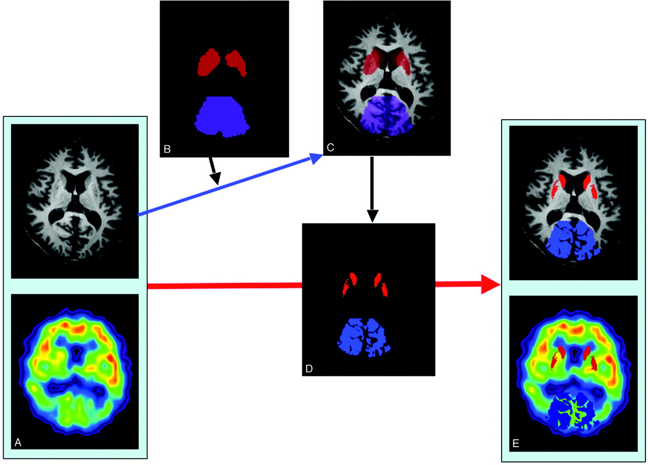
Schema for calculating regional structural volume and SPECT counts. An example of calculating striatal and occipital volume and SPECT counts is shown. A, The original MR imaging and SPECT images are coregistered. B−D, We transformed the striatal and occipital VOI templates to the individual brain (B); and by using these VOI templates on the individual MR image (C), we extracted individual regional segmented VOI templates (D) and calculated their volumes. E, Then, by using the extracted striatum and occipital lobe as VOIs for the individual coregistered SPECT image, we determined striatal and occipital lobe counts.

Fig 2.
  • Download figure
  • Open in new tab
  • Download powerpoint
Fig 2.

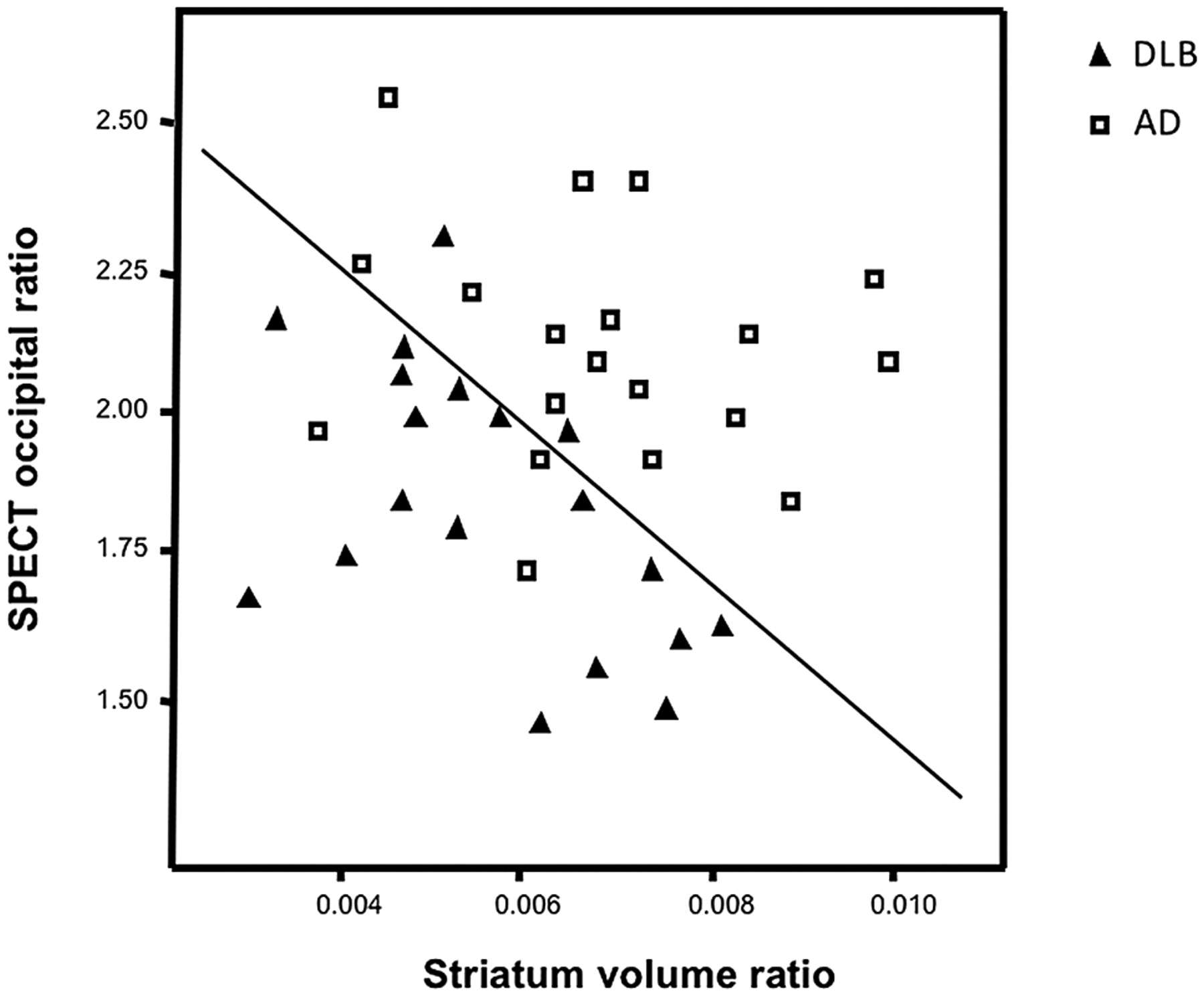
Distribution of SPECT occipital ratio and the striatal volume ratio of patients with DLB and AD. The square indicates AD; the triangle, DLB.

Linear discriminant analysis was performed on the regional ratio of volume and SPECT counts. Statistical tests were made at the 0.05 level. All statistical analyses were performed with the SPSS 17 (SPSS, Chicago, Illinois; and the JMP 8. (http://tech.its.iastate.edu/macosx/howtos/jmp8.shtml).

Results

The Table shows the mean volume and SPECT count ratios of the DLB and AD groups. Patients with DLB showed a significantly lower GM volume in the striatum compared with those with AD (P < .05). Patients with DLB showed significant cerebral blood flow reductions in the occipital lobe (P < .05). Figure 2 shows the distribution of the striatal volume ratio and the SPECT occipital ratio of both groups. We obtained the following linear discriminant analysis formula: S = (SPECT occipital ratio) × 4.934 + (striatal volume ratio) × 474.136 − 13.803, with S < 0 diagnosed as DLB, and S > 0 diagnosed as AD. DLB was correctly diagnosed in 17 of the 19 patients by using this approach. Similarly, AD was correctly diagnosed in 16 of 19 patients. Therefore, the sensitivity and specificity for discriminating DLB from AD were 89% and 84%, respectively.

View this table:
  • View inline
  • View popup

Mean volume ratio and SPECT counts ratio of the DLB and AD groups

Figure 3 shows the AUCs in the ROC analysis. For the combined striatal volume and occipital SPECT ratio, the AUC was 0.898; for the striatal volume ratio, the AUC was 0.679; and for the occipital SPECT ratio, the AUC was 0.798.

Fig 3.
  • Download figure
  • Open in new tab
  • Download powerpoint
Fig 3.

ROC curves to discriminate between DLB and AD. The measurement of the z-score in the combined striatal volume and occipital SPECT ratio yielded higher sensitivity and specificity (AUC = 0.898) (a) compared with the independent striatal volume ratio (AUC = 0.679) (b) or the independent occipital SPECT ratio (AUC = 0.798) (c).

Discussion

Several previous studies have reported that detecting occipital and parietotemporal hypoperfusion can be useful in distinguishing DLB from AD.18–21 However, hypoperfusion in the parietotemporal lobe has been observed in the brains of patients with both DLB and frontotemporal dementia,22–24 and brain perfusion SPECT in these 2 disorders can be similar to that seen in AD. A large degree of overlap also exists between AD and DLB in regional uptake patterns.25 Kemp et al26 reported that the appearance of the occipital perfusion decrease is of little use for differentiating DLB from other diseases. Overall, the contribution of occipital hypoperfusion to diagnostic accuracy remains unclear.27 It was, however, recently demonstrated that 123FP-CIT, a ligand that binds to the dopamine transporter, is useful for differentiating AD and DLB.28–32 O'Brien et al33 demonstrated the accuracy of 123I-FP-CIT SPECT in diagnosing patients with possible DLB. A presynaptic dopamine transporter imaging is useful for diagnosing patients with DLB, though it is used as an additional examination after perfusion SPECT study.

A recent systematic review of the use of hexamethylpropyleneamine oxime SPECT to distinguish AD from other dementias reported a sensitivity of approximately 70%.34 The authors of this article suggested that perfusion SPECT is unable to draw a clear line between AD and other dementias. Nevertheless, because all SPECT studies included in the study used perfusion SPECT alone, SPECT may still be helpful in the clinical diagnosis of DLB if used in conjunction with other neuroimaging techniques. PET has a higher sensitivity and a higher spatial resolution than SPECT, so it may be more suitable for distinguishing DLB and AD.35

Combining measures of metaiodobenzylguanidine cardiac uptake reduction and MMSE score-change patterns with SPECT reduction patterns has been found to increase the accuracy of differentiating DLB from AD.36,37 Moreover, MR imaging is widely available in general hospitals. We found that with a combination of MR imaging and SPECT, we were able to discriminate DLB and AD with high sensitivity and specificity. As shown in the ROC analysis, the AUC resulting from combined MR imaging and SPECT was higher than the AUC resulting from either technique alone. This finding shows that discriminative power can be increased by using MR imaging and SPECT in conjunction. Kitayama et al38 previously suggested the usefulness of a combined analysis of hippocampal perfusion and GM volume by using SPECT in conjunction with MR imaging for the early diagnosis of AD. The present study is the first showing a clear differentiation of DLB and AD by using combined MR imaging and SPECT. Our method needs SPECT and MR imaging studies, but dopamine-transporter SPECT alone does not give any cerebral perfusion state and a dopamine transporter study must be performed as an additional study after perfusion and MR imaging studies.

Several previous studies have produced results regarding striatal volume in the DLB-affected brain that conflict with the present findings.39–41 A voxel-based morphometry study with a large sample number of 72 patients with DLB and 72 patients with AD (28 confirmed by autopsy) did not report greater atrophy of the striatum in DLB than in AD.42 In a previous article, however, we demonstrated that striatal volume reduction was able to differentiate AD from DLB.14 Another study showed that patients with DLB exhibited a significantly smaller putaminal volume ratio than controls and patients with AD.43 Some pathophysiologic studies have reported morphologic changes associated with DLB. The lack of postmortem evidence of wallerian degeneration in the DLB brain44 and the presence of widespread spongiform changes and gliosis affecting long projection fibers are a part of DLB pathophysiology.45

Zaja-Milatovic et al46,47 reported evidence of spinodendritic degeneration of gamma-aminobutyric acidsergic medium spiny neurons in specific regions of the caudate nucleus. This may be a structural correlate of the impairments in executive function in patients with DLB, and such pathologic changes may explain the reduction of striatal volume in these patients. These previous findings are in accord with the present result of a significantly lower striatal volume in patients with DLB compared with those with AD. Additionally, patients with PD had significantly smaller subcortical nuclei than age- and sex-matched controls.48–50 The 68% of patients with DLB in our study had parkinsonism, and the striatal volume loss may depend on the parkinsonism related to Lewy body disease.

Several previous perfusion studies demonstrated that hippocampal volume and perfusion in patients with AD were significantly lower than those in patients with DLB.11,14,51,52 In the present study, no significant differences in hippocampal volume were found between patients with DLB and those with AD. However, this may be explained by the restriction of this study to patients with only mild DLB and AD. Furthermore, the lack of a hippocampal volume difference in the present study is in accord with the report of Barber et al53 that the presence of hippocampal atrophy is useful in detecting AD but less useful in differentiating dementias. Also, most of the patients with DLB in this study were elderly (mean age, 76.0 years), probably including those with the common type of DLB, and were affected in the hippocampus.

The limitation of this study is the small number of patients included, and validation of the discriminative model used in a different and larger cohort of patients is needed.

Conclusions

We demonstrated that a combined morphologic and functional approach by using MR imaging and SPECT may be a suitable approach for distinguishing DLB from AD with high specificity and sensitivity.

References

  1. 1.↵
    1. McKeith IG,
    2. Dickson DW,
    3. Lowe J,
    4. et al
    . Diagnosis and management of dementia with Lewy bodies: third report of the DLB Consortium. Neurology 2005; 65: 1863–72. Epub 2005 Oct 19
    CrossRef
  2. 2.↵
    1. Hansen L,
    2. Salmon D,
    3. Galasko D,
    4. et al
    . The Lewy body variant of Alzheimer's disease: a clinical and pathologic entity. Neurology 1990; 40: 1–8
  3. 3.↵
    1. Kosaka K
    . Diffuse Lewy body disease in Japan. J Neurol 1990; 237: 197–204
    CrossRefPubMed
  4. 4.↵
    1. Galasko D
    . Lewy bodies and dementia. Curr Neurol Neurosci Rep 2001; 1: 435–41
    CrossRefPubMed
  5. 5.↵
    1. Zesiewicz TA,
    2. Baker MJ,
    3. Dunne PB,
    4. et al
    . Diffuse Lewy body disease. Curr Treat Options Neurol 2001; 3: 507–18
    CrossRefPubMed
  6. 6.↵
    1. Doubleday EK,
    2. Snowden JS,
    3. Varma AR,
    4. et al
    . Qualitative performance characteristics differentiate dementia with Lewy bodies and Alzheimer's disease. J Neurol Neurosurg Psychiatry 2002; 72: 602–07
    Abstract/FREE Full Text
  7. 7.↵
    1. Mega MS,
    2. Lee L,
    3. Dinov ID,
    4. et al
    . Cerebral correlates of psychotic symptoms in Alzheimer's disease. J Neurol Neurosurg Psychiatry 2000; 69: 167–71
    Abstract/FREE Full Text
  8. 8.↵
    1. Ishii K,
    2. Imamura T,
    3. Sasaki M,
    4. et al
    . Regional cerebral glucose metabolism in dementia with Lewy bodies and Alzheimer's disease. Neurology 1998; 51: 125–30
    CrossRef
  9. 9.↵
    1. Ishii K,
    2. Yamaji S,
    3. Kitagaki H,
    4. et al
    . Regional cerebral blood flow difference between dementia with Lewy bodies and AD. Neurology 1999; 53: 413–16
    CrossRef
  10. 10.↵
    1. Lobotesis K,
    2. Fenwick JD,
    3. Phipps A,
    4. et al
    . Occipital hypoperfusion on SPECT in dementia with Lewy bodies but not AD. Neurology 2001; 56: 643–49
    CrossRef
  11. 11.↵
    1. Ransmayr G,
    2. Seppi K,
    3. Donnemiller E,
    4. et al
    . Striatal dopamine transporter function in dementia with Lewy bodies and Parkinson's disease. Eur J Nucl Med 2001; 28: 1523–28
    CrossRefPubMed
  12. 12.↵
    1. Piggott MA,
    2. Marshall EF,
    3. Thomas N,
    4. et al
    . Striatal dopaminergic markers in dementia with Lewy bodies, Alzheimer's and Parkinson's diseases: rostrocaudal distribution. Brain 1999; 122: 1449–68
    Abstract/FREE Full Text
  13. 13.↵
    1. Petrella JR,
    2. Coleman RE,
    3. Doraiswamy PM
    . Neuroimaging and early diagnosis of Alzheimer disease: a look to the future. Radiology 2003; 226: 315–36
    PubMed
  14. 14.↵
    1. Ishii K,
    2. Soma T,
    3. Kono AK,
    4. et al
    . Comparison of regional brain volume and glucose metabolism between patients with mild dementia with Lewy bodies and those with mild Alzheimer's disease. J Nucl Med 2007; 48: 704–11
    Abstract/FREE Full Text
  15. 15.↵
    1. McKhann G,
    2. Drachman D,
    3. Folstein M,
    4. et al
    . Clinical diagnosis of Alzheimer's disease: report of the NINCDS-ADRDA Work Group under the auspices of Department of Health and Human Services Task Force on Alzheimer's Disease. Neurology 1984; 34: 939–44
    CrossRefPubMed
  16. 16.↵
    1. McKeith IG,
    2. Galasko D,
    3. Kosaka,
    4. et al
    . Consensus guidelines for the clinical and pathologic diagnosis of dementia with Lewy bodies (DLB): report of the consortium on DLB international workshop. Neurology 1996; 47: 1113–24
    CrossRef
  17. 17.↵
    1. Cummings JL,
    2. Mega M,
    3. Gray K,
    4. et al
    . The Neuropsychiatric Inventory: comprehensive assessment of psychopathology in dementia. Neurology 1994; 44: 2308–14
    CrossRefPubMed
  18. 18.↵
    1. Defebvre LJ,
    2. Leduc V,
    3. Duhamel A,
    4. et al
    . Technetium HMPAO SPECT study in dementia with Lewy bodies, Alzheimer's disease and idiopathic Parkinson's disease. J Nucl Med 1999; 40: 956–62
    Abstract/FREE Full Text
  19. 19.↵
    1. Minoshima S,
    2. Foster NL,
    3. Sima AA,
    4. et al
    . Alzheimer's disease versus dementia with Lewy bodies: cerebral metabolic distinction with autopsy confirmation. Ann Neurol 2001; 50: 358–65
    CrossRefPubMed
  20. 20.↵
    1. Shimizu S,
    2. Hanyu H,
    3. Kanetaka H,
    4. et al
    . Differentiation of dementia with Lewy bodies from Alzheimer's disease using brain SPECT. Dement Geriatr Cogn Disord 2005; 20: 25–30
    CrossRefPubMed
  21. 21.↵
    1. Pasquier J,
    2. Michel BF,
    3. Brenot-Rossi I,
    4. et al
    . Value of (99m)Tc-ECD SPET for the diagnosis of dementia with Lewy bodies. Eur J Nucl Med Mol Imaging 2002; 29: 1342–48. Epub 2002 Jul 31
    CrossRefPubMed
  22. 22.↵
    1. Charpentier P,
    2. Lavenu I,
    3. Defebvre L,
    4. et al
    . Alzheimer's disease and frontotemporal dementia are differentiated by discriminant analysis applied to 99mTc HmPAO SPECT data. J Neurol Neurosurg Psychiatry 2000; 69: 661–63
    Abstract/FREE Full Text
  23. 23.↵
    1. McNeill R,
    2. Sare GM,
    3. Manoharan M,
    4. et al
    . Accuracy of single-photon emission computed tomography in differentiating frontotemporal dementia from Alzheimer's disease. J Neurol Neurosurg Psychiatry 2007; 78: 350–55
    Abstract/FREE Full Text
  24. 24.↵
    1. Sjögren M,
    2. Gustafson L,
    3. Wikkelsö C,
    4. et al
    . Frontotemporal dementia can be distinguished from Alzheimer's disease and subcortical white matter dementia by an anterior-to-posterior rCBF-SPET ratio. Dement Geriatr Cogn Disord 2000; 11: 275–85
    CrossRefPubMed
  25. 25.↵
    1. Colloby SJ,
    2. Fenwick JD,
    3. Williams ED,
    4. et al
    . A comparison of 99mTc-HMPAO SPET changes in dementia with Lewy bodies and Alzheimer's disease using statistical parametric mapping. Eur J Nucl Med Mol Imaging 2002; 29: 615–22. Epub 2002 Mar 5
    CrossRefPubMed
  26. 26.↵
    1. Kemp PM,
    2. Hoffmann SA,
    3. Tossici-Bolt L,
    4. et al
    . Limitations of the HMPAO SPECT appearances of occipital lobe perfusion in the differential diagnosis of dementia with Lewy bodies. Nucl Med Commun 2007; 28: 451–56
    CrossRefPubMed
  27. 27.↵
    1. O'Brien JT
    . Role of imaging techniques in the diagnosis of dementia. Br J Radiol 2007; 80: S71–77
    Abstract/FREE Full Text
  28. 28.↵
    1. McKeith I,
    2. O'Brien J,
    3. Walker Z,
    4. et al
    . Sensitivity and specificity of dopamine transporter imaging with 123I-FP-CIT SPECT in dementia with Lewy bodies: a phase III, multicentre study. Lancet Neurol 2007; 6: 305–13
    CrossRefPubMed
  29. 29.↵
    1. O'Brien JT,
    2. Colloby S,
    3. Fenwick J,
    4. et al
    . Dopamine transporter loss visualized with FP-CIT SPECT in the differential diagnosis of dementia with Lewy bodies. Arch Neurol 2004; 61: 919–25
    CrossRefPubMed
  30. 30.↵
    1. Colloby SJ,
    2. Firbank MJ,
    3. Pakrasi S,
    4. et al
    . A comparison of 99mTc-exametazime and 123I-FP-CIT SPECT imaging in the differential diagnosis of Alzheimer's disease and dementia with Lewy bodies. Int Psychogeriatr 2008; 20: 1124–40
    CrossRefPubMed
  31. 31.↵
    1. Colloby SJ,
    2. Williams ED,
    3. Burn DJ,
    4. et al
    . Progression of dopaminergic degeneration in dementia with Lewy bodies and Parkinson's disease with and without dementia assessed using 123I-FP-CIT SPECT. Eur J Nucl Med Mol Imaging 2005; 32: 1176–85. Epub 2005 Jun 2
    CrossRefPubMed
  32. 32.↵
    1. Tatsch K
    . Imaging of the dopaminergic system in differential diagnosis of dementia. Eur J Nucl Med Mol Imaging 2008; 35 (suppl 1): S51–57
    CrossRefPubMed
  33. 33.↵
    1. O'Brien JT,
    2. McKeith IG,
    3. Walker Z,
    4. et al
    . Diagnostic accuracy of 123I-FP-CIT SPECT in possible dementia with Lewy bodies. Br J Psychiatry 2009; 194: 34–39
    Abstract/FREE Full Text
  34. 34.↵
    1. Dougall NJ,
    2. Bruggink S,
    3. Ebmeier KP
    . Systematic review of the diagnostic accuracy of 99mTc-HMPAO-SPECT in dementia. Am J Geriatr Psychiatry 2004; 12: 554–70
    CrossRefPubMed
  35. 35.↵
    1. Ishii K,
    2. Hosaka K,
    3. Mori T,
    4. et al
    . Comparison of FDG-PET and IMP-SPECT in patients with dementia with Lewy bodies. Ann Nucl Med 2004; 18: 447–51.
    PubMed
  36. 36.↵
    1. Hanyu H,
    2. Shimizu S,
    3. Hirao K,
    4. et al
    . Comparative value of brain perfusion SPECT and [(123)I]MIBG myocardial scintigraphy in distinguishing between dementia with Lewy bodies and Alzheimer's disease. Eur J Nucl Med Mol Imaging 2006; 33: 248–53
    CrossRefPubMed
  37. 37.↵
    1. Hanyu H,
    2. Shimizu S,
    3. Hirao K,
    4. et al
    . Differentiation of dementia with Lewy bodies from Alzheimer's disease using Mini-Mental State Examination and brain perfusion SPECT. J Neurol Sci 2006; 250: 97–102
    CrossRefPubMed
  38. 38.↵
    1. Kitayama N,
    2. Matsuda H,
    3. Ohnishi T,
    4. et al
    . Measurements of both hippocampal blood flow and hippocampal gray matter volume in the same individuals with Alzheimer's disease. Nucl Med Commun 2001; 22: 473–77
    CrossRefPubMed
  39. 39.↵
    1. Barber R,
    2. McKeith I,
    3. Ballard C,
    4. et al
    . Volumetric MRI study of the caudate nucleus in patients with dementia with Lewy bodies, Alzheimer's disease, and vascular dementia. J Neurol Neurosurg Psychiatry 2002; 72: 406–07
    Abstract/FREE Full Text
  40. 40.↵
    1. Almeida OP,
    2. Burton EJ,
    3. McKeith I,
    4. et al
    . MRI study of caudate nucleus volume in Parkinson's disease with and without dementia with Lewy bodies and Alzheimer's disease. Dement Geriatr Cogn Disord 2003; 16: 57–63
    CrossRefPubMed
  41. 41.↵
    1. Rombouts SA,
    2. Barkhof F,
    3. Witter MP,
    4. et al
    . Unbiased whole-brain analysis of gray matter loss in Alzheimer's disease. Neurosci Lett 2000; 285: 231–33
    CrossRefPubMed
  42. 42.↵
    1. Whitwell JL,
    2. Weigand SD,
    3. Shiung MM,
    4. et al
    . Focal atrophy in dementia with Lewy bodies on MRI: a distinct pattern from Alzheimer's disease. Brain 2007; 130: 708–19
    Abstract/FREE Full Text
  43. 43.↵
    1. Cousins DA,
    2. Burton EJ,
    3. Burn D,
    4. et al
    . Atrophy of the putamen in dementia with Lewy bodies but not Alzheimer's disease: an MRI study. Neurology 2003; 61: 1191–95
    CrossRef
  44. 44.↵
    1. Katsuse O,
    2. Iseki E,
    3. Marui W,
    4. et al
    . Developmental stages of cortical Lewy bodies and their relation to axonal transport blockage in brains of patients with dementia with Lewy bodies. J Neurol Sci 2003; 211: 29–35
    CrossRefPubMed
  45. 45.↵
    1. Higuchi M,
    2. Tashiro M,
    3. Arai H,
    4. et al
    . Glucose hypometabolism and neuropathological correlates in brains of dementia with Lewy bodies. Exp Neurol 2000; 162: 247–56
    CrossRefPubMed
  46. 46.↵
    1. Zaja-Milatovic S,
    2. Keene CD,
    3. Montine KS,
    4. et al
    . Selective dendritic degeneration of medium spiny neurons in dementia with Lewy bodies. Neurology 2006; 66: 1591–93
    CrossRef
  47. 47.↵
    1. Zaja-Milatovic S,
    2. Milatovic D,
    3. Schantz AM,
    4. et al
    . Dendritic degeneration in neostriatal medium spiny neurons in Parkinson disease. Neurology 2005; 64: 545–47
    CrossRef
  48. 48.↵
    1. Lisanby SH,
    2. McDonald WM,
    3. Massey EW,
    4. et al
    . Diminished subcortical nuclei volumes in Parkinson's disease by MR imaging. J Neural Transm Suppl 1993; 40: 13–21
    PubMed
  49. 49.↵
    1. Krabbe K,
    2. Karlsborg M,
    3. Hansen A,
    4. et al
    . Increased intracranial volume in Parkinson's disease. J Neurol Sci 2005; 239: 45–52
    CrossRefPubMed
  50. 50.↵
    1. Geng DY,
    2. Li YX,
    3. Zee CS
    . Magnetic resonance imaging-based volumetric analysis of basal ganglia nuclei and substantia nigra in patients with Parkinson's disease. Neurosurgery 2006; 58: 256–62
    CrossRefPubMed
  51. 51.↵
    1. Jack CR Jr.,
    2. Petersen RC,
    3. O'Brien PC,
    4. et al
    . MR-based hippocampal volumetry in the diagnosis of Alzheimer's disease. Neurology 1992; 42: 183–88
    CrossRef
  52. 52.↵
    1. Pennanen C,
    2. Kivipelto M,
    3. Tuomainen S,
    4. et al
    . Hippocampus and entorhinal cortex in mild cognitive impairment and early AD. Neurobiol Aging 2004; 25: 303–10
    CrossRefPubMed
  53. 53.↵
    1. Barber R,
    2. Gholkar A,
    3. Scheltens P,
    4. et al
    . Medial temporal lobe atrophy on MRI in dementia with Lewy bodies. Neurology 1999; 52: 1153–58
    CrossRef
  • Received June 14, 2009.
  • Accepted after revision August 18, 2009.
  • Copyright © American Society of Neuroradiology
View Abstract
PreviousNext
Back to top

In this issue

American Journal of Neuroradiology: 31 (4)
American Journal of Neuroradiology
Vol. 31, Issue 4
1 Apr 2010
  • Table of Contents
  • Index by author
Advertisement
Print
Download PDF
Email Article

Thank you for your interest in spreading the word on American Journal of Neuroradiology.

NOTE: We only request your email address so that the person you are recommending the page to knows that you wanted them to see it, and that it is not junk mail. We do not capture any email address.

Enter multiple addresses on separate lines or separate them with commas.
Differential Diagnosis of Dementia with Lewy Bodies and Alzheimer Disease Using Combined MR Imaging and Brain Perfusion Single-Photon Emission Tomography
(Your Name) has sent you a message from American Journal of Neuroradiology
(Your Name) thought you would like to see the American Journal of Neuroradiology web site.
CAPTCHA
This question is for testing whether or not you are a human visitor and to prevent automated spam submissions.
Cite this article
H. Goto, K. Ishii, T. Uemura, N. Miyamoto, T. Yoshikawa, K. Shimada, S. Ohkawa
Differential Diagnosis of Dementia with Lewy Bodies and Alzheimer Disease Using Combined MR Imaging and Brain Perfusion Single-Photon Emission Tomography
American Journal of Neuroradiology Apr 2010, 31 (4) 720-725; DOI: 10.3174/ajnr.A1926

Citation Manager Formats

  • BibTeX
  • Bookends
  • EasyBib
  • EndNote (tagged)
  • EndNote 8 (xml)
  • Medlars
  • Mendeley
  • Papers
  • RefWorks Tagged
  • Ref Manager
  • RIS
  • Zotero
0 Responses
Respond to this article
Share
Bookmark this article
Differential Diagnosis of Dementia with Lewy Bodies and Alzheimer Disease Using Combined MR Imaging and Brain Perfusion Single-Photon Emission Tomography
H. Goto, K. Ishii, T. Uemura, N. Miyamoto, T. Yoshikawa, K. Shimada, S. Ohkawa
American Journal of Neuroradiology Apr 2010, 31 (4) 720-725; DOI: 10.3174/ajnr.A1926
del.icio.us logo Twitter logo Facebook logo Mendeley logo
  • Tweet Widget
  • Facebook Like
  • Google Plus One
Purchase

Jump to section

  • Article
    • Abstract
    • Abbreviations
    • Materials and Methods
    • Results
    • Discussion
    • Conclusions
    • References
  • Figures & Data
  • Info & Metrics
  • Responses
  • References
  • PDF

Related Articles

  • No related articles found.
  • PubMed
  • Google Scholar

Cited By...

  • No citing articles found.
  • Crossref (27)
  • Google Scholar

This article has been cited by the following articles in journals that are participating in Crossref Cited-by Linking.

  • EFNS‐ENS Guidelines on the diagnosis and management of disorders associated with dementia
    S. Sorbi, J. Hort, T. Erkinjuntti, T. Fladby, G. Gainotti, H. Gurvit, B. Nacmias, F. Pasquier, B. O. Popescu, I. Rektorova, D. Religa, R. Rusina, M. Rossor, R. Schmidt, E. Stefanova, J. D. Warren, P. Scheltens
    European Journal of Neurology 2012 19 9
  • Neuroimaging characteristics of dementia with Lewy bodies
    Elijah Mak, Li Su, Guy B Williams, John T O’Brien
    Alzheimer's Research & Therapy 2014 6 2
  • Are Major Dementias Triggered by Poor Blood Flow to the Brain? Theoretical Considerations
    Jack c. de la Torre, Patrizia Mecocci
    Journal of Alzheimer's Disease 2017 57 2
  • Brain perfusion in dementia with Lewy bodies and Alzheimer’s disease: an arterial spin labeling MRI study on prodromal and mild dementia stages
    Daniel Roquet, Marion Sourty, Anne Botzung, Jean-Paul Armspach, Frédéric Blanc
    Alzheimer's Research & Therapy 2016 8 1
  • Progressive cortical thinning and subcortical atrophy in dementia with Lewy bodies and Alzheimer&#039;s disease
    Elijah Mak, Li Su, Guy B. Williams, Rosie Watson, Michael J. Firbank, Andrew M. Blamire, John T. O'Brien
    Neurobiology of Aging 2015 36 4
  • Automatic Volumetry of the Cerebrospinal Fluid Space in Idiopathic Normal Pressure Hydrocephalus
    Kazunari Ishii, Tsutomu Soma, Kenichi Shimada, Haruhiko Oda, Akira Terashima, Ryota Kawasaki
    Dementia and Geriatric Cognitive Disorders Extra 2013 3 1
  • The diagnosis and evaluation of dementia and mild cognitive impairment with emphasis on SPECT perfusion neuroimaging
    Theodore A. Henderson
    CNS Spectrums 2012 17 4
  • A semi-quantitative method for correlating brain disease groups with normal controls using SPECT: Alzheimer&#039;s disease versus vascular dementia
    Yin J. Chen, Georg Deutsch, Ramadass Satya, Hong-Gang Liu, James M. Mountz
    Computerized Medical Imaging and Graphics 2013 37 1
  • ACR Appropriateness Criteria Dementia and Movement Disorders
    Franz J. Wippold, Douglas C. Brown, Daniel F. Broderick, Judah Burns, Amanda S. Corey, Tejaswini K. Deshmukh, Annette C. Douglas, Kathryn Holloway, Bharathi D. Jagadeesan, Jennifer S. Jurgens, Tabassum A. Kennedy, Nandini D. Patel, Joel S. Perlmutter, Joshua M. Rosenow, Konstantin Slavin, Ratham M. Subramaniam
    Journal of the American College of Radiology 2015 12 1
  • Tau PET and relative cerebral blood flow in dementia with Lewy bodies: A PET study
    E.E. Wolters, M. van de Beek, R. Ossenkoppele, S.S.V. Golla, S.C.J. Verfaillie, E.M. Coomans, T Timmers, D. Visser, H. Tuncel, F. Barkhof, R. Boellaard, A.D. Windhorst, W.M. van der Flier, Ph. Scheltens, A.W. Lemstra, B.N.M. van Berckel
    NeuroImage: Clinical 2020 28

More in this TOC Section

  • Usefulness of Quantitative Susceptibility Mapping for the Diagnosis of Parkinson Disease
  • White Matter Alterations in the Brains of Patients with Active, Remitted, and Cured Cushing Syndrome: A DTI Study
  • Qualitative and Quantitative Analysis of MR Imaging Findings in Patients with Middle Cerebral Artery Stroke Implanted with Mesenchymal Stem Cells
Show more BRAIN

Similar Articles

Advertisement

Indexed Content

  • Current Issue
  • Accepted Manuscripts
  • Article Preview
  • Past Issues
  • Editorials
  • Editor's Choice
  • Fellows' Journal Club
  • Letters to the Editor
  • Video Articles

Cases

  • Case Collection
  • Archive - Case of the Week
  • Archive - Case of the Month
  • Archive - Classic Case

Special Collections

  • AJNR Awards
  • ASNR Foundation Special Collection
  • Most Impactful AJNR Articles
  • Photon-Counting CT
  • Spinal CSF Leak Articles (Jan 2020-June 2024)

More from AJNR

  • Trainee Corner
  • Imaging Protocols
  • MRI Safety Corner

Multimedia

  • AJNR Podcasts
  • AJNR Scantastics

Resources

  • Turnaround Time
  • Submit a Manuscript
  • Submit a Video Article
  • Submit an eLetter to the Editor/Response
  • Manuscript Submission Guidelines
  • Statistical Tips
  • Fast Publishing of Accepted Manuscripts
  • Graphical Abstract Preparation
  • Imaging Protocol Submission
  • Evidence-Based Medicine Level Guide
  • Publishing Checklists
  • Author Policies
  • Become a Reviewer/Academy of Reviewers
  • News and Updates

About Us

  • About AJNR
  • Editorial Board
  • Editorial Board Alumni
  • Alerts
  • Permissions
  • Not an AJNR Subscriber? Join Now
  • Advertise with Us
  • Librarian Resources
  • Feedback
  • Terms and Conditions
  • AJNR Editorial Board Alumni

American Society of Neuroradiology

  • Not an ASNR Member? Join Now

© 2025 by the American Society of Neuroradiology All rights, including for text and data mining, AI training, and similar technologies, are reserved.
Print ISSN: 0195-6108 Online ISSN: 1936-959X

Powered by HighWire