
Article Figures & Data
Figures
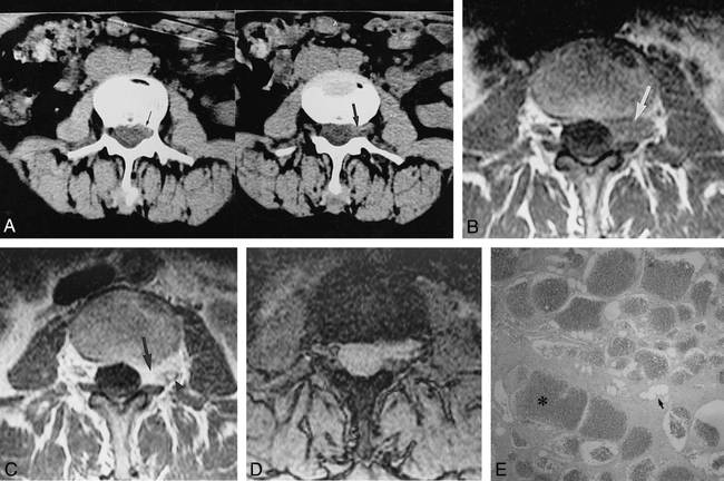
- fig 1.
Case 1: 51-year-old woman with low back pain and right-sided sciatica for 6 months.
A, CT scan of the lumbar spine shows a left-sided ventrolateral extradural mass isodense with respect to the intervertebral disk extending through the left foramen (wide arrow, right) and slightly eroding the posterior wall of the L3 vertebral body (thin arrow, left).
B, Transverse T1-weighted (660/15/3) image at L3–L4 level confirms the presence of an extradural soft-tissue mass (arrow), which is isointense relative to the intervertebral disk and extends laterally through the left intervertebral foramen.
C, Contrast-enhanced T1-weighted (570/15/3) image at the same level shows intense and homogeneous enhancement of the extradural lesion (arrow). The left L3 nerve root is slightly displaced posteriorly by the infiltrating mass (arrowhead).
D, Gradient-echo (660/12/1; 25°) image shows the lateroforaminal mass to be isointense with respect to the contents of the adjacent dural sac.
E, Photomicrograph shows the typical findings of cavernous hemangioma. Note many dilated blood-filled vessels lined with flattened endothelium (asterisk), without muscular layer, immersed in stroma that contains adipose tissue (arrow). No fresh or organized interstitial hemorrhage was observed (hematoxylin-eosin, original magnification ×40).
- fig 2.
Case 2: 16-year-old boy with left leg pain for 1 year.
A and B, Sagittal MR images show a well-defined ovoid extradural mass (arrow) with intermediate signal intensity on T1-weighted (500/15/3) image (A) and high signal intensity on T2-weighted (5000/90/2) image (B).
C, Transverse T1-weighted (660/15/3) image through L5 level confirms the location of the left ventrolateral extradural mass (arrow). At surgery, a cavernous hemangioma was found and resected.
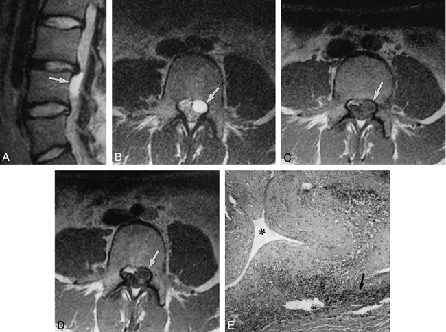
- fig 3.
Case 3: 19-year-old woman with 3-month history of left-sided radiculopathy and pollakiuria.
A, Left parasagittal T2-weighted (5000/90/2) image shows the ovoid shape of the mass (arrow), which is hyperintense with respect to CSF in the thecal sac. Note the bulging disk at L3–L4 level.
B, Transverse T2-weighted (5000/90/2) image shows a well-demarcated uniformly hyperintense lesion (arrow) without remodeling of the posterior vertebral body.
C, Transverse T1-weighted (802/12/4) image at the superior aspect of L4 shows an extradural mass occupying the left lateral recess (arrow) whose signal intensity is slightly greater than that of the contents of the adjacent dural sac.
D, Contrast-enhanced T1-weighted (802/12/4) image shows no significant changes in the signal intensity of the extradural lesion.
E, Pathologic findings of arteriovenous hemangioma included a large vascular element (asterisk) with thickened wall in association with interstitial hemosiderin deposits (arrow) and hemorrhagic foci (hematoxylin-eosin, original magnification ×100).


