
Article Figures & Data
Figures
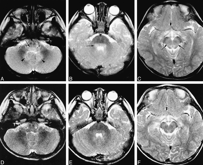
- fig 1.
Male patient, 4 years old. Acute EV 71 encephalitis. Patient presented with HFMD on June 10, 1998. Two days later, patient developed somnolence, tachycardia, and tachypnea. All MR images are T2-weighted images (3567/120/6 [TR/TE/excitations]).
A, Hyperintense lesions in the posterior portion of the medulla oblongata (arrow) and the bilateral dentate nuclei (arrowheads) of the cerebellum.
B, Hyperintense lesions in the posterior portion of the pons (arrow).
C, Hyperintense lesions in central-most portion of the midbrain (arrows).
D–F, Patient completely recovered without any sequelae. Follow-up MR imaging on July 29, 1998. The hyperintense lesions in the medulla, pons, midbrain, and dentate nuclei had disappeared. (The mild high-signal intensity of the posterior portion of the pons is normal in infants, possibly because of its under-myelinated status.)
- fig 2.
Female patient, 10 months old. Chronic stage of EV 71 encephalomyelitis. Patient presented with HFMD on June 20, 1998. Two days later, patient developed somnolence, tachycardia, tachypnea, and coma. Patient recovered very slowly, awaking in September. Patient remained ventilator- and oxygen-dependent. MR imaging was performed on September 22, 1998. All images are T2-weighted images (3560/120/6 [TR/TE/excitations]).
A, In sagittal sections, the lesions appear as a linear high signal in the posterior portions of the pons and medulla oblongata (arrows) and in the whole cervical spinal cord (white arrows).
B, Two symmetrical, well-defined hyperintense lesions in the cervical spinal cord (arrows), corresponding to the locations of ventral horns of the spinal cord.
- fig 3.
C (T1-weighted image) and D (T2-weighted image), Discrete lesions also were seen in the posterior aspect of the pons (arrows) and dentate nuclei (arrowheads). The nuclei of V, VI, VII, IX nerves were destroyed.
E and F (T1-weighted images), Most of the midbrain was destroyed, including the red nuclei, substantia nuclei, the medial lemniscus, and the nuclei of III and IV nerves.
G (T1-weighted image), Lesions are noted in the bilateral thalami (short arrows) and the putamina (long arrows).
H (T1-weighted image), Two symmetrical lesions within the cervical spinal cord (arrows), corresponding to the locations of the ventral horns.
Female patient, 22 months old. Chronic stage of EV 71 encephalomyelitis. Patient presented with HFMD on July 17, 1998. Two days later, she developed a sudden consciousness change, tachycardia, tachypnea, then coma. MR imaging was performed on October 16, 1998, while the patient was in a vegetative state and respirator-dependent. T2-weighted images (3567/120/4 [TR/TE/exciations]) and T1-weighted images (553/20/2) were performed.
A and B, Very low-signal intensity lesions on T1-weighted image (A) and bright on T2-weighted image (B) in the posterior portion of the medulla oblongata (arrows). The dorsal nucleus of the vagus nerve, the nuclei of the solitary tract, and the medial longitudinal fasciculus were destroyed. There were fluid accumulations in the bilateral mastoids, reflecting mastoiditis.


Tables

MR imaging lesion sites in EV 71 encephalomyelitis

In this issue


Jump to section
Related Articles
Cited By...
- A Single Mutation in the VP1 of Enterovirus 71 Is Responsible for Increased Virulence and Neurotropism in Adult Interferon-Deficient Mice
- MRI Findings in Children with Acute Flaccid Paralysis and Cranial Nerve Dysfunction Occurring during the 2014 Enterovirus D68 Outbreak
- Walking unsteadily: a case of acute cerebellar ataxia
- A Non-Mouse-Adapted Enterovirus 71 (EV71) Strain Exhibits Neurotropism, Causing Neurological Manifestations in a Novel Mouse Model of EV71 Infection
- Enterovirus 71: Emerging Central Nervous System Pathogen?!
- Survival after pulmonary edema due to enterovirus 71 encephalitis
- Acute Flaccid Paralysis in Infants and Young Children with Enterovirus 71 Infection: MR Imaging Findings and Clinical Correlates