
Article Figures & Data
Figures
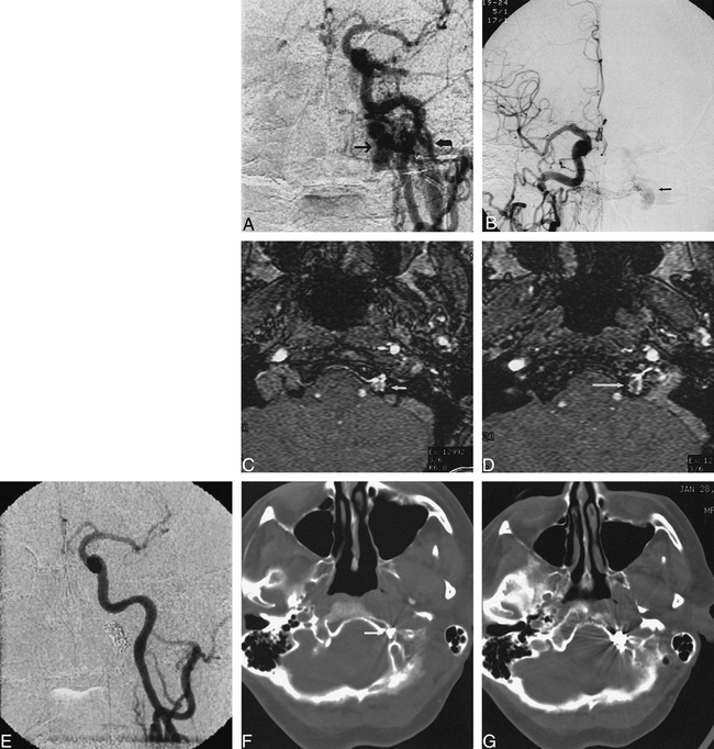
- fig 1.
Case 1.
A, Anteroposterior view of left common carotid artery demonstrates a DAVF at skull base. Fistula was located in superior aspect of dilated anterior condylar vein (arrow), medial to jugular bulb (curved arrow).
B, Anteroposterior view of right common carotid artery demonstrates fistula (arrow) medial to jugular bulb. This view was used to direct transvenous embolization to exact site of the fistula.
C, MR angiography source image from three-dimensional time-of-flight sequence (54/9/1 [TR/TE/excitations]) at level of hypoglossal canal shows abnormal flow signal in left hypoglossal canal (arrow).
D, MR angiography source image from three-dimensional time-of-flight sequence (54/9/1) at level of junction of sigmoid sinus and jugular bulb demonstrates abnormal intraosseous flow (arrow) medial to jugular bulb.
E, Anteroposterior view of right common carotid artery following transvenous coil embolization confirms occlusion of fistula.
F, CT scan following transvenous embolization demonstrates portion of coil mass in hypoglossal canal (arrow).
G, CT scan after embolization shows coil mass to be partially intraosseous and located medial to jugular bulb.
- fig 2.
Case 2.
A, Anteroposterior view of right common carotid artery shows dural fistula in region of right jugular bulb. Note poor opacification of intracranial circulation owing to steal. Retrograde filling of the right transverse, sigmoid, straight, and superior sagital sinuses is visible.
B, Anteroposterior view of left common carotid artery demonstrates skull base DAVF located medial to jugular bulb. Fistula is located in superior aspect of dilated anterior condylar vein (arrow).
C, MR angiography source image from three-dimensional time-of-flight (42/9/1) sequence shows abnormal flow in hypoglossal canal (arrow) compatible with DAVF of anterior condylar vein.
D, MR angiography source image at level of jugular bulb shows abnormal intraosseous flow (arrow) medial to jugular bulb.
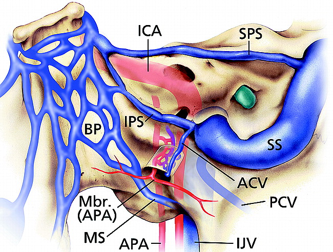
- fig 3.
Illustration of normal veins and venous sinuses of skull base demonstrates anterior condylar vein originating at junction of inferior petrosal sinus and jugular bulb and then coursing through hypoglossal (anterior condylar) canal. ICA, internal carotid artery; APA, ascending pharyngeal artery; Mbr (APA), meningeal branch of the ascending pharyngeal artery; ACV, anterior condylar vein; PCV, posterior condylar vein; BP, basilar plexus; SPS, superior petrosal sinus; IPS, inferior petrosal sinus; MS, marginal sinus; SS, sigmoid sinus; IJV, internal jugular vein. (Printed with permission from Mayfield Clinic.)


In this issue


Jump to section
Related Articles
Cited By...
- Endovascular treatment strategy, technique, and outcomes for dural arteriovenous fistulas of the marginal sinus region
- Transvenous coil embolization with intra-operative cone beam CT assistance in the treatment of hypoglossal canal dural arteriovenous fistulae
- Unique percutaneous direct puncture technique for occlusion of a hypoglossal canal dural arteriovenous fistula
- Republished: Endovascular treatment of posterior condylar canal dural arteriovenous fistula
- Endovascular treatment of posterior condylar canal dural arteriovenous fistula
- Acute subarachnoid hemorrhage in posterior condylar canal dural arteriovenous fistula: imaging features with endovascular management
- Acute subarachnoid hemorrhage in posterior condylar canal dural arteriovenous fistula: imaging features with endovascular management
- Onyx embolization of anterior condylar confluence dural arteriovenous fistula
- Onyx embolization of anterior condylar confluence dural arteriovenous fistula
- Intraosseous Cranial Dural Arteriovenous Fistula Treated with Transvenous Embolization