
Article Figures & Data
Figures
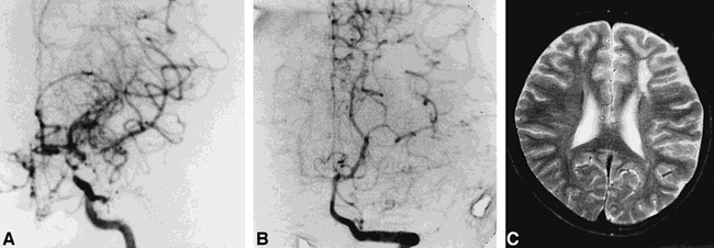
- fig 1.
12-year-old girl with an initial manifestation of transient motor weakness in right upper and lower extremities.
A, Left carotid angiogram (anteroposterior view) shows stenoocclusive changes at the terminal part of the ICA and the proximal part of the ACA and MCA. Moyamoya vessels at the base of the brain and a partial disappearance of cortical branches of the ACA and MCA are also evident (ICA stage III). A right carotid angiogram showed ICA stage III and the right PCA was also well opacified without stenoocclusive changes (PCA stage 1) (not shown).
B, Left vertebral angiogram (Towne projection) shows left PCA with no stenoocclusive changes (PCA stage 1). Good leptomeningeal collaterals to the anterior circulation are present.
C, Axial T2-weighted (3500/90/1) MR image reveals cerebral infarction in the left AWS but no abnormalities in the right.
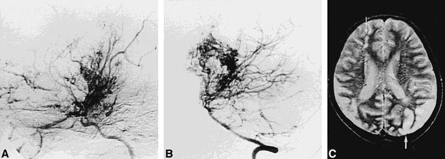
- fig 2.
3-year-old boy with an initial manifestation of transient bilateral motor weakness followed by left hemiparesis and mild right hemiparesis.
A, Anteroposterior view of a right carotid angiogram shows stenoocclusive changes at the terminal part of the ICA and the proximal part of the ACA and MCA. Well-developed moyamoya vessels around the terminal part of the ICA and a partial disappearance of cortical branches of the ACA and MCA are also evident (ICA stage III).
B, The right PCA is also opacified on lateral view of right carotid angiogram. The right PCA shows mild stenosis in its ambient segment (arrow) with delayed opacification of the right parietooccipital artery (arrowhead) (PCA stage 2). Leptomeningeal collaterals were poor at a later phase (not shown). Vertebral angiography did not fill the right PCA, and the left PCA showed no stenoocclusive changes (PCA stage 1) (not shown). A left carotid angiogram showed ICA stage II (not shown).
C, Axial T2-weighted (2500/90/1) MR image shows infarction in the right frontal (ant-MCA) (arrow) and parietal regions (post-MCA).
- fig 3.
8-year-old girl who initially presented with transient left-sided motor weakness. Examination revealed decreased visual acuity.
A, Lateral view of a right carotid angiogram shows partial disappearance of cortical branches of the ACA and MCA with well-developed moyamoya vessels at the base of the brain (ICA stage III). A left carotid angiogram also showed ICA stage III (not shown).
B, Lateral view of left vertebral angiogram shows advanced stenosis of bilateral PCAs with well-developed PCA moyamoya. Cortical branches of the PCA are partially opacified (PCA stage 3, bilaterally). Anastomoses between the PCA moyamoya and medullary arteries were well defined at a later phase (not shown).
C, Axial T2-weighted (2500/90/1) MR image reveals infarctions in the right AWS (thin arrow), bilateral post-MCAs, and left PWS (thick arrow). Infarctions in the right ant-MCA and the right PWS were also visible (not shown). No infarction in the PCA territory is evident on either side.
- fig 4.
35-year-old man who initially presented with right hemiparesis and speech disturbance at 3 years of age. Moyamoya disease was diagnosed at the same age on the basis of cerebral angiographic findings.
A, Lateral view of a left carotid angiogram shows complete occlusion of the ICA just distal to the origin of the ophthalmic artery (ICA stage VI). The ophthalmic artery is enlarged and provides collateral circulation mainly to the ACA distribution. The basal perforators are slightly dilated. A right carotid angiogram also showed ICA stage VI (not shown).
B, Lateral view of right vertebral angiogram discloses severe stenoocclusive changes of bilateral PCAs with no opacification of cortical branches (PCA stage 4).
C and D, Axial T2-weighted (3000/100/1) MR images reveal infarctions in the AWS, ant-MCA, post-MCA, PWS, and PCA territories on both sides.


Tables
 TABLE 1:
Angiographic ICA staging of stenoocclusive lesions in patients with moyamoya disease*
- TABLE 2:
Angiographic PCA staging of stenoocclusive lesions in patients with moyamoya disease
- TABLE 3:
Relationship between angiographic ICA stage and PCA stage in patients with moyamoya disease
- TABLE 4:
Relationship between ICA stage and degree of leptomeningeal collaterals from the PCA in patients with moyamoya disease
- TABLE 5:
Relationship between ICA stage and sites of infarction in patients with moyamoya disease
- TABLE 6:
Relationship between PCA stage and number of infarcted regions in patients with moyamoya disease
- TABLE 7:
Relationship between PCA stage and sites of infarction in patients with moyamoya disease


In this issue


Jump to section
Related Articles
Cited By...
- Predictive role of heterozygous p.R4810K of RNF213 in the phenotype of Chinese moyamoya disease
- Clinical and Angiographic Features and Stroke Types in Adult Moyamoya Disease
- Letter by Mugikura and Takahashi Regarding Article, "Perfusion Characteristics of Moyamoya Disease: An Anatomically and Clinically Oriented Analysis and Comparison"
- Response to Letter Regarding Article, "Perfusion Characteristics of Moyamoya Disease: An Anatomically and Clinically Oriented Analysis and Comparison"
- Moyamoya Disease in China: Its Clinical Features and Outcomes
- Posterior Circulation and High Prevalence of Ischemic Stroke among Young Pediatric Patients with Moyamoya Disease: Evidence of Angiography-Based Differences by Age at Diagnosis
- The different infarct patterns between adulthood-onset and childhood-onset moyamoya disease
- The Leptomeningeal "Ivy Sign" on Fluid-Attenuated Inversion Recovery MR Imaging in Moyamoya Disease: A Sign of Decreased Cerebral Vascular Reserve?
- Changing ischaemic lesion patterns in adult moyamoya disease
- Predominant Involvement of Ipsilateral Anterior and Posterior Circulations in Moyamoya Disease