
Article Figures & Data
Figures
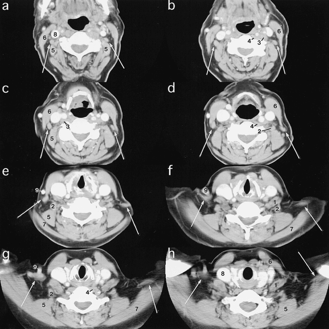
- fig 1.
A–H, CT scans obtained from superior (a) to inferior (h) in a subject with a left levator claviculae muscle (arrows). At the most superior level at which it is seen, the muscle is located lateral to the transverse process of C3 and immediately anterior and lateral to the levator scapulae (5). Its medial margin is posterolateral to the longus capitis muscle (3). As the muscle descends inferiorly, it moves laterally, lying anterior and lateral to the levator scapulae (5). The muscle moves from medial to the sternocleidomastoid muscle (6) to posterior to it. Just above its insertion, the levator claviculae muscle lies close to the trapezius (7).
Key to Abbreviations
1 Anterior scalene muscle
2 Middle and/or posterior scalene muscle
3 Longus capitis muscle
4 Longus colli muscle
5 Levator scapulae muscle
6 Sternocleidomastoid muscle
7 Trapezius muscle
8 Internal jugular vein
9 External jugular vein
n Lymph node
→ Levator claviculae muscle
- fig 2.
A–H, CT scans obtained from superior (a) to inferior (h) in the subject with bilateral levator claviculae muscles (arrows). The muscles have a similar course to the levator claviculae muscles in fig 1. On the right, the muscle lies anterior to the levator scapulae (5) and anteromedial to the trapezius (7), and appears to insert near the middle of the clavicle. On the left, the muscle has a more lateral course and insertion


